Hubbry Logo
CAD/CAM dentistryCAD/CAM dentistryMain
Open search
CAD/CAM dentistry
Community hub
CAD/CAM dentistry
logo
8 pages, 0 posts
0 subscribers
Be the first to start a discussion here.
CAD/CAM dentistry
CAD/CAM dentistry
from Wikipedia

Chrome-cobalt disc with bridges and crowns manufactured using WorkNC Dental CAD/CAM

CAD/CAM dentistry is a field of dentistry and prosthodontics using CAD/CAM (computer-aided-design and computer-aided-manufacturing) to improve the design and creation of dental restorations,[1][2] especially dental prostheses, including crowns, crown lays, veneers, inlays and onlays, fixed dental prostheses (bridges), dental implant supported restorations, dentures (removable or fixed), and orthodontic appliances. CAD/CAM technology allows the delivery of a well-fitting, aesthetic, and a durable prostheses for the patient.[3] CAD/CAM complements earlier technologies used for these purposes by any combination of increasing the speed of design and creation; increasing the convenience or simplicity of the design, creation, and insertion processes; and making possible restorations and appliances that otherwise would have been infeasible. Other goals include reducing unit cost and making affordable restorations and appliances that otherwise would have been prohibitively expensive. However, to date, chairside CAD/CAM often involves extra time on the part of the dentist, and the fee is often at least two times higher than for conventional restorative treatments using lab services.

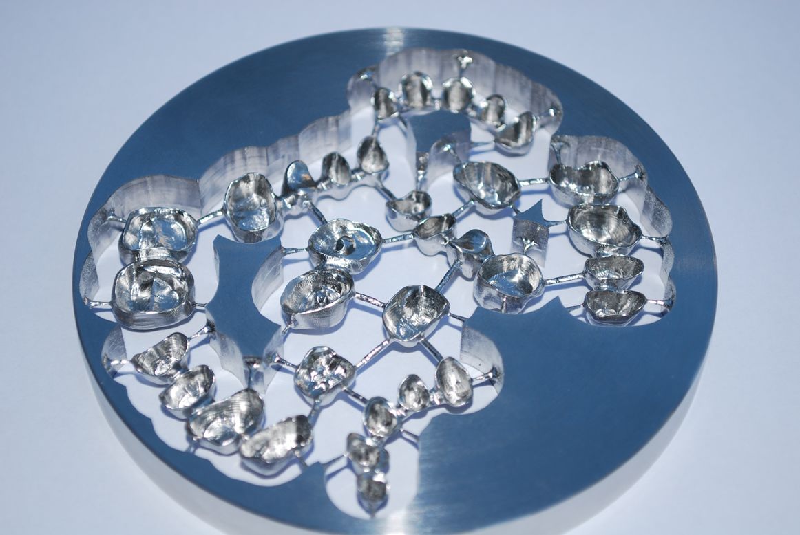
Like other CAD/CAM fields, CAD/CAM dentistry uses subtractive processes (such as CNC milling) [4] and additive processes (such as 3D printing) to produce physical instances from 3D models.

Some mentions of "CAD/CAM" and "milling technology" in dental technology have loosely treated those two terms as if they were interchangeable, largely because before the 2010s, most CAD/CAM-directed manufacturing was CNC cutting, not additive manufacturing, so CAD/CAM and CNC were usually coinstantiated; but whereas this loose/imprecise usage was once somewhat close to accurate, it no longer is, as the term "CAD/CAM" does not specify the method of production except that whatever method is used takes input from CAD/CAM,[5] and today additive and subtractive methods are both widely used.

Application of CAD/CAM in dentistry

[edit]

Computer-aided design (CAD) and computer-aided manufacture (CAM) is a process where non-digital data is captured, converted into a digital format, edited as necessary, and subsequently converted back into a physical form with the exact dimensions and materials specified during the digital design process, usually by either 3D printing or milling.[6] This set of stages is known as a "digital workflow".[7]

CAD/CAM may be used to provide a machine-led means of fabricating dental prostheses that are used to restore or replace teeth. This is an alternative to the traditional process of prosthesis fabrication using physical techniques, in which the dentist makes an impression of the site that is to be restored. This is then transported to the laboratory where a study model is made. On that model, an imitation of the final design is made using wax – known as a wax up – which represents the size and shape of the finished dental prosthesis. The wax is then encased in an investment mold, burned out and replaced with the desired material as part of lost wax casting.[8] CAD/CAM makes such procedures unnecessary for the impression is recorded digitally and the manufacture of the appliance is accompanied by additive (3D printing) or subtractive (milling) means.

Examples of dental prostheses that can be manufactured using this system include:[8]

History

[edit]

Although CAD/CAM dentistry was used in the mid-1980s, early efforts were considered a cumbersome novelty, requiring an inordinate amount of time to produce a viable product. This inefficiency prevented its use within dental offices and limited it to labside use (that is, used within dental laboratories). As adjunctive techniques, software, and materials improved, the chairside use of CAD/CAM (use within dental offices/surgeries) increased.[9] For example, the commercialization of Cerec by Sirona made CAD/CAM available to dentists who formerly would not have had avenues for using it.

The first CAD/CAM system used in dentistry was produced in the 1970s by Professor François Duret [10][11] and colleagues. The process contains a number of steps. Firstly, an optical impression of the intraoral abutment is obtained by scanning with an intra-oral digitizer. The digitized information is transferred to the monitor where a 3D graphic design is produced. The restoration can then be designed on the computer. The final restoration is then milled from a block. Professor Duret and colleagues subsequently developed the 'sopha system' however this was not widely used, perhaps lacking the accuracy, materials and computer capabilities required in dentistry.[12] The second generation of CADCAM attempted to develop this system further, but struggled to obtain occlusal morphology using an intra oral scanner, so prepared a stone model first before digitising the model.

Development of a various digitizers followed: a laser beam with a position sensitive detector sensor, a contact probe and a laser with a charged coupled device camera. Due to development of more sophisticated CAD/CAM systems both metal and ceramic restorations could be produced.[12]

Mormann and colleagues later developed a CADCAM system named CEREC, which they used to produce a type of dental restoration called an inlay. The inlay preparation is scanned using an intra-oral camera. A compact machine used chairside allowed design of the restoration from a ceramic block.[12] The major advantage of this system was the chair side approach allowing same-day restorations.[13] However, this technique was limited in that it couldn't be used for contouring or occlusal patterns. The CEREC system is used widely across the world, and studies have shown long term clinical success.[12]

The Procera system was developed by Anderson and colleagues. They used CADCAM to develop composite veneers. The Procera system later developed as a processing centre connected to satellite digitisers worldwide to produce all ceramic frameworks. This system is used around the world today.[13]

Difference from conventional restoration

[edit]

Chairside CAD/CAM restoration typically creates and lutes(bonds) the prosthesis the same day. Conventional prostheses, such as crowns, have temporaries placed for one to several weeks while a dental laboratory or in-house dental lab produces the restoration.[14] The patient returns later to have the temporaries removed and the laboratory-made crown cemented or bonded in place. An in-house CAD/CAM system enables the dentist to create a finished inlay in as little as one hour.[15] CAD/CAM systems use an optical camera to take a virtual impression by creating a 3D image which is imported into a software program and results in a computer-generated cast on which the restoration is designed.[16]

Bonded veneer CAD/CAM restorations are more conservative in their preparation of the tooth. As bonding is more effective on tooth enamel than the underlying dentin, care is taken not to remove the enamel layer. Though one-day service is a benefit that is typically claimed by dentists offering chairside CAD/CAM services, the dentist's time is commonly doubled and the fee is therefore doubled.

Process

[edit]

All CAD/CAM systems consist of a computer aided design (CAD) and computer aided manufacture (CAM) stage and the key stages can broadly be summarised as the following:

  • Optical/contat scanning that captures the intraoral or extraoral condition of the patient.
  • Use of software that can turn the captured images into a digital model to upon which a dental prosthesis can be designed and prepared for fabrication.
  • Instruction to devices that can facilitate the conversion of the design into a product by way of 3D printing or milling depending on the CAD/CAM system used.[5]
Screen shot from a CEREC CAD system, 2006

For a single unit prosthesis, after decayed or broken areas of the tooth are corrected by the dentist, an optical impression is made of the prepared tooth and the surrounding teeth. These images are then turned into a digital model by proprietary software within which the prosthesis is created virtually. The software sends this data to a milling machine where the prosthesis is milled. Stains and glazes can be added to the surfaces of the milled ceramic crown or bridge to correct the otherwise monochromatic appearance of the restoration. The restoration is then adjusted in the patient's mouth and luted or bonded in place.

Integrating optical scan data with cone beam computed tomography datasets within implantology software also enables surgical teams to digitally plan implant placement and fabricate a surgical guide for precise implementation of that plan. Combining CAD/CAM software with 3D images from a 3D imaging system means greater safety and security from any kind of intraoperative mistakes.

Computer-aided design (CAD)

[edit]

To design and manufacture a dental prosthesis, the physical space which it will replace within the mouth has to be converted into a digital format. To do this, a digital impression must be taken. This will convert the space into a digital image which must then be converted into a file extension that can be read by the CAD software system being used.[13]    

Once in a digital form, the structures within the mouth will be displayed as a 3D image. Using CAD software, the size and shape of the restoration can be virtually altered, thus replacing the wax up stage present in the traditional approach.[12][17]  

Digital impressions

[edit]

Digital impressions are a means of recording the shape of a patient's dental structures by using scanners. In CAD/CAM's infancy, desktop scanners were used which digitised study models or Dental impressions - indirect representations of the patient's dentition.[18] These devices are also known as extra oral scanners and can be contact or non-contact.[17]

Contact scanners use stylus profilometers that are placed against and run along the contours of an object. The contact of the stylus against the object is represented digitally as a set of co-ordinates (point cloud), which is analyzed by an onboard mathematical algorithm to build up a 3D image of the object (mesh).[7][19]  

Non-contact scanners capture the shape of dental structures by using optics, such as light-emitting diodes. Light is emitted from the scanner which hits the object and then reflects into an onboard sensor, usually a charge couple device (CCD) or a position sensing detector (PSD).[12] These reflections allow the scanner to build up a 3D image of the object as with contact scanners [7]  Extraoral non-contact scanners can obtain this information by different means, namely: structured light, laser light and confocal microscopy.[17]  Contact scanners are more accurate than non-contact scanners but are rarely used anymore because they are slow and their imaging is unnecessarily detailed, ten times what is required for the success of a dental prosthesis.[17]

Intra-oral scanners are a form of non-contact scanners that have grown in popularity due to their ability to digitize a patient's dentition directly in the mouth, avoiding the need for either a physical impression or a plaster study model, as is the case with extraoral scanners. This allows the fabrication of dental prostheses to be a completely digital process from the very first stage. Older scanners require a contrast powder to be placed on all the structures which were to be scanned whereas newer products do not require such a step.[17]  

Intra-oral scanners interpret reflected light to produce a 3D image representing the patient's teeth, using systems including:[20]

  • Confocal laser scanner microscopy
  • Triangulation
  • Optical coherent tomography  
  • Accordion fringe interferometry
  • Active wavefront sampling 

The file extension most recognised by CAD software is an STL file. This file type records and describes an object's geometry as a series of connected triangles, the density of which, depends upon the "resolution and the mathematical algorithm that was used to create the data".[17] Most available scanners will produce STL files however some produce proprietary file types that can only be interpreted by select CAD software.[17][20]

CAD Software

[edit]

CAD software visualises the digital impression captured by extra or intra oral scanners and provides numerous design tools. Popular software packages include Dental System, DentalCAD and CEREC.[17] Some of the most common ways in which the virtual dental prosthesis can be edited are as follows:  

  • The size and shape of restorations can be adjusted.
  • The shape of teeth is often adjusted using dental burs prior to scanning to accommodate a dental prosthesis such as a crown. This is called a preparation and the edge of this is known as the margin. Margins need to be demarcated so that the dental prosthesis finishes flush with the rest of the tooth to reduce the chances of plaque build-up under the prosthesis. Margins can be detected automatically which would normally have to be delineated by a technician visually. They can also be adjusted manually.
  • The path of insertion axis can be determined automatically which dictates the direction the dental prosthesis must move to fit into the tooth/mouth.  
  • Measurements can be made between points on the digital model which can help inform the technician if any modifications to the tooth are needed to accommodate the dental prosthesis. The material must be thick enough to provide adequate strength but also not so thick as to cause the restored tooth to contact the opposing tooth before all other teeth in the arch – this would prop the patient's mouth open and prevent them from being able to bite normally.

Materials used in computer-aided manufacture

[edit]

CAD/CAM is a rapidly evolving field, hence the materials in use are always changing. Materials that can be manufactured using CADCAM software currently include metals, porcelain, lithium disilicate, zirconia and resin materials.  CAD/CAM restorations are milled from solid blocks of ceramic or composite-resin. If pre-sintered ceramic ingots are used, subsequent sintering to reduce porosity is required and the CAD-CAM technology needs to account for any casting shrinkage during this process. Glass-based restorations can also be manufactured using CAD-CAM. Similar to ceramics, milling of glass ingots occurs and molten glass infiltration is used to reduce porosity.[21] The advantage of materials manufactured by CADCAM is the consistency in quality of restoration when mass produced.

Metals

[edit]

Metals such as CoCr and titanium can be manufactured using CADCAM software. Precious metals cannot be machined for a variety of reasons, including expense. Pre-sintered CoCr blocks are available, and requires sintering after to achieve the desired mechanical properties. This method replaces the more traditional lost-wax technique.[22]

Ceramics

[edit]

Feldspathic and leucite-reinforced ceramics

[edit]

The microstructure of feldspathic and leucite reinforced ceramics is a glassy matrix with crystalline loads. It has low flexural strength, very good optical properties and an advantageous bonding abilities. A major advantage is its good aesthetics, with a variety of shades available and high translucency. However, it is a fragile material and is susceptible to damage by occlusal forces.[22]

Lithium disilicate, zirconium oxide and lithium silicate ceramics

[edit]

Lithium disilicate, zirconium oxide and lithium silicate ceramics also have a biphasic structure with crystalline particles dispersed in a glass matrix. They have a high flexural strength, good optical properties and ability to bond. It produces highly aesthetic restorations in a variety of shades is useful as well as its high mechanical strength.[22]

Zirconia

[edit]

Zirconia has a polycrystalline structure. It has a high flexural strength. However, both its optical properties and ability to bond are weak. Its main advantage is its mechanical strength.[21] CAD-CAM processing means that polycrystalline zirconia can be utilised for copings and frameworks. Its superior mechanical properties means it can be used for long-span bridgework, cores can be produced in thinner layers and can be utilised in posterior fixed partial dentures.[21] However, the aesthetics of zirconia restorations are not as good as other types of ceramic.

Resin materials

[edit]

Three resin materials are available: resin composite, PMMA, and Nano-ceramics. PMMA is made of polymethylmethacrylate polymers with no filler. However, resin composite is composed of inorganic filler in a resin matrix. Similarly, nano-ceramic is nanoparticles embedded in a resin matrix. All three materials have a weak flexural strength and disadvantageous optical properties. However, the ability to bond is very effective. An advantage of these materials is the ability to manufacture them quickly through fast milling, so are great to used for direct composite repairs. However, the aesthetic quality of these materials limit their utility.[21]

Advantages and drawbacks

[edit]

CAD/CAM has improved the quality of prostheses in dentistry and standardised the production process. It has increased productivity and the opportunity to work with new materials with a high level of accuracy.[5]

Though CAD/CAM is a major technological advancement, it is important that dentists' technique is suited to CAD/CAM milling. This includes: correct tooth preparation with a continuous preparation margin (which is recognisable to the scanner e.g. in the form of a chamfer); avoiding the use of shoulderless preparations and parallel walls and the use of rounded incisor and occlusal edges to prevent the concentration of tension.[5]

Crowns and bridges require a precise fit on tooth abutments or stumps. Fit accuracy varies according to the CAD/CAD system utilized and from user to user. Some systems are designed to attain higher standards of accuracy than others and some users are more skilled than others. As of 2014, 20 new systems were expected to become available by 2020.[23]

Further research is needed to evaluate CAD/CAM technology compared to the other attachment systems (such as ball, magnetic and telescopic systems), as an option for attaching overdentures to implants.[24]

Advantages of CAD/CAM

[edit]

The advantages CAD/CAM provides when compared with the traditional laboratory and chairside led techniques are that it 1) allows for use of materials otherwise unavailable in the laboratory; 2) provides cheaper alternatives when compared with conventional materials; 3) decreases labour cost and time for dental technicians and 4) standardises the quality of restorations.[8][13]

Ceramic materials in particular, can be highly time-consuming to work with. To make a ceramic dental prosthesis by hand, the technician has to meticulously build up porcelain powder and sinter it onto the surface of a coping. With CAD/CAM, labour times are significantly reduced, with CAD systems with some reviews reporting that only 5–6 minutes of technician input is required to produce a dental prosthesis.[13] In this way, the cost of production is reduced because labour costs are lower. Furthermore, CAD/CAM systems mill prosthesis from blocks of material which are mass manufactured, again reducing costs for the dental offices and laboratories when compared with traditional techniques.[13] These blocks are made so that any internal porosities have been removed which are difficult to eliminate during conventional fabrication.[13]

CAD/CAM has also found great merit with regards to reducing the shrinkage which occurs when ceramics are heated during sintering – a process required to give the ceramic restoration adequate strength so it can be used successfully within the mouth. It is difficult to account for this phenomenon in a dental laboratory using traditional techniques.[13] CAM can reduce shrinkage by two different methods. The first is to produce a prosthesis just greater than the desired size. This means that on firing, the prosthesis will shrink to the original intended size.[13] The second is by milling the prosthesis from a block that has already been fully sintered, which eliminates shrinkage but causes increased wear on cutting tools because the block is stronger than when partially sintered.[13]

Benefits of intraoral scanning

[edit]

The advent of intraoral scanners affords additional advantages when compared with the traditional physical workflow, particularly for dentists. In the traditional method, dental impressions must be taken, and the materials used to facilitate this are vulnerable to distortion over time which can decrease the accuracy of the eventual dental prosthesis. These inaccuracies are compounded by subsequent steps such as the fabrication of study models based on the impressions. Intra-oral scanners rapidly digitise what they scan which removes the risk of distortion/damage to the date.[25] Furthermore, dental impressions are often discomforting for patients, particularly those who have a strong gag reflex due to the bulk of material needed to capture a patient's entire dentition.[25] Intra-oral scanners reduce this element.

Intraoral scanning saves considerable time in post processing when compared with conventional dental impressions because the 3D model can be instantly emailed to a dental laboratory, whereas with the conventional technique, the impression must be disinfected and physically transported to the laboratory which is a longer process.[25]

Disadvantages of CAD/CAM

[edit]
  • Learning curve: With any new technology there is a steep learning curve. With time and experience operators will need to understand how to work the equipment and software used for CAD/CAM technology. Initially it can be difficult to adopt a new digital workflow when operators were comfortable with using their long-standing process in dentistry. This would also mean staff would need to be trained to feel comfortable using CAD/CAM systems. 
  • Cost: digital dentistry requires a large financial investment, including buying and maintaining equipment as well as software updates.[26] However, in the long run the investment will pay off as it can save money on expenses such as laboratory fees and single use impression equipment.[27]  
  • Errors in occlusion assessment: Compared to the conventional technique of making complete dentures, CAD/CAM has a few disadvantages.[28] The systems do not accurately assess element of balanced occlusion. As the denture teeth are not set on the denture base with the assistance of an articulator, there is difficulty in achieving a balanced occlusion. Hence, human assessment is still required, and the teeth will have to be clinically remounted to achieve balanced occlusion.[29]
  • Environmental impact: resin particles are produced during the milling process, which adds to plastic pollution.[27]

Future prospects

[edit]

Digital dentistry is growing at an accelerating rate, and CAD/CAM systems will continue to evolve and improve.[30][31]

References

[edit]
Revisions and contributorsEdit on WikipediaRead on Wikipedia
from Grokipedia
CAD/CAM dentistry encompasses the integration of computer-aided design (CAD) and computer-aided manufacturing (CAM) technologies in the planning and fabrication of dental restorations and prostheses, enabling precise digital workflows from intraoral scanning to milling or 3D printing. This approach revolutionized restorative procedures by allowing for the creation of customized items such as crowns, inlays, onlays, bridges, veneers, implant abutments, and dentures with high accuracy and minimal manual intervention. Key processes involve digital impression capture via intraoral scanners, virtual design using specialized software, and subtractive (milling) or additive (3D printing) production, often facilitating chairside treatments completed in a single visit. The origins of CAD/CAM in dentistry trace back to the 1970s, pioneered by François Duret, building on industrial applications from the 1960s, with pioneering systems like CEREC introduced in 1985 by Mörmann and Brandestini for chairside ceramic restorations. Early developments addressed challenges such as rising metal costs and allergies, leading to innovations in metal-free materials; notable milestones include the Procera system (1990s) for titanium copings and the adoption of zirconia in the 2000s for enhanced strength. Over time, advancements in scanning resolution, AI-assisted design, and material science have expanded its scope beyond fixed prosthodontics to orthodontics (e.g., clear aligners) and maxillofacial prosthetics. Common materials in CAD/CAM dentistry include feldspathic and leucite-reinforced ceramics for aesthetic anterior restorations, lithium disilicate (flexural strength 360–400 MPa) for versatile crowns and bridges, high-strength zirconia (>1000 MPa) for posterior durability, and hybrid ceramics or PMMA resins for temporary or provisional uses. These materials offer superior translucency, color stability, and fracture resistance compared to traditional lab-fabricated options, with indications tailored to clinical needs—such as single-unit crowns for lithium disilicate or multi-unit bridges for zirconia. In removable prosthodontics, CAD/CAM milling of pre-polymerized PMMA reduces fabrication sessions from five (conventional) to two or three, improving fit, retention, and patient comfort while minimizing errors. The primary advantages of CAD/CAM dentistry include enhanced precision (reducing marginal gaps to <100 μm), time efficiency for same-day delivery, improved aesthetics, and better long-term outcomes, with studies showing survival rates of 90–95% for restorations over 5–10 years. It also supports digital archiving for future modifications and reduces cross-contamination risks through contactless workflows. However, challenges persist, such as high initial equipment costs (often exceeding $100,000 for chairside systems), a learning curve for clinicians, and limited long-term in vivo data, particularly in developing regions. Despite these, as of 2025, ongoing innovations in 3D printing and AI integration continue to broaden its clinical adoption.

Overview

Definition and Principles

CAD/CAM dentistry refers to the integration of computer-aided design (CAD) and computer-aided manufacturing (CAM) technologies to create dental restorations and prosthetics, such as crowns, bridges, and veneers, through digital processes that enhance precision and efficiency compared to traditional methods. In this system, CAD involves the virtual modeling of restorations using specialized software to generate three-dimensional (3D) designs based on digital scans of the patient's oral anatomy, while CAM automates the physical production of these designs from biocompatible materials like ceramics, zirconia, or resins. This approach eliminates many manual steps, reducing errors and enabling customized, anatomically accurate outcomes. The core principles of CAD/CAM dentistry revolve around the seamless integration of digital data acquisition, algorithmic design, and automated fabrication to achieve high levels of precision, repeatability, and biocompatibility. The process begins with 3D scanning to capture detailed topographical data of teeth or impressions, which is then processed by software algorithms that facilitate virtual restoration design, ensuring optimal fit, occlusion, and aesthetics. Manufacturing occurs via subtractive methods, such as computer numerical control (CNC) milling that removes excess material from solid blocks, or additive techniques like 3D printing that layer materials sequentially; these methods prioritize materials with proven biocompatibility, such as zirconia with flexural strengths of 500-1200 MPa, to ensure long-term durability in the oral environment. Precision is a hallmark, with vertical misfits often limited to 2.5-3.2 μm and rotational discrepancies under 3°, while repeatability allows consistent production across multiple units without variability from human technique. The fundamental components form a cohesive hardware-software ecosystem: intraoral scanners or optical sensors acquire digital impressions without physical molds, design software (e.g., CAD programs) enables clinicians or technicians to model restorations virtually, and fabrication devices like milling machines or 3D printers execute the CAM instructions to produce the final prosthesis. This ecosystem has evolved from standalone laboratory-based systems, which required outsourcing and multiple visits, to integrated chairside setups that combine all elements in the dental office, facilitating same-day restorations and improving patient satisfaction through reduced turnaround times.

Applications

CAD/CAM technology in dentistry primarily facilitates the fabrication of indirect restorations, enabling precise, patient-specific designs that improve fit, aesthetics, and longevity compared to traditional methods. Key applications include single crowns, which are milled from digital scans for marginal adaptation and reduced chair time; bridges, allowing multi-unit spans with seamless connector designs; inlays and onlays for conservative posterior restorations that preserve tooth structure; veneers for anterior aesthetic enhancements with minimal preparation; implant abutments customized to optimize emergence profiles and soft tissue support; and full-mouth rehabilitations, where integrated digital planning ensures occlusal harmony across multiple units. These uses leverage CAD software for virtual modeling, followed by CAM-driven milling or printing, resulting in restorations that exhibit superior mechanical properties and patient satisfaction. In orthodontics, CAD/CAM supports the production of clear aligners and retainers through digital planning, where intraoral scans inform sequential tooth movement simulations in software like ClinCheck or 3Shape. This approach enables the fabrication of thermoformed or directly 3D-printed appliances using biocompatible resins, offering predictable force application and reduced treatment visits. Retainers, similarly designed post-treatment, maintain alignment with high precision, minimizing relapse risks. For implantology, CAD/CAM excels in creating custom surgical guides from CBCT-integrated models, guiding precise implant placement to avoid vital structures and enhance prosthetic outcomes. Provisional restorations, milled chairside, provide immediate function and aesthetics during osseointegration, supporting soft tissue contouring. These applications reduce surgical errors and improve implant success rates through digitally verified positioning. In prosthodontics, particularly for edentulous patients, CAD/CAM produces denture bases and frameworks with enhanced retention and stability, often using milled PMMA for porosity-free results. Frameworks for removable partial dentures are designed virtually to distribute forces evenly, improving comfort over cast metal alternatives. As of 2025, emerging applications include full digital dentures, where CAD/CAM workflows with virtual try-ins and milled bases reduce adjustments and enable rapid replication from archived files, benefiting complex cases like those in elderly patients. Orthodontic appliances, such as lingual retainers and functional devices, are increasingly fabricated via CAD/CAM for customized biomechanics and material efficiency.

Historical Development

Early Innovations

The origins of CAD/CAM dentistry trace back to the early 1970s, when French dentist François Duret conceived the idea of using optical impressions to create digital models of dental structures, aiming to replace traditional physical molds with computerized processes. In 1973, Duret completed his thesis titled "Empreinte Optique," which explored holographic and optical imaging techniques for capturing dental anatomy, laying the theoretical foundation for automated prosthesis design. This work marked the first application of computer-aided technologies to restorative dentistry, focusing on precision and reproducibility in crown fabrication. By the early 1980s, Duret advanced these concepts into practical innovations, filing initial patents in 1980 for methods of prosthesis production using CAD/CAM systems and presenting the first prototype of a CAD/CAM-generated dental crown in September 1983 at a French dental congress. This prototype, part of the Sopha BioConcept system, utilized subtractive milling to shape restorations from ceramic blocks based on digitized impressions, demonstrating the feasibility of computer-controlled manufacturing for inlays and crowns. Concurrently, in 1985, Swiss researchers Werner Mörmann and Marco Brandestini introduced the CEREC 1 system, the first commercially available chairside CAD/CAM unit, which employed an intraoral optoelectronic camera for 3D data acquisition and subtractive milling of feldspathic ceramic for posterior inlays. The system's debut treatment occurred on September 19, 1985, at the University of Zurich, shifting initial fabrication from dental laboratories to the operatory. During the 1980s and 1990s, key milestones included the evolution of software from basic 2D modeling in CEREC 1, which relied on sectional images for simple inlay designs, to 3D reconstruction in CEREC 2 launched in 1994, enabling full crown and veneer creation with improved occlusal analysis. Another significant development was the Procera system, introduced in the late 1980s by Matts Andersson and commercialized in the 1990s, which used CAD/CAM for high-precision fabrication of titanium copings and later ceramic structures, addressing needs for durable, biocompatible frameworks in fixed prosthodontics. This progression facilitated the transition from laboratory-dependent workflows—where digitized models were sent for milling—to more integrated chairside processes, though early systems like Duret's Sopha remained primarily lab-oriented. Subtractive milling became the dominant manufacturing method, using diamond burs to carve restorations from pre-sintered blocks, offering superior fit accuracy of 50-100 micrometers compared to conventional techniques. Despite these breakthroughs, early CAD/CAM systems faced significant limitations, including prohibitively high costs for equipment and materials, which restricted adoption to well-funded practices and laboratories until the 2000s. Additionally, the steep learning curve for software operation and the need for manual occlusal adjustments, coupled with limited material options like feldspathic porcelain, confined applications to single-unit restorations rather than complex prosthetics. Fabrication remained largely lab-based for most users due to the bulkiness and expense of chairside units, slowing widespread clinical integration.

Modern Advancements

In the 2000s, CAD/CAM dentistry saw widespread adoption of intraoral scanners, marking a significant shift from traditional impression techniques to digital data acquisition, alongside the introduction of high-strength materials like zirconia for enhanced durability in posterior restorations. The iTero intraoral scanner, introduced in early 2007 by Align Technology, represented a pivotal advancement, enabling powder-free, high-resolution optical impressions that integrated seamlessly with orthodontic and restorative workflows. This period also featured a transition to open-system architectures, exemplified by the iTero system's compatibility with third-party software like Invisalign, which promoted interoperability and reduced reliance on proprietary closed systems. These developments enhanced clinical efficiency by minimizing physical impressions and associated errors, laying the groundwork for broader digital integration in dental practices. The 2010s brought the integration of additive manufacturing, or 3D printing, into CAD/CAM workflows, expanding fabrication options beyond subtractive milling to include resins and metals for prosthetics and surgical guides. Technologies such as stereolithography and digital light processing enabled precise layering of biocompatible materials, with early applications in producing temporary restorations and models as early as 2012. Hybrid CAD/CAM approaches emerged, combining additive and subtractive methods to optimize production; for instance, 3D printing for initial prototyping followed by milling for final metal frameworks, which improved material utilization and customization in implant dentistry. These innovations addressed limitations in traditional milling, such as waste generation, and supported more complex geometries unattainable with single-modality systems. By the 2020s, AI-assisted design tools revolutionized CAD/CAM processes, particularly through automated margin detection that streamlines crown and bridge preparation. Systems like 3Shape's Automate, introduced in 2021, use machine learning algorithms to identify preparation margins from intraoral scans with minimal manual input, reducing design time by up to 70% while maintaining clinician oversight for occlusion and contacts. Cloud-based collaboration platforms, such as Dentsply Sirona's DS Core launched in 2022, facilitated real-time data sharing between clinics and labs, enabling remote design reviews and manufacturing coordination via secure, AI-enhanced ecosystems. Intraoral scanner accuracy also advanced, achieving trueness levels of 15-20 microns for full-arch impressions, supported by enhanced optics and AI-driven mesh processing that corrects distortions in real time. These modern advancements have profoundly impacted clinical practice, reducing chair time from multiple days across visits to a single appointment of several hours for restorations like crowns. The global dental CAD/CAM market, valued at USD 2.17 billion in 2024, is projected to reach USD 4.61 billion by 2032, driven by these efficiency gains and digital adoption in emerging markets.

Comparison to Conventional Dentistry

Key Differences

CAD/CAM dentistry distinguishes itself from conventional analog methods primarily through enhanced accuracy in capturing and reproducing dental structures. Digital intraoral scanners used in CAD/CAM workflows achieve marginal gaps of approximately 60 μm for crowns, significantly lower than the 78 μm observed with conventional impression techniques, leading to better pre-cementation fit. Studies on full-arch impressions show mixed results, with conventional methods exhibiting trueness around 16-20 μm but precision sometimes limited by material distortions exceeding 100 μm in complex cases; CAD/CAM systems often achieve comparable or better precision (below 50 μm in some studies) through optical scanning, though trueness can vary. This precision minimizes adjustments during placement and improves long-term restoration success. Time efficiency represents another fundamental difference, as CAD/CAM enables same-day restorations via in-office scanning, design, and milling, often completing the process in under two hours excluding milling time. In contrast, conventional dentistry requires multiple patient visits—typically two or three—spanning one to three weeks, including impression taking, laboratory fabrication, and fitting. Although in vitro comparisons indicate that full chairside CAD/CAM workflows may take 43-58 minutes compared to 10 minutes for conventional impression alone, the elimination of lab turnaround and interim temporaries substantially reduces overall treatment duration and patient inconvenience. Customization is markedly advanced in CAD/CAM, employing parametric design software that generates patient-specific restorations tailored to individual anatomy, occlusion, and aesthetics, far surpassing the reliance on standardized prefabricated molds or manual wax-ups in conventional approaches. This digital parametric modeling allows iterative adjustments in virtual environments for optimal fit and function. The cost structure of CAD/CAM involves a substantial upfront investment in equipment and software, often ranging from $75,000 to $200,000, but yields lower per-unit costs—saving approximately $175 in lab fees per restoration—once amortized over high-volume use. Conventional methods, by comparison, avoid initial capital outlay but accumulate ongoing laboratory charges for each case, potentially increasing long-term expenses for practices handling frequent indirect restorations. As of 2025, full chairside systems typically cost $40,000–$100,000, with subscription models (e.g., $100–$230/month) influencing ROI, which remains 12–24 months in high-volume practices. Quality control in CAD/CAM benefits from automated digital verification processes, such as 3D scanning and similarity measures, which ensure consistent morphological accuracy and detect deviations in real-time, mitigating the artisan variability and human error prevalent in manual conventional workflows.

Transition Factors

The transition to CAD/CAM dentistry from conventional methods involves several practical considerations that influence adoption rates in clinical practices. Key factors include the need for specialized training, infrastructure investments, economic viability, regulatory compliance, and patient perceptions, each presenting both barriers and facilitators to implementation. Training requirements represent a primary barrier, as both dentists and dental technicians must acquire proficiency in CAD/CAM software for design tasks and hardware operation for scanning and milling. Specialized courses, often conducted by experienced dentists and technicians, focus on digital workflows using platforms like 3Shape or exocad, enabling users to integrate these tools into daily practice. In educational settings, curricula in countries such as Japan and South Korea incorporate CAD/CAM modules, but national examinations still emphasize traditional skills, necessitating post-graduation upskilling through clinical training or vendor-provided programs to bridge the gap. This ongoing education is essential for accurate digital impression capture and restoration fabrication, with hands-on sessions typically lasting from several days to weeks depending on prior experience. Infrastructure demands further complicate the shift, requiring dental practices to equip with intraoral scanners, milling machines, and compatible computing systems to support end-to-end digital workflows. Scanners, such as powder-free models compatible with STL file formats, capture oral data without traditional trays, while in-office mills process materials like zirconia or composites on-site, often necessitating dedicated space and utilities like compressed air. IT integration is critical, involving HIPAA-compliant networks for secure file transmission to laboratories, cybersecurity measures, and software interoperability to avoid proprietary silos that hinder collaboration. Modular setups, including laptops and backup systems, allow scalability for small practices, but initial configurations can cost $60,000–$100,000, emphasizing the need for vendor support in installation and maintenance. Economic factors play a pivotal role in adoption, with return on investment (ROI) calculations balancing high upfront costs against long-term savings from reduced laboratory outsourcing and improved efficiency. For instance, in-office milling systems can achieve payback periods of 12–24 months through eliminated lab fees and shorter chairside times, particularly in high-volume practices producing 20 or more restorations monthly, where annual net profits may exceed $60,000 after covering material and leasing costs of approximately $40 per unit and $1,800–$2,500 monthly. These gains are amplified in settings with steady patient flow, as digital processes enable same-day deliveries, enhancing practice revenue without proportional increases in overhead. Regulatory aspects have facilitated broader acceptance since the 2010s, with the FDA classifying optical impression systems for CAD/CAM restorations as Class II devices under 21 CFR 872.3661 and exempting them from premarket notification requirements to streamline approvals. Increased FDA scrutiny on CAD/CAM-milled implants and prosthetics began in the early 2010s, prompting laboratories to ensure compliance through validated software and materials, which has standardized digital workflows while maintaining safety standards. Patient acceptance serves as a key facilitator, driven by the shift to digital impressions that minimize discomfort compared to conventional methods using viscous trays and materials. Systematic reviews indicate significantly higher satisfaction (standardized mean difference of 0.55) and reduced discomfort (SMD of -2.24), including lower nausea and anxiety, leading to greater willingness for repeat treatments and recommendations. This comfort improvement, alongside faster procedures, encourages patient uptake of CAD/CAM options, indirectly supporting practice transitions by boosting case acceptance rates.

Workflow

Data Acquisition

Data acquisition in CAD/CAM dentistry involves capturing precise digital representations of the patient's oral anatomy as the foundational step in the digital workflow, enabling subsequent design and fabrication of restorations. This process primarily utilizes optical scanning technologies to generate three-dimensional (3D) surface models, replacing traditional physical impressions and minimizing errors associated with material distortion. Intraoral scanning employs handheld devices, such as those based on confocal microscopy, to directly capture 3D images of teeth, gums, and surrounding structures within the mouth. Confocal laser scanning, a widely adopted technique, projects a focused light beam to measure distances at multiple focal planes, reconstructing high-resolution surface topography without physical contact. These scanners offer significant advantages over conventional alginate impressions, including reduced patient discomfort, elimination of impression material shrinkage, and faster acquisition times, with studies demonstrating comparable or superior accuracy in ideal conditions. For instance, intraoral scanners avoid distortions from polymerization or tray inaccuracies inherent in physical molds, enhancing overall workflow efficiency. Extraoral scanning complements intraoral methods by digitizing physical models or impressions created in the laboratory using desktop scanners, which employ similar optical principles like structured light or laser triangulation to produce virtual casts. These lab-based systems are particularly useful for verifying or augmenting intraoral data, achieving clinically acceptable trueness values below 50 μm in controlled settings. Key techniques in scanning include powder-free approaches, as in confocal systems, which rely on inherent surface reflectivity for imaging without additional coatings, contrasting with older powder-based methods that apply titanium oxide to improve contrast but risk uneven application and residue. Modern intraoral scanners also provide real-time visualization on integrated screens, allowing immediate feedback and adjustments, while advanced algorithms enable stitchless imaging by seamlessly merging overlapping frames without manual intervention. Accuracy in data acquisition is critical, with intraoral scanners typically achieving resolutions of 5-20 microns and precision errors under 15 μm for short-span scans, though full-arch captures may deviate up to 100 μm due to factors like soft tissue movement or saliva interference. Common errors include distortions from patient motion or wet environments, which can compromise model fidelity if not mitigated during scanning. By 2025, AI-enhanced processing has emerged to address these challenges, with algorithms automatically detecting and reducing artifacts such as glare or tissue deformation in scan data, improving overall trueness in preliminary evaluations. These integrations, seen in systems like AI-driven intraoral scanners, facilitate cleaner datasets for downstream applications without extending chairside time.

Design Process

The design process in CAD/CAM dentistry transforms intraoral scan data into detailed digital blueprints for restorations, such as crowns and bridges, using computer-aided design (CAD) software to ensure precision, esthetics, and functionality. This phase follows data acquisition and focuses on virtual modeling, where clinicians or technicians manipulate 3D models to replicate natural tooth anatomy while accounting for occlusion and fit. Proprietary software like exocad and 3Shape leads the market due to their intuitive interfaces, dental-specific libraries, and seamless integration with scanners and mills, supporting workflows from single crowns to full-arch designs. In contrast, open-source options such as Blender equipped with dental add-ons (e.g., Blenderfordental) enable cost-free customization but require more manual setup for specialized tasks like implant guides. Virtual articulators, a key feature in tools like exocad, simulate jaw movements by importing physical articulator data and adjusting parameters such as condylar inclination (up to 60°) and Bennett angle (up to 15°), allowing dynamic occlusion analysis to prevent interferences during mastication. The core design steps commence with margin detection, where algorithms automatically identify the finish line on the prepared tooth, ideally a 360° chamfer of 1 mm width for optimal milling accuracy. Next, selection from pre-built tooth libraries provides anatomical bases, such as shape and size variants for incisors or molars, which are adapted to the patient's scan. Anatomical modeling then refines the restoration, incorporating reductions like 1.5 mm facial and 2 mm incisal for anterior teeth or 1.5 mm axial for posteriors to maintain strength and esthetics. Finally, fit verification simulates milling paths to check internal adaptations, identifying overmilling risks via virtual swirl marks and ensuring passive seating with tolerances under 100 μm. By 2025, artificial intelligence (AI) has significantly automated aspects of the design process, with systems like 3Shape Automate generating crown proposals—including optimal occlusal thickness and marginal integrity—in as little as 90 seconds using deep learning trained on millions of scans. These AI tools segment teeth, propose shapes with morphological trueness comparable to manual methods (global deviation RMSE of 79.8 μm), and suggest functional parameters, though they process only 93% of cases without intervention and excel in routine esthetics over complex multi-unit restorations. Upon completion, the design yields STL files as the primary output—a triangulated mesh format representing the restoration's surface geometry for compatibility with computer-aided manufacturing (CAM) systems and subtractive or additive fabrication. Iterative editing refines esthetics (e.g., shade matching) and function (e.g., proximal contacts) through layer-by-layer adjustments, often revisiting occlusion simulations before export. A primary challenge in this process is the learning curve for handling complex geometries, where proprietary software like 3Shape shortens training to weeks via streamlined interfaces, while open-source tools demand months of practice to avoid meshing errors or imprecise modeling. This steep proficiency requirement can limit adoption in smaller practices, emphasizing the need for targeted education on virtual tools.

Manufacturing Process

The manufacturing process in CAD/CAM dentistry encompasses the computer-aided manufacturing (CAM) phase, where digital designs are translated into physical restorations through automated fabrication techniques. This stage involves generating tool paths from CAD files to produce precise dental prosthetics, such as crowns, bridges, and implant frameworks, using either subtractive or additive methods. The choice of method depends on material properties and clinical requirements, ensuring high accuracy with tolerances as low as 10 μm in tool positioning. Subtractive manufacturing, the predominant approach, employs computer numeric controlled (CNC) milling machines to carve restorations from solid blanks of metals or ceramics. Five-axis milling systems are widely used, allowing simultaneous multi-directional tool movements to create complex geometries and smooth surfaces, which is essential for undercuts in dental prosthetics like acrylic denture bases. Dry milling processes presintered materials to avoid heat-induced damage, resulting in quicker production but requiring compensation for 25–30% shrinkage during final firing; wet milling, involving coolant fluids, is preferred for dense ceramics like zirconia to minimize surface defects of 15–60 μm and enhance finish quality. Additive manufacturing, or 3D printing, builds restorations layer by layer and is increasingly applied for resins and temporary prosthetics due to its ability to produce intricate designs efficiently. Stereolithography (SLA) uses a laser to cure photopolymer resins point-by-point, while digital light processing (DLP) projects entire layers via light projection for faster curing, both achieving high precision for temporary crowns, bridges, inlays, and onlays with filler contents of 30–50 wt%. These vat photopolymerization techniques dominate dental applications, offering customization for patient-specific temporaries. Post-processing refines the fabricated restorations to achieve clinical functionality and esthetics. For ceramics milled in a presintered state, sintering in a furnace densifies the structure, often taking 15 minutes for zirconia to attain high flexural strength while accounting for shrinkage. Subsequent polishing reduces surface roughness to improve longevity and reduce plaque accumulation, with techniques varying by material to achieve smoothness comparable to natural enamel. Staining and glazing enhance color matching and translucency, particularly for lithium disilicate, where post-sintering adjustments minimize shade shifts from high-translucency blocks. Chairside systems, such as CEREC, enable in-office milling for single-visit restorations, integrating scanning, design, and fabrication to reduce patient appointments and achieve marginal fits superior to some conventional methods, with 5-year survival rates of 97%. In contrast, centralized laboratories handle complex cases by receiving digital files for outsourced milling, allowing access to advanced equipment but requiring multiple visits. By 2025, hybrid systems combining subtractive milling with additive printing have emerged, optimizing workflows for intricate structures like bioactive grafts and custom abutments through integrated CNC and vat photopolymerization, improving precision and material versatility in implantology.

Materials

Metals

Metallic materials play a crucial role in CAD/CAM dentistry, particularly for structural components requiring durability and load-bearing capacity, such as frameworks for removable partial dentures and implant-supported prostheses. These alloys are selected for their ability to withstand masticatory forces while integrating seamlessly with digital workflows, enabling precise milling or additive manufacturing from prefabricated blocks or powders. Common types include cobalt-chromium (Co-Cr) alloys, titanium and its alloys, and noble metal alloys like gold-based compositions, each offering distinct advantages in biocompatibility and mechanical performance. Cobalt-chromium alloys are widely used due to their high strength, hardness, and excellent corrosion resistance, which ensure long-term stability in the oral environment. These non-precious base metals exhibit superior biocompatibility, making them suitable for patients with nickel sensitivities when formulated nickel-free, and they provide robust support for frameworks and clasps in partial dentures. Titanium alloys, such as Ti-6Al-4V, offer an exceptional strength-to-weight ratio, low density, and outstanding biocompatibility, promoting osseointegration for implant abutments and full-arch hybrid prostheses. Their natural oxide layer enhances corrosion resistance, while radiolucency facilitates radiographic evaluation without obscuring underlying structures. Noble metals, including gold alloys, are prized for their malleability, tarnish resistance, and biocompatibility, allowing for precise adaptation in crowns, bridges, and inlays where marginal integrity is paramount. In CAD/CAM fabrication, these metals are primarily processed through subtractive milling from solid discs or blocks, which yields homogeneous structures with minimal internal defects and high precision for complex geometries. Hybrid approaches combine milling with traditional casting, particularly for Co-Cr, to optimize fit while leveraging the alloy's castability. Titanium's fatigue resistance and low weight support its use in long-span bridges as frameworks, where it resists deformation under repeated occlusal loads, with occlusal surfaces typically veneered for wear protection; clinical studies report prosthetic survival rates exceeding 95% over 10 years for implant-supported designs. Co-Cr alloys similarly demonstrate high wear resistance, outperforming some alternatives in high-load scenarios due to their hardness. Despite their mechanical advantages, metallic materials in CAD/CAM dentistry are limited by inherent opacity, which compromises esthetics in visible areas and often necessitates veneering with porcelain or resin to mimic natural tooth translucency. This additional layering can introduce risks of debonding or chipping at the metal-ceramic interface if not properly managed. As of 2025, advancements in metal additive manufacturing, particularly laser powder bed fusion for titanium alloys, have improved the production of custom abutments with micron-level precision and enhanced osseointegration through tailored porosity. These innovations reduce peri-implant complications and enable patient-specific designs for hybrid dentures, achieving fit deviations under 50 microns.

Ceramics

Ceramic materials play a pivotal role in CAD/CAM dentistry due to their ability to combine aesthetic appeal with functional durability, particularly for indirect restorations such as crowns and veneers. These materials are primarily glass-based or polycrystalline, offering superior biocompatibility and optical properties that mimic natural dentition, making them ideal for anterior and posterior applications where esthetics are paramount. Key subtypes of ceramics used in CAD/CAM systems include feldspathic porcelain, leucite-reinforced glass ceramics, lithium disilicate, and high-strength zirconia variants. Feldspathic porcelain, known for its fine-grained structure, provides exceptional translucency suitable for veneers and thin facings in anterior restorations. Leucite-reinforced glass ceramics enhance strength while maintaining light-scattering properties that replicate the natural opalescence of teeth, with flexural strengths typically around 150 MPa, making them appropriate for inlays, onlays, and single crowns. Lithium disilicate ceramics offer a balance of translucency and higher flexural strength (approximately 360-500 MPa), enabling their use in both anterior and posterior crowns where bondability to resin cements is crucial for retention. High-strength zirconia, a polycrystalline ceramic, provides the highest flexural strength in the range of 900-1500 MPa among these subtypes, though its earlier opacity has been improved in translucent formulations for anterior use without compromising structural integrity. Hybrid ceramics, such as polymer-infiltrated ceramic networks (e.g., VITA Enamic), combine a ceramic skeleton infiltrated with polymers to achieve flexural strengths of 150-200 MPa, offering shock absorption and lifelike aesthetics for crowns, inlays, and onlays in load-bearing areas. Fabrication of these ceramics in CAD/CAM workflows typically involves milling from pre-sintered blocks, followed by a crystallization or sintering step to achieve final density and properties; this process is particularly suited for producing precise veneers and crowns with minimal material waste. For instance, lithium disilicate blocks are milled in a partially crystallized state (lithium metasilicate) for machinability, then heat-treated to convert to the stronger disilicate phase, ensuring smooth margins and accurate fit. Zirconia follows a similar pre-sintered milling approach, with post-processing sintering to densify the material, often referenced in the broader manufacturing process for its role in enhancing mechanical performance. Recent advancements as of 2025 include multilayered zirconia ceramics with composition gradients to achieve natural esthetic transitions, such as varying translucency and color shades within a single restoration, improving mimicry of vital tooth gradients for anterior applications. These multilayered blocks reduce the need for manual veneering, streamlining CAD/CAM production while enhancing optical depth. Ceramics exhibit excellent biocompatibility, characterized by low plaque affinity due to their smooth, non-porous surfaces post-polishing, which minimizes bacterial adhesion compared to metallic alternatives. Their natural tooth-like appearance, stemming from inherent translucency and color stability, further supports soft tissue health and long-term gingival adaptation in clinical settings.

Polymers

Polymeric materials play a crucial role in CAD/CAM dentistry, particularly for provisional restorations and flexible frameworks, offering advantages in biocompatibility, lightweight design, and ease of fabrication compared to more rigid alternatives. These materials are primarily used for short-term applications due to their mechanical limitations, enabling rapid production of temporary crowns, bridges, and diagnostic aids through milling or additive manufacturing processes. In CAD/CAM workflows, polymers like acrylics and resins facilitate chairside customization, reducing patient wait times and supporting transitional phases in treatment planning. Key types of polymers include polymethyl methacrylate (PMMA), also known as acrylics, which are widely employed for temporary restorations such as provisional crowns and denture bases due to their established use in prosthetic applications. Composite resins, consisting of ceramic-filled polymer matrices, serve as versatile options for inlays, onlays, and veneers, providing improved esthetics and repairability in CAD/CAM systems. Flexible polyetheretherketone (PEEK) is utilized for frameworks in removable partial dentures (RPDs), offering high biocompatibility and elasticity for patient-specific designs fabricated via direct milling of blanks. These polymers exhibit favorable properties for CAD/CAM processing, including ease of milling and 3D printing, high polishability for smooth surfaces, and cost-effectiveness relative to ceramics or metals, making them suitable for high-volume provisional work. Their flexural strength typically ranges from 50 to 200 MPa, adequate for short-term use but insufficient for long-term load-bearing without reinforcement. In CAM applications, 3D printing techniques such as vat photopolymerization enable the production of precise surgical guides and orthodontic aligners from these materials, enhancing accuracy in implant placement and alignment corrections. Recent innovations as of 2025 include bioactive resins incorporating fluoride-releasing mechanisms, which promote remineralization and antibacterial effects in provisional restorations by gradually releasing ions like fluoride and calcium to mimic natural tooth responses. These advancements address limitations in traditional polymers, potentially extending their utility in caries-prone patients through controlled ion elution. Despite these benefits, polymers in CAD/CAM dentistry are susceptible to drawbacks such as wear from occlusal forces and color instability when exposed to staining agents like coffee or red wine, which can lead to surface degradation and aesthetic changes over time. These issues necessitate careful patient selection and maintenance protocols to mitigate premature failure in provisional settings.

Benefits and Limitations

Advantages

CAD/CAM dentistry enhances the precision of dental restorations, achieving marginal fits often below 100 microns, with some systems demonstrating gaps as low as 19-27 microns, which minimizes gaps that could lead to bacterial infiltration and subsequent failures. This accuracy reduces the need for remakes, as digital workflows allow for immediate verification and adjustments during design, leading to fewer postoperative corrections compared to traditional methods. Furthermore, the improved fit contributes to greater longevity of restorations, with lithium disilicate CAD/CAM crowns exhibiting survival rates of 90.7% to 96.6% over five years, outperforming many conventional materials due to consistent material properties and reduced stress concentrations. The efficiency of CAD/CAM systems is particularly evident in chairside delivery, enabling single-visit restorations that eliminate the need for laboratory involvement and cut the number of patient appointments by approximately 47%, from an average of 5.7 to 3 sessions for complex cases like implant overdentures. This streamlined process saves clinical time, with intraoral scanning taking up to 23 minutes less than conventional impressions for single units and 13 minutes less for full arches, allowing dentists to handle more cases without compromising quality. By integrating design and fabrication in-office, CAD/CAM fosters independence from external labs, accelerating turnaround and enhancing workflow control. Patients benefit from the non-invasive nature of intraoral scanning in CAD/CAM procedures, which avoids the discomfort of traditional alginate or silicone impressions, reducing gag reflex, nausea, and overall procedure time while improving satisfaction and preference rates. Economically, despite initial equipment investments, CAD/CAM yields long-term savings through lower material waste, fewer remakes, and reduced visit frequency, with digital workflows cutting overall costs by 14.2% for implant-supported overdentures compared to conventional approaches. This standardization ensures consistent quality across restorations, minimizing variability from manual techniques. Specific to intraoral scanning, real-time visual feedback during acquisition allows clinicians and patients to review and correct scans instantly, improving accuracy and communication without physical models. Additionally, these digital methods enhance hygiene by eliminating impression materials that can harbor contaminants, reducing aerosol generation, and enabling easy sterilization of scanner tips, thereby lowering infection risks in clinical settings.

Disadvantages

One significant barrier to the adoption of CAD/CAM dentistry is the high initial cost of equipment and ongoing maintenance, with complete systems such as chairside mills and scanners often costing $80,000 to $150,000 or more, making them financially unfeasible for low-volume or small practices. Maintenance expenses, including software updates and milling bur replacements, further escalate operational costs, deterring widespread implementation in resource-limited settings. Technical challenges in CAD/CAM systems include software glitches and connectivity issues that can disrupt workflows, such as integration failures between scanners and design software. Scan inaccuracies are particularly problematic in areas with deep subgingival margins, where factors like moisture, insufficient tissue retraction, and poor preparation design lead to incomplete or distorted digital impressions. Additionally, a steep learning curve requires extensive training—often several months—for clinicians to master scanning, design, and milling processes, resulting in initial inefficiencies and errors. Material limitations in CAD/CAM dentistry encompass the inherent brittleness of certain ceramics, such as feldspathic and leucite-reinforced types, which exhibit low fracture toughness (typically 0.8–1.5 MPa⋅m^{1/2}) and are prone to chipping or subsurface defects during milling and under occlusal stress. This brittleness restricts their use in high-load posterior restorations, where higher-strength alternatives like zirconia are preferred despite their opacity. Polymers, including PMMA-based blocks, face constraints in color stability and texture replication, showing susceptibility to staining and wear that compromises aesthetics and limits longevity to typically 1–2 years for long-term provisional uses. Accessibility issues highlight a digital divide, particularly in underserved or developing regions, where financial barriers and limited infrastructure prevent equitable adoption of CAD/CAM technologies. As of 2025, the global market is valued at USD 3.1 billion, but growth is uneven, exacerbating challenges in low-resource areas. These systems' dependency on reliable power supplies and IT infrastructure exacerbates challenges in areas with inconsistent electricity or internet, hindering their use in rural or low-resource dental practices. Data privacy risks associated with cloud-based CAD/CAM platforms persist, where patient scan data transmission raises potential for breaches and unauthorized access despite encryption protocols. Over-reliance on automation in these systems can also lead to undetected errors in design or fabrication if clinician oversight is inadequate, amplifying risks in complex cases.

Future Prospects

Emerging Technologies

Artificial intelligence (AI) is expanding in CAD/CAM dentistry to include predictive analytics for treatment planning and automated diagnostics, enabling more precise and personalized patient outcomes beyond current design applications. Predictive models analyze patient data, such as imaging and genetic factors, to forecast treatment success rates and optimize restoration designs, with potential to improve outcomes and reduce revisions in simulated scenarios. Automated diagnostics leverage machine learning to detect subtle pathologies like early caries or occlusal discrepancies from intraoral scans, achieving detection accuracies exceeding 95% in recent studies. These advancements build on existing AI tools but project toward real-time integration in CAD software for dynamic adjustments during virtual planning. Advanced manufacturing techniques, particularly multi-material 3D printing, are emerging to produce monolithic restorations that combine ceramics, metals, and polymers in a single structure, enhancing durability and aesthetics without layering interfaces. This approach allows for functionally graded materials that mimic natural tooth gradients, improving fracture resistance by 30-50% compared to traditional monolithic zirconia. Clinical trials indicate that multi-material prints achieve marginal fits under 100 microns, supporting long-term stability for full-arch prosthetics. Such innovations streamline CAD/CAM workflows by enabling direct printing from digital designs, reducing production time to hours. Integration of augmented reality (AR) and virtual reality (VR) facilitates virtual try-ins, allowing patients and clinicians to simulate restorations in real-time overlays on live video feeds or immersive environments. AR systems project prosthetic designs onto the patient's anatomy via headsets, enabling adjustments for fit and shade with sub-millimeter precision during consultations. Blockchain technology complements this by providing secure, decentralized data sharing for collaborative CAD files across dental networks, ensuring tamper-proof records and compliance with privacy regulations like HIPAA. Pilot implementations suggest improved data security in multi-provider workflows. Nanotechnology innovations include self-healing ceramics that incorporate microcapsules releasing repair agents upon cracking, extending restoration lifespan in high-stress areas like molars. These materials autonomously seal microcracks up to 50 microns, maintaining structural integrity over 5-10 years in vitro tests. Antimicrobial coatings, often silver or zinc oxide nanoparticles embedded in CAD/CAM resins, inhibit biofilm formation on restorations, reducing secondary caries risk by 70% compared to uncoated surfaces. Such coatings integrate seamlessly into 3D-printed frameworks, promoting biocompatibility without altering mechanical properties. Clinical frontiers feature robotic-assisted placement systems that execute precise implant insertions guided by CAD models, achieving angular deviations below 2 degrees and linear accuracies under 0.5 mm. These robots reduce surgeon fatigue and enable minimally invasive procedures in complex anatomies. Full-mouth digital workflows encompass end-to-end processes from scanning to milling or printing, converting edentulous arches to implant-supported prosthetics in single visits with fit discrepancies minimized to 50 microns. This streamlines CAD/CAM for comprehensive rehabilitations, enhancing predictability and patient satisfaction. The global market for CAD/CAM dentistry is projected to grow from USD 2.4 billion in 2025 to USD 5.65 billion by 2034, exhibiting a compound annual growth rate (CAGR) of 10.01% (2026-2034) per Fortune Business Insights, with alternative projections including USD 3.1 billion in 2025 to USD 6.1 billion by 2034 at 8% CAGR per Global Market Insights and USD 3.10 billion in 2025 to USD 7.48 billion by 2034 at 10.29% CAGR per Precedence Research. This expansion is driven by factors such as the rising prevalence of dental disorders and an aging global population, with the World Health Organization estimating untreated dental caries affecting 2.5 billion people and severe periodontal disease affecting 1 billion people globally, alongside projections of the global population aged 60 years and older doubling to 2.1 billion by 2050 (WHO) or aged 65+ reaching 1.6 billion (UN). In regions like Japan and North America, the aging demographic particularly accelerates demand for precise, restorative solutions enabled by CAD/CAM technologies. Adoption of CAD/CAM systems in dental practices has surged, with intraoral scanners—a key component of digital workflows—reaching 57% penetration in U.S. practices by 2025, while CAD/CAM milling is widespread in over 90% of dental laboratories but remains growing in in-office settings. Globally, adoption shows disparities, with North America and Europe leading at higher rates due to advanced infrastructure, compared to slower uptake in Asia-Pacific and Latin America due to economic and training barriers. Integration trends emphasize enhanced collaboration through cloud-based platforms, such as Medit ClinicCAD, which enable seamless data sharing between dentists and labs for real-time design reviews and workflow optimization. Interoperability is supported by standards like ISO 18618:2022, an XML-based format for transferring dental case and CAD/CAM data, alongside common file formats such as STL and PLY, facilitating cross-system compatibility without proprietary lock-in. These developments promote ecosystem-wide efficiency, reducing turnaround times by up to 75% in collaborative workflows. Sustainability benefits arise from CAD/CAM's shift to digital processes, which minimize material waste compared to analog methods—traditional impressions generate significant disposable waste, while digital scanning eliminates this, reducing environmental impact in restorative procedures. Additionally, the adoption of eco-friendly materials, such as recyclable polymers and biocompatible ceramics, aligns with broader green dentistry initiatives, lowering the carbon footprint of production and disposal. Policy influences are bolstering growth through expanded reimbursements for digital procedures; in the U.S., evolving Current Dental Terminology (CDT) codes for CAD/CAM restorations and digital impressions, updated in 2025, facilitate insurance coverage, with favorable policies in North America promoting reimbursements for integrated digital workflows compared to analog alternatives. Government incentives in Europe and Asia further encourage adoption by subsidizing training and equipment for sustainable digital transitions.

References

Add your contribution
Related Hubs
User Avatar
No comments yet.