Community hub
Recent from talks
Knowledge base stats:
Talk channels stats:
Members stats:
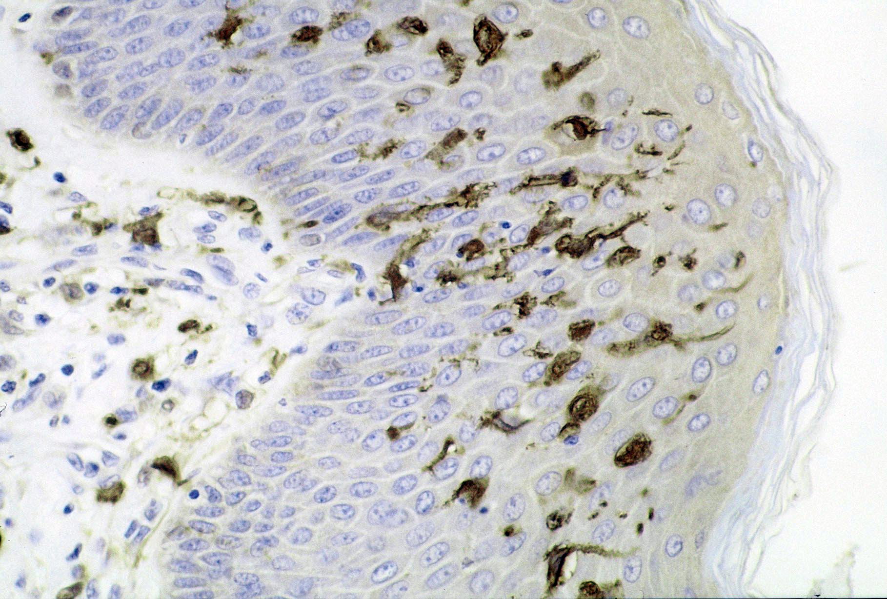
Dendritic cell
A dendritic cell (DC) is an antigen-presenting cell (also known as an accessory cell) of the mammalian immune system. A DC's main function is to process antigen material and present it on the cell surface to the T cells of the immune system. They act as messengers between the innate and adaptive immune systems.
Dendritic cells are present in tissues that are in contact with the body's external environment, such as the skin, and the inner lining of the nose, lungs, stomach and intestines. They can also be found in an immature and mature state in the blood. Once activated, they migrate to the lymph nodes, where they interact with T cells and B cells to initiate and shape the adaptive immune response. At certain development stages they grow branched projections, the dendrites, that give the cell its name (δένδρον or déndron being Greek for 'tree'). While similar in appearance to the dendrites of neurons, these are structures distinct from them. Immature dendritic cells are also called veiled cells, as they possess large cytoplasmic 'veils' rather than dendrites.[citation needed]
Dendritic cells were first described by Paul Langerhans (hence Langerhans cells) in the late nineteenth century. The term dendritic cells was coined in 1973 by Ralph M. Steinman and Zanvil A. Cohn. For discovering the central role of dendritic cells in the adaptive immune response, Steinman was awarded the Albert Lasker Award for Basic Medical Research in 2007 and the Nobel Prize in Physiology or Medicine in 2011.
The morphology of dendritic cells results in a very large surface-to-volume ratio. That is, the dendritic cell has a very large surface area compared to the overall cell volume.
The most common division of dendritic cells is conventional dendritic cells (a.k.a. myeloid dendritic cells) vs. plasmacytoid dendritic cell (most likely of lymphoid lineage) as described in the table below:
The markers BDCA-2, BDCA-3, and BDCA-4 can be used to discriminate among the types.
Lymphoid and myeloid DCs evolve from lymphoid and myeloid precursors, respectively, and thus are of hematopoietic origin. By contrast, follicular dendritic cells (FDC) are probably of mesenchymal rather than hematopoietic origin and do not express MHC class II, but are so named because they are located in lymphoid follicles and have long "dendritic" processes.
The blood DCs are typically identified and enumerated in flow cytometry. Three types of DCs have been defined in human blood: the CD1c+ myeloid DCs, the CD141+ myeloid DCs and the CD303+ plasmacytoid DCs. This represents the nomenclature proposed by the nomenclature committee of the International Union of Immunological Societies. Dendritic cells that circulate in blood do not have all the typical features of their counterparts in tissue, i.e. they are less mature and have no dendrites. Still, they can perform complex functions including chemokine-production (in CD1c+ myeloid DCs), cross-presentation (in CD141+ myeloid DCs), and IFNalpha production (in CD303+ plasmacytoid DCs).
Hub AI
Dendritic cell AI simulator
(@Dendritic cell_simulator)
Dendritic cell
A dendritic cell (DC) is an antigen-presenting cell (also known as an accessory cell) of the mammalian immune system. A DC's main function is to process antigen material and present it on the cell surface to the T cells of the immune system. They act as messengers between the innate and adaptive immune systems.
Dendritic cells are present in tissues that are in contact with the body's external environment, such as the skin, and the inner lining of the nose, lungs, stomach and intestines. They can also be found in an immature and mature state in the blood. Once activated, they migrate to the lymph nodes, where they interact with T cells and B cells to initiate and shape the adaptive immune response. At certain development stages they grow branched projections, the dendrites, that give the cell its name (δένδρον or déndron being Greek for 'tree'). While similar in appearance to the dendrites of neurons, these are structures distinct from them. Immature dendritic cells are also called veiled cells, as they possess large cytoplasmic 'veils' rather than dendrites.[citation needed]
Dendritic cells were first described by Paul Langerhans (hence Langerhans cells) in the late nineteenth century. The term dendritic cells was coined in 1973 by Ralph M. Steinman and Zanvil A. Cohn. For discovering the central role of dendritic cells in the adaptive immune response, Steinman was awarded the Albert Lasker Award for Basic Medical Research in 2007 and the Nobel Prize in Physiology or Medicine in 2011.
The morphology of dendritic cells results in a very large surface-to-volume ratio. That is, the dendritic cell has a very large surface area compared to the overall cell volume.
The most common division of dendritic cells is conventional dendritic cells (a.k.a. myeloid dendritic cells) vs. plasmacytoid dendritic cell (most likely of lymphoid lineage) as described in the table below:
The markers BDCA-2, BDCA-3, and BDCA-4 can be used to discriminate among the types.
Lymphoid and myeloid DCs evolve from lymphoid and myeloid precursors, respectively, and thus are of hematopoietic origin. By contrast, follicular dendritic cells (FDC) are probably of mesenchymal rather than hematopoietic origin and do not express MHC class II, but are so named because they are located in lymphoid follicles and have long "dendritic" processes.
The blood DCs are typically identified and enumerated in flow cytometry. Three types of DCs have been defined in human blood: the CD1c+ myeloid DCs, the CD141+ myeloid DCs and the CD303+ plasmacytoid DCs. This represents the nomenclature proposed by the nomenclature committee of the International Union of Immunological Societies. Dendritic cells that circulate in blood do not have all the typical features of their counterparts in tissue, i.e. they are less mature and have no dendrites. Still, they can perform complex functions including chemokine-production (in CD1c+ myeloid DCs), cross-presentation (in CD141+ myeloid DCs), and IFNalpha production (in CD303+ plasmacytoid DCs).