Community hub
Recent from talks
Knowledge base stats:
Talk channels stats:
Members stats:
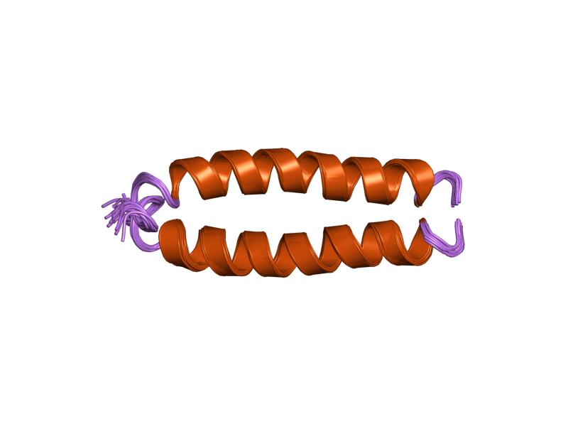
Fusion protein
Fusion proteins or chimeric proteins (literally, made of parts from different sources) are proteins created through the joining of two or more genes that originally coded for separate proteins. Translation of this fusion gene results in a single or multiple polypeptides with functional properties derived from each of the original proteins. Recombinant fusion proteins are created artificially by recombinant DNA technology for use in biological research or therapeutics. Chimeric or chimera usually designate hybrid proteins made of polypeptides having different functions or physico-chemical patterns. Chimeric mutant proteins occur naturally when a complex mutation, such as a chromosomal translocation, tandem duplication, or retrotransposition creates a novel coding sequence containing parts of the coding sequences from two different genes. Naturally occurring fusion proteins are commonly found in cancer cells, where they may function as oncoproteins. The bcr-abl fusion protein is a well-known example of an oncogenic fusion protein, and is considered to be the primary oncogenic driver of chronic myelogenous leukemia.
Some fusion proteins combine whole peptides and therefore contain all functional domains of the original proteins. However, other fusion proteins, especially those that occur naturally, combine only portions of coding sequences and therefore do not maintain the original functions of the parental genes that formed them.
Many whole gene fusions are fully functional and can still act to replace the original peptides. Some, however, experience interactions between the two proteins that can modify their functions. Beyond these effects, some gene fusions may cause regulatory changes that alter when and where these genes act. For partial gene fusions, the shuffling of different active sites and binding domains have the potential to result in new proteins with novel functions.
The fusion of fluorescent tags to proteins in a host cell is a widely popular technique used in experimental cell and biology research in order to track protein interactions in real time. The first fluorescent tag, green fluorescent protein (GFP), was isolated from Aequorea victoria and is still used frequently in modern research. More recent derivations include photoconvertible fluorescent proteins (PCFPs), which were first isolated from Anthozoa. The most commonly used PCFP is the Kaede fluorescent tag, but the development of Kikume green-red (KikGR) in 2005 offers a brighter signal and more efficient photoconversion. The advantage of using PCFP fluorescent tags is the ability to track the interaction of overlapping biochemical pathways in real time. The tag will change color from green to red once the protein reaches a point of interest in the pathway, and the alternate colored protein can be monitored through the duration of pathway. This technique is especially useful when studying G-protein coupled receptor (GPCR) recycling pathways. The fates of recycled G-protein receptors may either be sent to the plasma membrane to be recycled, marked by a green fluorescent tag, or may be sent to a lysosome for degradation, marked by a red fluorescent tag.
The purpose of creating fusion proteins in drug development is to impart properties from each of the "parent" proteins to the resulting chimeric protein. Several chimeric protein drugs are currently available for medical use.
Many chimeric protein drugs are monoclonal antibodies whose specificity for a target molecule was developed using mice and hence were initially "mouse" antibodies. As non-human proteins, mouse antibodies tend to evoke an immune reaction if administered to humans. The chimerization process involves engineering the replacement of segments of the antibody molecule that distinguish it from a human antibody. For example, human constant domains can be introduced, thereby eliminating most of the potentially immunogenic portions of the drug without altering its specificity for the intended therapeutic target. Antibody nomenclature indicates this type of modification by inserting -xi- into the non-proprietary name (e.g., abci-xi-mab). If parts of the variable domains are also replaced by human portions, humanized antibodies are obtained. Although not conceptually distinct from chimeras, this type is indicated using -zu- such as in dacli-zu-mab. See the list of monoclonal antibodies for more examples.
In addition to chimeric and humanized antibodies, there are other pharmaceutical purposes for the creation of chimeric constructs. Etanercept, for example, is a TNFα blocker created through the combination of a tumor necrosis factor receptor (TNFR) with the immunoglobulin G1 Fc segment. TNFR provides specificity for the drug target and the antibody Fc segment is believed to add stability and deliverability of the drug. Additional chimeric proteins used for therapeutic applications include:
A recombinant fusion protein is a protein created through genetic engineering of a fusion gene. This typically involves removing the stop codon from a cDNA sequence coding for the first protein, then appending the cDNA sequence of the second protein in frame through ligation or overlap extension PCR. That DNA sequence will then be expressed by a cell as a single protein. The protein can be engineered to include the full sequence of both original proteins, or only a portion of either.
Hub AI
Fusion protein AI simulator
(@Fusion protein_simulator)
Fusion protein
Fusion proteins or chimeric proteins (literally, made of parts from different sources) are proteins created through the joining of two or more genes that originally coded for separate proteins. Translation of this fusion gene results in a single or multiple polypeptides with functional properties derived from each of the original proteins. Recombinant fusion proteins are created artificially by recombinant DNA technology for use in biological research or therapeutics. Chimeric or chimera usually designate hybrid proteins made of polypeptides having different functions or physico-chemical patterns. Chimeric mutant proteins occur naturally when a complex mutation, such as a chromosomal translocation, tandem duplication, or retrotransposition creates a novel coding sequence containing parts of the coding sequences from two different genes. Naturally occurring fusion proteins are commonly found in cancer cells, where they may function as oncoproteins. The bcr-abl fusion protein is a well-known example of an oncogenic fusion protein, and is considered to be the primary oncogenic driver of chronic myelogenous leukemia.
Some fusion proteins combine whole peptides and therefore contain all functional domains of the original proteins. However, other fusion proteins, especially those that occur naturally, combine only portions of coding sequences and therefore do not maintain the original functions of the parental genes that formed them.
Many whole gene fusions are fully functional and can still act to replace the original peptides. Some, however, experience interactions between the two proteins that can modify their functions. Beyond these effects, some gene fusions may cause regulatory changes that alter when and where these genes act. For partial gene fusions, the shuffling of different active sites and binding domains have the potential to result in new proteins with novel functions.
The fusion of fluorescent tags to proteins in a host cell is a widely popular technique used in experimental cell and biology research in order to track protein interactions in real time. The first fluorescent tag, green fluorescent protein (GFP), was isolated from Aequorea victoria and is still used frequently in modern research. More recent derivations include photoconvertible fluorescent proteins (PCFPs), which were first isolated from Anthozoa. The most commonly used PCFP is the Kaede fluorescent tag, but the development of Kikume green-red (KikGR) in 2005 offers a brighter signal and more efficient photoconversion. The advantage of using PCFP fluorescent tags is the ability to track the interaction of overlapping biochemical pathways in real time. The tag will change color from green to red once the protein reaches a point of interest in the pathway, and the alternate colored protein can be monitored through the duration of pathway. This technique is especially useful when studying G-protein coupled receptor (GPCR) recycling pathways. The fates of recycled G-protein receptors may either be sent to the plasma membrane to be recycled, marked by a green fluorescent tag, or may be sent to a lysosome for degradation, marked by a red fluorescent tag.
The purpose of creating fusion proteins in drug development is to impart properties from each of the "parent" proteins to the resulting chimeric protein. Several chimeric protein drugs are currently available for medical use.
Many chimeric protein drugs are monoclonal antibodies whose specificity for a target molecule was developed using mice and hence were initially "mouse" antibodies. As non-human proteins, mouse antibodies tend to evoke an immune reaction if administered to humans. The chimerization process involves engineering the replacement of segments of the antibody molecule that distinguish it from a human antibody. For example, human constant domains can be introduced, thereby eliminating most of the potentially immunogenic portions of the drug without altering its specificity for the intended therapeutic target. Antibody nomenclature indicates this type of modification by inserting -xi- into the non-proprietary name (e.g., abci-xi-mab). If parts of the variable domains are also replaced by human portions, humanized antibodies are obtained. Although not conceptually distinct from chimeras, this type is indicated using -zu- such as in dacli-zu-mab. See the list of monoclonal antibodies for more examples.
In addition to chimeric and humanized antibodies, there are other pharmaceutical purposes for the creation of chimeric constructs. Etanercept, for example, is a TNFα blocker created through the combination of a tumor necrosis factor receptor (TNFR) with the immunoglobulin G1 Fc segment. TNFR provides specificity for the drug target and the antibody Fc segment is believed to add stability and deliverability of the drug. Additional chimeric proteins used for therapeutic applications include:
A recombinant fusion protein is a protein created through genetic engineering of a fusion gene. This typically involves removing the stop codon from a cDNA sequence coding for the first protein, then appending the cDNA sequence of the second protein in frame through ligation or overlap extension PCR. That DNA sequence will then be expressed by a cell as a single protein. The protein can be engineered to include the full sequence of both original proteins, or only a portion of either.