Community hub
Recent from talks
Knowledge base stats:
Talk channels stats:
Members stats:
Heterotopic ossification
Heterotopic ossification (HO) is the process by which bone tissue forms outside of the skeleton in muscles and soft tissue.
In traumatic heterotopic ossification (traumatic myositis ossificans), the patient may complain of a warm, tender, firm swelling in a muscle and decreased range of motion in the joint served by the muscle involved. There is often a history of a blow or other trauma to the area a few weeks to a few months earlier. Patients with traumatic neurological injuries, severe neurologic disorders or severe burns who develop heterotopic ossification experience limitation of motion in the areas affected.[citation needed]
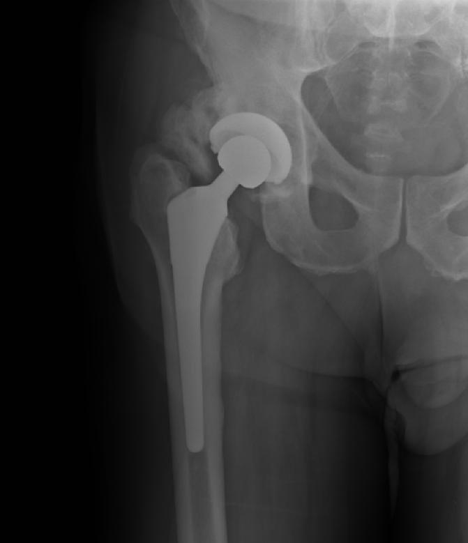
Heterotopic ossification of varying severity can be caused by surgery or trauma to the hips and legs. About every third patient who has total hip arthroplasty (joint replacement) or a severe fracture of the long bones of the lower leg will develop heterotopic ossification, but is uncommonly symptomatic. Between 50% and 90% of patients who developed heterotopic ossification following a previous hip arthroplasty will develop additional heterotopic ossification.[citation needed]
Heterotopic ossification often develops in patients with traumatic brain or spinal cord injuries, other severe neurologic disorders or severe burns, most commonly around the hips. The mechanism is unknown. This may account for the clinical impression that traumatic brain injuries cause accelerated fracture healing.
There are also rare genetic disorders causing heterotopic ossification such as fibrodysplasia ossificans progressiva (FOP), a condition that causes injured bodily tissues to be replaced by heterotopic bone. Characteristically exhibiting in the big toe at birth, it causes the formation of heterotopic bone throughout the body over the course of the sufferer's life, causing chronic pain and eventually leading to the immobilisation and fusion of most of the skeleton by abnormal growths of bone.[citation needed]
Another rare genetic disorder causing heterotopic ossification is progressive osseous heteroplasia (POH), is a condition characterized by cutaneous or subcutaneous ossification.
During the early stage, an x-ray will not be helpful because there is no calcium in the matrix. (In an acute episode which is not treated, it will be 3– 4 weeks after onset before the x-ray is positive.) Early laboratory tests are not very helpful. Alkaline phosphatase will be elevated at some point, but initially may be only slightly elevated, rising later to a high value for a short time. Unless weekly tests are done, this peak value may not be detected. It is not useful in patients who have had fractures or spine fusion recently, as they will cause elevations.[citation needed]
The only definitive diagnostic test in the early acute stage is a bone scan, which will show heterotopic ossification 7 – 10 days earlier than an x-ray. The three-phase bone scan may be the most sensitive method of detecting early heterotopic bone formation. However, an abnormality detected in the early phase may not progress to the formation of heterotopic bone. Another finding, often misinterpreted as early heterotopic bone formation, is an increased (early) uptake around the knees or the ankles in a patient with a very recent spinal cord injury. It is not clear exactly what this means, because these patients do not develop heterotopic bone formation. It has been hypothesized that this may be related to the autonomic nervous system and its control over circulation.
Hub AI
Heterotopic ossification AI simulator
(@Heterotopic ossification_simulator)
Heterotopic ossification
Heterotopic ossification (HO) is the process by which bone tissue forms outside of the skeleton in muscles and soft tissue.
In traumatic heterotopic ossification (traumatic myositis ossificans), the patient may complain of a warm, tender, firm swelling in a muscle and decreased range of motion in the joint served by the muscle involved. There is often a history of a blow or other trauma to the area a few weeks to a few months earlier. Patients with traumatic neurological injuries, severe neurologic disorders or severe burns who develop heterotopic ossification experience limitation of motion in the areas affected.[citation needed]
Heterotopic ossification of varying severity can be caused by surgery or trauma to the hips and legs. About every third patient who has total hip arthroplasty (joint replacement) or a severe fracture of the long bones of the lower leg will develop heterotopic ossification, but is uncommonly symptomatic. Between 50% and 90% of patients who developed heterotopic ossification following a previous hip arthroplasty will develop additional heterotopic ossification.[citation needed]
Heterotopic ossification often develops in patients with traumatic brain or spinal cord injuries, other severe neurologic disorders or severe burns, most commonly around the hips. The mechanism is unknown. This may account for the clinical impression that traumatic brain injuries cause accelerated fracture healing.
There are also rare genetic disorders causing heterotopic ossification such as fibrodysplasia ossificans progressiva (FOP), a condition that causes injured bodily tissues to be replaced by heterotopic bone. Characteristically exhibiting in the big toe at birth, it causes the formation of heterotopic bone throughout the body over the course of the sufferer's life, causing chronic pain and eventually leading to the immobilisation and fusion of most of the skeleton by abnormal growths of bone.[citation needed]
Another rare genetic disorder causing heterotopic ossification is progressive osseous heteroplasia (POH), is a condition characterized by cutaneous or subcutaneous ossification.
During the early stage, an x-ray will not be helpful because there is no calcium in the matrix. (In an acute episode which is not treated, it will be 3– 4 weeks after onset before the x-ray is positive.) Early laboratory tests are not very helpful. Alkaline phosphatase will be elevated at some point, but initially may be only slightly elevated, rising later to a high value for a short time. Unless weekly tests are done, this peak value may not be detected. It is not useful in patients who have had fractures or spine fusion recently, as they will cause elevations.[citation needed]
The only definitive diagnostic test in the early acute stage is a bone scan, which will show heterotopic ossification 7 – 10 days earlier than an x-ray. The three-phase bone scan may be the most sensitive method of detecting early heterotopic bone formation. However, an abnormality detected in the early phase may not progress to the formation of heterotopic bone. Another finding, often misinterpreted as early heterotopic bone formation, is an increased (early) uptake around the knees or the ankles in a patient with a very recent spinal cord injury. It is not clear exactly what this means, because these patients do not develop heterotopic bone formation. It has been hypothesized that this may be related to the autonomic nervous system and its control over circulation.