Hubbry Logo
search
logo
1866800

Heterotopic ossification

logo
Community Hub0 Subscribers
Read side by side
from Wikipedia
Heterotopic ossification
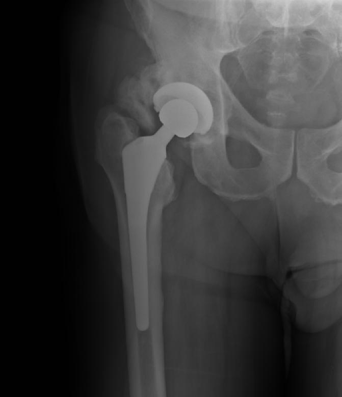
Heterotopic ossification around the hip joint in a patient who has undergone hip arthroplasty

Heterotopic ossification (HO) is the process by which bone tissue forms outside of the skeleton in muscles and soft tissue.[1]

Symptoms

[edit]

In traumatic heterotopic ossification (traumatic myositis ossificans), the patient may complain of a warm, tender, firm swelling in a muscle and decreased range of motion in the joint served by the muscle involved. There is often a history of a blow or other trauma to the area a few weeks to a few months earlier. Patients with traumatic neurological injuries, severe neurologic disorders or severe burns who develop heterotopic ossification experience limitation of motion in the areas affected.[citation needed]

Causes

[edit]

Heterotopic ossification of varying severity can be caused by surgery or trauma to the hips and legs. About every third patient who has total hip arthroplasty (joint replacement) or a severe fracture of the long bones of the lower leg will develop heterotopic ossification, but is uncommonly symptomatic. Between 50% and 90% of patients who developed heterotopic ossification following a previous hip arthroplasty will develop additional heterotopic ossification.[citation needed]

Heterotopic ossification often develops in patients with traumatic brain or spinal cord injuries, other severe neurologic disorders or severe burns, most commonly around the hips. The mechanism is unknown. This may account for the clinical impression that traumatic brain injuries cause accelerated fracture healing.[2]

There are also rare genetic disorders causing heterotopic ossification such as fibrodysplasia ossificans progressiva (FOP), a condition that causes injured bodily tissues to be replaced by heterotopic bone. Characteristically exhibiting in the big toe at birth, it causes the formation of heterotopic bone throughout the body over the course of the sufferer's life, causing chronic pain and eventually leading to the immobilisation and fusion of most of the skeleton by abnormal growths of bone.[citation needed]

Another rare genetic disorder causing heterotopic ossification is progressive osseous heteroplasia (POH), is a condition characterized by cutaneous or subcutaneous ossification.

Diagnosis

[edit]
Heterotopic ossification of the elbow, after comminuted fracture and arthroplasty.
Heteropic ossification of the elbow, after comminuted fracture and arthroplasty.

During the early stage, an x-ray will not be helpful because there is no calcium in the matrix. (In an acute episode which is not treated, it will be 3– 4 weeks after onset before the x-ray is positive.) Early laboratory tests are not very helpful. Alkaline phosphatase will be elevated at some point, but initially may be only slightly elevated, rising later to a high value for a short time. Unless weekly tests are done, this peak value may not be detected. It is not useful in patients who have had fractures or spine fusion recently, as they will cause elevations.[citation needed]

The only definitive diagnostic test in the early acute stage is a bone scan, which will show heterotopic ossification 7 – 10 days earlier than an x-ray. The three-phase bone scan may be the most sensitive method of detecting early heterotopic bone formation. However, an abnormality detected in the early phase may not progress to the formation of heterotopic bone. Another finding, often misinterpreted as early heterotopic bone formation, is an increased (early) uptake around the knees or the ankles in a patient with a very recent spinal cord injury. It is not clear exactly what this means, because these patients do not develop heterotopic bone formation. It has been hypothesized that this may be related to the autonomic nervous system and its control over circulation.[3]

When the initial presentation is swelling and increased temperature in a leg, the differential diagnosis includes thrombophlebitis. It may be necessary to do both a bone scan and a venogram to differentiate between heterotopic ossification and thrombophlebitis, and it is even possible that both could be present simultaneously. In heterotopic ossification, the swelling tends to be more proximal and localized, with little or no foot/ankle edema, whereas in thrombophlebitis the swelling is usually more uniform throughout the leg.[4]

Treatment

[edit]

There is no clear form of treatment. Originally, bisphosphonates were expected to be of value after hip surgery but there has been no convincing evidence of benefit, despite having been used prophylactically.[5]

Depending on the growth's location, orientation and severity, surgical removal may be possible.

Surgical removal of a Heterotopic Ossification fusing the right Humerus and Radius following a severe TBI and complete fracture of the Ulna.

Radiation Therapy.

Elbow heterotopic ossification radiation therapy field, status post surgery.

Prophylactic radiation therapy for the prevention of heterotopic ossification has been employed since the 1970s. A variety of doses and techniques have been used. Generally, radiation therapy should be delivered as close as practical to the time of surgery. A dose of 7-8 Gray in a single fraction within 24–48 hours of surgery has been used successfully. Treatment volumes include the peri-articular region, and can be used for hip, knee, elbow, shoulder, jaw or in patients after spinal cord trauma.

Single dose radiation therapy is well tolerated and is cost effective, without an increase in bleeding, infection or wound healing disturbances.[6]

Other possible treatments.

Certain antiinflammatory agents, such as indomethacin, ibuprofen and aspirin, have shown some effect in preventing recurrence of heterotopic ossification after total hip replacement. [7]

Conservative treatments such as passive range of motion exercises or other mobilization techniques provided by physical therapists or occupational therapists may also assist in preventing HO. A review article looked at 114 adult patients retrospectively and suggested that the lower incidence of HO in patients with a very severe TBI may have been due to early intensive physical and occupational therapy in conjunction with pharmacological treatment.[8] Another review article also recommended physiotherapy as an adjunct to pharmacological and medical treatments because passive range of motion exercises may maintain range at the joint and prevent secondary soft tissue contractures, which are often associated with joint immobility.[9]

See also

[edit]

References

[edit]
[edit]
Revisions and contributorsEdit on WikipediaRead on Wikipedia
from Grokipedia
Heterotopic ossification (HO) is the abnormal formation of mature, lamellar bone within extraskeletal soft tissues, such as muscles and tendons, where bone does not normally develop.[1] This condition arises from a dysregulated process of endochondral or intramembranous ossification, often triggered by trauma, surgery, or neurological insults, and can lead to significant morbidity through joint stiffness and restricted mobility.[2] HO is classified into acquired (traumatic or neurogenic, the latter also known as para-osteo-arthropathy (POA) especially in periarticular locations associated with neurological conditions such as spinal cord injury or paraplegia) and genetic forms, with the latter including rare disorders like fibrodysplasia ossificans progressiva (FOP).[3][4] The incidence of HO varies by context, affecting up to 90% of high-risk total hip arthroplasty cases and 20-30% of spinal cord injury patients, typically manifesting 3-12 weeks post-inciting event with symptoms including pain, swelling, warmth, and reduced range of motion.[1] Risk factors encompass older age, male sex, prolonged immobility, and spasticity, while pathogenesis involves inflammation, mesenchymal stem cell differentiation driven by bone morphogenetic proteins (BMPs), and contributions from immune cells like macrophages.[2] In neurogenic HO, common after traumatic brain injury or stroke, ectopic bone often forms around large joints like the hip and elbow, potentially causing nerve compression or ankylosis in severe cases.[3] Diagnosis relies on imaging, with triple-phase bone scintigraphy detecting early activity as soon as 2-3 weeks post-onset, followed by plain radiographs showing mature ossification after 4-6 weeks; computed tomography or magnetic resonance imaging further delineates extent and maturity.[1] Laboratory markers such as elevated alkaline phosphatase may support suspicion but are nonspecific.[2] Prophylaxis is key in at-risk populations, utilizing nonsteroidal anti-inflammatory drugs (e.g., indomethacin), bisphosphonates, or low-dose radiation therapy to inhibit formation, while mature HO may require surgical resection after 12-18 months to restore function.[3] Targeted therapies, including the FDA-approved palovarotene for FOP (as of 2023), inhibit BMP pathways in genetic forms, though most cases remain asymptomatic or self-limiting.[2][5]

Overview

Definition

Heterotopic ossification (HO) is a pathological process defined as the formation of mature, lamellar bone in extraskeletal soft tissues, such as muscles, tendons, and ligaments, where bone formation does not normally occur.[1] This ectopic bone is histologically identical to normal skeletal bone, consisting of organized trabeculae and marrow elements, and can lead to significant morbidity by restricting joint mobility.[2] HO must be distinguished from dystrophic calcification, which involves the amorphous deposition of calcium salts in necrotic or damaged tissues without the formation of structured bone matrix or hematopoietic elements.[6] While dystrophic calcification may occasionally progress to ossification, the two processes are histologically and radiographically distinct, with HO featuring true bone development rather than mere mineral accumulation.[7] The condition was first described in 1883 by German physician Bernhard Riedel in a case of post-traumatic bone formation.[8] Further recognition came during World War I, when French physicians Maurice Dejerine and Pierre Ceillier documented HO in soldiers with spinal cord injuries, terming it "para-osteo-arthropathy" (POA). The combining form "para-osteo" indicates the periarticular location (near joints) of the abnormal bone formation, and POA refers to neurogenic heterotopic ossification involving the formation of bone in soft tissues adjacent to joints, commonly associated with neurological conditions such as spinal cord injury or paraplegia.[3][4] The basic mechanism of HO involves aberrant activation of osteogenic pathways, primarily through endochondral ossification—where a cartilaginous anlage forms and is subsequently replaced by bone—or, less commonly, intramembranous ossification, which generates bone directly from mesenchymal precursors without a cartilage intermediate.[9] These processes mirror normal skeletal development but occur in inappropriate soft tissue sites, often around major joints like the hips and elbows.[10]

Epidemiology

Heterotopic ossification (HO) is a common complication following certain orthopedic surgeries and traumas, with varying incidence rates depending on the underlying condition. After total hip arthroplasty, the overall incidence ranges from 10% to 30%, though it can reach up to 90% in high-risk patients such as those with prior HO or ankylosing spondylitis.[1] In patients with spinal cord injury, the incidence is reported between 16% and 53%, while in those with traumatic brain injury, it varies from 8% to 20% for clinically significant cases.[11][12] Demographic patterns show a higher prevalence in males, with a sex ratio of approximately 3:2, and in young adults, particularly those under 30 years old following trauma or surgery.[2] Genetic forms, such as fibrodysplasia ossificans progressiva, are extremely rare, affecting about 1 in 2 million individuals worldwide, with no specific ethnic or geographic predisposition.[13] Temporal trends indicate an increase in HO reports since 2020, linked to prolonged immobilization in critically ill COVID-19 patients, where prevalence reached up to 19% in those requiring extended mechanical ventilation.[14] Specific high-risk groups include burn patients, with an overall incidence of 0.2% to 4% but exceeding 50% in cases of major burns involving more than 30% total body surface area, and combat-injured individuals, where rates approach 65% in high-energy blast-related orthopedic traumas.[15][16][17]

Pathophysiology

Mechanisms

Heterotopic ossification (HO) develops through a well-characterized three-phase process involving inflammation, osteogenesis, and maturation, primarily driven by dysregulated signaling in mesenchymal progenitor cells.[2] This ectopic bone formation typically occurs via endochondral ossification, where cartilage intermediates precede bone deposition, though intramembranous pathways can contribute in some contexts.[18] The inflammatory phase is initiated by local trauma or injury to soft tissues, such as muscle or connective tissue, which disrupts the tissue microenvironment and prompts the release of pro-inflammatory cytokines including interleukin-6 (IL-6) and bone morphogenetic proteins (BMPs), particularly BMP-2 and BMP-4.[15] These cytokines create a permissive niche by recruiting circulating and resident mesenchymal stem cells (MSCs), as well as immune cells like macrophages and mast cells, which amplify the inflammatory response through further cytokine production and substance P release.[2] This phase, lasting days to weeks, establishes the foundational cellular pool for subsequent bone formation.[18] During the osteogenic phase, recruited MSCs differentiate into chondrocytes or osteoblasts under the influence of the BMP signaling pathway, which activates Smad1/5/8 transcription factors to promote chondrogenesis in hypoxic conditions and osteogenesis via Runx2 expression.[15] Overexpression of BMPs and their receptors, such as ALK2 (encoded by ACVR1), enhances this differentiation, often leading to endochondral ossification where a cartilaginous anlage forms before mineralization.[2] In rare genetic forms like fibrodysplasia ossificans progressiva (FOP), mutations in ACVR1 (e.g., R206H) cause hypersensitivity to ligands like activin A, resulting in constitutive BMP pathway activation and progressive HO.[18] The maturation phase involves vascularization of the forming bone matrix, driven by hypoxia-inducible factor 1-alpha (HIF-1α) and vascular endothelial growth factor (VEGF), which supports remodeling into mature lamellar bone with hematopoietic elements over 6-12 months.[15] This remodeling integrates neural and vascular networks, stabilizing the ectopic bone while reducing inflammation, and is marked by peripheral maturation zones.[2]

Classification

Heterotopic ossification (HO) is primarily classified into two major categories: acquired and genetic forms, distinguished by their etiology and clinical presentation. Acquired HO arises secondary to identifiable triggers such as trauma, surgery, or neurological injury, whereas genetic HO results from inherited mutations leading to progressive ectopic bone formation without external provocation. This dichotomy aids in differentiating management approaches and prognostic implications.[15] Acquired HO is further subdivided into traumatic and neurogenic subtypes. Traumatic acquired HO occurs following musculoskeletal injuries, fractures, burns, or invasive procedures like joint arthroplasty, with bone formation typically confined to periarticular soft tissues. Neurogenic acquired HO, also known as para-osteo-arthropathy (POA), which refers to abnormal bone formation in periarticular soft tissues associated with neurological conditions such as spinal cord injury or paraplegia, develops after central nervous system insults such as traumatic brain injury or spinal cord injury, often involving more extensive and symmetric ossification due to dysregulated neural signaling.[19][15][4] Genetic forms of HO are rare and include fibrodysplasia ossificans progressiva (FOP) and progressive osseous heteroplasia (POH). FOP, caused by activating mutations in the ACVR1 gene, manifests as episodic, progressive HO in soft tissues, often triggered by minor trauma or spontaneously, leading to cumulative disability through joint ankylosis. POH, resulting from inactivating mutations in the GNAS gene, begins with dermal ossification in infancy and advances to deeper subcutaneous and muscular layers, characterized by non-inflammatory, plaque-like bone formation. Severity and extent of HO are assessed using joint-specific grading systems that incorporate radiographic findings and clinical functional impairment, allowing differentiation between mild, asymptomatic ossification and severe, motion-restricting disease. The Brooker classification, introduced for HO around the hip following total hip arthroplasty, is widely used and divides cases into four radiographic grades: Grade I (isolated islands of bone within soft tissues, no impingement); Grade II (bone spurs from the pelvis or femur with at least 1 cm between opposing surfaces); Grade III (bone spurs with less than 1 cm between surfaces, causing partial bridging); and Grade IV (complete ankylosis of the hip joint). This system correlates radiographic progression with functional outcomes, though it primarily emphasizes imaging over clinical metrics. For the elbow, the Hastings and Graham classification integrates both clinical and radiographic data into six subclasses to evaluate location and impact on range of motion. Class I denotes radiographic evidence of ossification without functional deficit. Class IIA indicates ossification limiting flexion-extension, while Class IIB limits pronation-supination. Class IIIA involves ankylosis in flexion-extension, IIIB in pronation-supination, and IIIC in both axes, reflecting severe joint fusion. These systems highlight a distinction between purely radiographic grading, which quantifies bone volume and position, and clinical grading, which prioritizes measurable limitations in joint mobility and daily function.[20][15] Emerging literature from the 2020s has identified post-infectious HO as a potential subtype within acquired forms, particularly following severe viral infections such as COVID-19 pneumonia, where prolonged immobilization and inflammatory responses may precipitate ectopic bone in soft tissues like the hip or shoulder. Case reports and reviews document this phenomenon in critically ill patients, suggesting an inflammatory-immune mediated pathway akin to neurogenic HO, though further studies are needed to formalize its classification.[21]

Etiology

Acquired forms

Acquired heterotopic ossification arises from environmental, traumatic, or medical interventions that disrupt normal tissue architecture, leading to ectopic bone formation in soft tissues. Unlike genetic forms, these cases are typically triggered by acute events and are more prevalent in individuals with significant injury or procedural histories.[12] Traumatic causes represent a primary category, often following musculoskeletal injuries such as fractures, severe burns, or orthopedic surgeries like joint replacements. For instance, heterotopic ossification develops in 15-90% of patients after total hip arthroplasty depending on prophylaxis and risk factors, with clinically significant cases (Brooker grades III-IV) occurring in 5-10%.[22] Post-fracture heterotopic ossification is linked to direct soft tissue damage and hematoma formation, while burns covering more than 30% of total body surface area increase risk due to extensive inflammation and immobilization, with an incidence of approximately 3-5%.[23][24] Neurogenic forms, also known as para-osteo-arthropathy (POA), occur secondary to central nervous system insults, including spinal cord injury (commonly resulting in paraplegia), traumatic brain injury, or stroke, where disrupted neural signaling promotes osteogenic differentiation in periarticular tissues.[4] The incidence after spinal cord injury ranges from 20-30%, particularly in complete injuries at cervical or thoracic levels, and often affects the hips, knees, and elbows within 1-4 months post-injury.[25] Traumatic brain injury carries a 11-22% risk, escalating with injury severity such as prolonged coma or high injury severity scores, while stroke is associated with a lower incidence of 0.5-1.2%, predominantly in hemiplegic limbs.[19][26] Iatrogenic factors contribute through medical interventions that induce local trauma or systemic stress, such as prolonged immobilization in intensive care settings or invasive procedures including surgeries and mechanical ventilation. Extended bed rest exceeding two weeks, common in critical illness, elevates risk by fostering muscle atrophy and venous stasis, with cases reported in up to 5% of immobilized patients.[27] Surgical delays beyond 48 hours from injury or repeat procedures further amplify this by prolonging inflammatory exposure.[28] Several modifiers influence the likelihood and severity of acquired heterotopic ossification, including the extent of initial injury, timing of interventions, and underlying joint conditions. Greater injury severity, measured by high injury severity scores or extensive tissue damage, correlates with up to 11% higher incidence, while delayed surgery heightens risk through sustained hematoma and fibrosis.[29] Patients with hypertrophic osteoarthritis face elevated odds post-arthroplasty due to preexisting osteoproliferative tendencies in the joint capsule.[2]

Genetic forms

Genetic forms of heterotopic ossification encompass rare inherited disorders characterized by progressive ectopic bone formation due to specific genetic mutations, often involving dysregulation of the bone morphogenetic protein (BMP) signaling pathway.[30] These conditions differ from acquired forms by their lifelong, systemic progression without requiring external triggers for initial onset, though trauma can exacerbate them.[31] The prototypical genetic disorder is fibrodysplasia ossificans progressiva (FOP), an ultrarare autosomal dominant condition caused by a heterozygous missense mutation in the ACVR1 gene (encoding activin A receptor type I, also known as ALK2), most commonly the R206H variant.[30] This mutation leads to aberrant activation of the BMP pathway, resulting in episodic flare-ups of heterotopic ossification in soft tissues, often triggered by minor trauma, intramuscular injections, or surgery, with malformations such as short halluces present at birth.[32] FOP affects approximately 1 in 2 million individuals worldwide, with nearly all cases arising from de novo mutations.[30] Progressive osseous heteroplasia (POH) represents another distinct genetic entity, characterized by paternal inheritance of inactivating mutations in the GNAS gene, which encodes the alpha subunit of the stimulatory G protein (Gsα).[33] These mutations disrupt G protein-coupled signaling, leading to heterotopic ossification that begins in the dermis and subcutaneous tissues during infancy or early childhood, progressively invading deeper skeletal muscles and potentially causing severe immobility.[34] Unlike FOP, POH lacks congenital malformations and flare-up episodes but shares a similar inexorable progression, with an estimated prevalence of less than 1 in 1 million.[35] Other genetic variants include historical designations such as myositis ossificans congenita, now largely recognized as synonymous with or early descriptions of FOP, though rare allelic variants in ACVR1 or GNAS can present with overlapping but milder phenotypes.[36] There is currently no curative treatment for these disorders; management emphasizes avoidance of known triggers like invasive procedures to minimize progression.[30] Recent advances in the 2020s have focused on targeted therapies for FOP, including clinical trials of ALK2 inhibitors such as palovarotene, a retinoic acid receptor gamma (RARγ) agonist that attenuates BMP/ACVR1 signaling and reduces new heterotopic bone volume.[37] The phase 3 MOVE trial (NCT03312634) demonstrated that palovarotene significantly lowered annualized heterotopic ossification rates in adults and children, leading to its FDA approval in 2023 as the first disease-modifying therapy for FOP.[38] Ongoing research explores additional selective ALK2 inhibitors, like BLU-782, in early-phase trials to further refine treatment options.[39]

Clinical features

Symptoms

Heterotopic ossification is often asymptomatic and discovered incidentally on imaging.[19] When symptomatic, it often manifests initially with patient-reported localized pain at the affected site during the early inflammatory phase, typically emerging 2 to 12 weeks after an inciting event such as trauma.[1][19] Patients frequently describe a sensation of warmth and noticeable swelling in the area, which can resemble the discomfort of an infection and prompt concerns about underlying inflammation.[3][2] Tenderness upon touch is commonly reported, with pain intensifying during movement or pressure.[40] As the condition progresses into the maturation phase, individuals experience escalating stiffness and a progressive reduction in range of motion, often leading to the development of joint contractures that hinder routine tasks like walking or grasping objects.[2][1] Patients report a hardening sensation under the skin as the ectopic bone forms, accompanied by worsening pain that limits joint function over weeks to months.[40][3] Systemic symptoms can include low-grade fever or general malaise during the acute inflammatory stage, reflecting the body's response to the aberrant bone formation.[1][3] Site-specific complaints vary by location; for hip involvement, patients often describe limping and difficulty bearing weight due to painful stiffness during ambulation.[40] In cases affecting the elbow, individuals report challenges with limited flexion, such as trouble bending the arm to perform daily activities like eating or dressing.[19][2]

Physical examination findings

During physical examination of heterotopic ossification (HO), clinicians typically identify localized signs in the soft tissues surrounding affected joints, often prompted by patient reports of pain or restricted movement. The examination begins with inspection and palpation of the involved area, revealing early inflammatory changes that progress to more defined bony structures over time. Common sites include the hips, elbows, and knees, where findings vary by stage of ossification.[1][3] A hallmark finding is a palpable mass in the soft tissues, initially presenting as a firm, tender nodule with a cartilaginous consistency that evolves into a solid, bony prominence within 4-7 weeks. This mass is often hard and well-demarcated, particularly in the muscles of the pectoralis major, biceps, or thigh, and may mimic deep vein thrombosis due to associated swelling. Tenderness upon palpation is prominent in the early proliferative phase, reflecting underlying inflammation.[3][41][1] Joint involvement manifests as decreased range of motion (ROM), with stiffness or contractures limiting flexion and extension, especially around the hip or elbow. In advanced stages, crepitus may be elicited during passive movement due to irregular bony surfaces, and complete ankylosis can occur, fusing the joint and preventing any mobility. These mechanical restrictions are exacerbated by spasticity in patients with underlying neurologic conditions.[19][41][3] Inflammatory signs are evident early, including erythema and increased local temperature over the affected area, accompanied by soft tissue swelling and possible joint effusion. These features, along with tenderness, indicate an active process resembling cellulitis or infection, though a low-grade fever may also be noted systemically.[1][19][41] Neurological findings may include muscle weakness or sensory alterations if the ectopic bone impinges on nearby nerves, such as in post-spinal cord injury cases where peripheral neuropathy develops from compression or ischemia. This can lead to nerve scarring, fibrosis, or exacerbated spasticity, altering reflexes and motor function in the affected limb.[19][41][3]

Diagnosis

Clinical assessment

The clinical assessment of heterotopic ossification (HO) begins with a thorough history-taking to identify potential triggers and risk factors. Patients should be queried about recent trauma, such as fractures or high-energy injuries, surgical interventions like total hip or knee arthroplasty, or neurological events including spinal cord injury, traumatic brain injury, or stroke, as these are common precipitants in acquired forms of HO.[1][42] For suspected genetic forms, such as fibrodysplasia ossificans progressiva (FOP), a detailed family history is essential, given its autosomal dominant inheritance pattern often linked to ACVR1 gene mutations, which can be confirmed by genetic testing. Clinical features such as malformations of the great toes also aid diagnosis.[42][1] Additional historical elements include prolonged immobility, spasticity, pressure ulcers, deep vein thrombosis, tracheostomy, or burns, which elevate risk in neurogenic or traumatic contexts.[1] Differential diagnosis is crucial to distinguish HO from other conditions presenting with soft tissue swelling or pain, particularly in the early stages. Key considerations include infections such as cellulitis or osteomyelitis, malignancies like osteosarcoma or soft tissue sarcoma, and orthopedic complications like fracture non-union or hematoma.[1][43] In genetic cases, progressive osseous heteroplasia or other ectopic bone disorders must be ruled out through clinical correlation.[42] Risk stratification during assessment relies on integrating historical details with established grading systems to gauge severity and guide further evaluation. For instance, in post-hip arthroplasty patients, historical context can inform application of the Brooker classification, which categorizes HO from grade I (small bone islands) to grade IV (complete ankylosis), helping predict functional impact.[1] Factors like injury severity, patient age, and prior episodes further refine this assessment.[42] Early clinical suspicion is vital, as HO often manifests within 2-4 weeks post-injury or surgery, allowing for timely intervention to mitigate progression.[1] Physical examination may complement this by noting localized swelling or warmth, though definitive findings are detailed elsewhere.[1]

Imaging and laboratory tests

Diagnosis of heterotopic ossification (HO) relies on a combination of imaging modalities and laboratory tests to confirm the presence of ectopic bone formation and assess its stage, typically following clinical suspicion. Radiography remains the initial and most accessible imaging tool, demonstrating ossification in soft tissues approximately 3-6 weeks after onset, often exhibiting a characteristic zonal pattern with peripheral mature cortical bone surrounding a central radiolucent or less dense area of immature bone.[44] This zonal architecture, described histologically and radiographically, reflects the progressive maturation from central fibroproliferative tissue to peripheral ossification, aiding in differentiation from other soft tissue masses.[45] Advanced imaging techniques provide complementary details for early detection and characterization. Computed tomography (CT) excels in delineating the extent and maturity of ectopic bone, particularly useful for visualizing mature cortical details and the zonal pattern with central fatty marrow in later stages, though it is not the primary diagnostic modality due to radiation exposure.[1] Magnetic resonance imaging (MRI) is valuable for identifying early soft tissue changes, such as edema and muscle enlargement with heterogeneous high T2 signal intensity, before mineralization occurs, but its role is limited by cost and lack of specificity for routine HO diagnosis.[44] Ultrasound can detect early soft tissue abnormalities and is useful for monitoring, though not as sensitive as bone scintigraphy for confirming ossification.[46] Bone scintigraphy using technetium-99m diphosphonate is the most sensitive method for early detection, showing increased uptake as early as 2-3 weeks post-injury via triple-phase scanning, which helps confirm active ossification before radiographic visibility and differentiates HO from infection or thrombosis.[1] Laboratory tests support diagnosis by indicating active bone formation, though they are nonspecific. Serum alkaline phosphatase (ALP) levels often elevate during the active phase of HO, rising up to 2 weeks after initiation and peaking at 3.5 times normal by 10 weeks, with levels exceeding 250 IU/L correlating with significant ossification in high-risk patients such as those post-total hip arthroplasty.[47] Inflammatory markers like erythrocyte sedimentation rate (ESR) and C-reactive protein (CRP) may be elevated in the early inflammatory phase but typically normalize unless secondary infection is present, providing limited diagnostic utility without imaging correlation.[1] Biopsy is rarely performed for HO diagnosis due to the risk of inducing further ectopic bone formation through tissue trauma, particularly in genetic forms like fibrodysplasia ossificans progressiva where it is contraindicated. When necessary, histopathological examination reveals the zonal pattern with outer hypercellular zones transitioning to woven and lamellar bone, but this invasive approach is reserved for cases mimicking malignancy to avoid exacerbating the condition.[2]

Management

Prevention strategies

Prevention strategies for heterotopic ossification (HO) primarily target high-risk patients, such as those undergoing hip arthroplasty or sustaining trauma to the acetabular region, where acquired forms predominate.[2] These measures aim to mitigate the inflammatory and osteogenic processes that initiate ectopic bone formation, with evidence supporting a multimodal approach tailored to individual risk factors.[48] Pharmacologic prophylaxis remains a cornerstone, particularly with nonsteroidal anti-inflammatory drugs (NSAIDs). Indomethacin, administered at 25 mg three times daily for 6 weeks following surgery, effectively reduces HO incidence after hip arthroplasty and acetabular fracture repair, achieving rates comparable to radiation therapy in preventing clinically significant ossification.[2] Other NSAIDs, such as ibuprofen, diclofenac, and celecoxib, have shown similar benefits in select cohorts, with celecoxib at 200 mg daily preventing recurrence after elbow procedures.[2] Bisphosphonates, like etidronate at 20 mg/kg daily for up to 2 weeks, are used in select cases such as burn or head injury patients, where they may limit HO progression, though evidence for outright prevention is inconsistent and suggests potential delay rather than elimination of ossification.[2] Radiation therapy provides a targeted option for high-risk scenarios, especially post-hip surgery. A single low-dose of 700 cGy (7 Gy), delivered within 24-72 hours postoperatively, suppresses mesenchymal cell differentiation into osteoblasts, reducing HO occurrence to as low as 10-25% in affected patients compared to higher rates without intervention.[2] This modality is particularly efficacious for hip-related procedures, with preoperative administration also viable but less commonly employed due to logistical challenges.[49] Rehabilitation protocols emphasize early intervention to counteract immobilization, a known risk amplifier. Passive range-of-motion (ROM) exercises and gradual mobilization, initiated soon after the inciting event, help maintain joint function and appear to lower HO risk in trauma and surgical contexts, though aggressive ROM in burn patients may paradoxically increase incidence.[2] These strategies are often combined with pharmacologic or radiation measures for optimal outcomes.[48] In surgical contexts, particularly revision procedures for prior HO, extended excision margins during debridement of ectopic bone or necrotic tissue—such as the gluteus minimus in acetabular repairs—reduce recurrence risk by removing potential nidi of osteogenic precursors.[48] This approach is reserved for intraoperative identification of high-risk anatomy and is typically adjunctive to other prophylactics.[2]

Treatment options

Treatment of established heterotopic ossification (HO) primarily aims to alleviate symptoms, preserve function, and address mature ectopic bone formation through a combination of conservative, pharmacological, and invasive modalities.[1] Conservative management focuses on symptom relief and functional maintenance, particularly for patients with mild to moderate HO. Physical therapy, including gentle active and passive range-of-motion exercises, is employed to prevent joint ankylosis and maintain mobility once HO is radiographically confirmed, though aggressive manipulation should be avoided to prevent exacerbation.[1] Analgesics such as nonsteroidal anti-inflammatory drugs (NSAIDs), for example indomethacin at 75 mg/day for 3 weeks in spinal cord injury patients, provide pain control and may help arrest ongoing bone formation.[1] Bisphosphonates, notably etidronate, are used to inhibit progression by suppressing osteoclast activity; typical regimens include 20 mg/kg/day for 2 weeks followed by 10 mg/kg/day for 10 weeks in spinal cord injury cases or 20 mg/kg/day for 3 months post-total hip arthroplasty.[1] While these agents may delay rather than fully halt ossification, they offer a non-invasive option for early intervention.[2] Radiation therapy serves as an adjuvant to reduce recurrence after surgical intervention, particularly in high-risk cases like post-traumatic or post-arthroplasty HO. Postoperative external beam radiation, delivered as a single dose of 700–800 cGy within 72 hours of surgery, effectively inhibits further bone formation by targeting proliferating fibroblasts and osteoprogenitor cells.[1] Doses ranging from 400–800 cGy have shown efficacy, with 700 cGy demonstrating superior outcomes compared to lower doses in preventing recurrence following hip arthroplasty.[2] Surgical excision remains the definitive treatment for symptomatic, mature HO causing significant functional impairment, such as joint restriction or neurovascular compression. The procedure is typically performed 6–18 months after HO onset, once isotopic uptake on bone scans normalizes, indicating maturation and reduced recurrence risk from immature, highly vascular tissue.[1][2] Excision can substantially improve range of motion and daily activities, but recurrence rates range from 20–50% without adjunctive therapies like radiation or NSAIDs, influenced by factors such as incomplete resection or ongoing inflammation.[2][50] Emerging therapies target the molecular pathways driving HO, offering promise for both acquired and genetic forms like fibrodysplasia ossificans progressiva (FOP). Bone morphogenetic protein (BMP) inhibitors, such as LDN-193189 and ALK3-Fc fusion proteins, block BMP signaling and SMAD1/5/8 phosphorylation, reducing ectopic bone volume in preclinical FOP models.[50] Palovarotene, a retinoic acid receptor gamma agonist, was approved by Health Canada in 2022 and by the U.S. FDA on August 16, 2023, for reducing the volume of new heterotopic ossification in adults and children aged 8 years and older (females) or 10 years and older (males) with FOP, based on completed phase 3 trials (NCT03312634, NCT05027802) showing reduced new HO lesions (e.g., 62% reduction in annualized volume).[51][52] For garetosmab (REGN2477), an anti-activin A antibody, the phase 2 trial (NCT03188666) is completed, and the phase 3 OPTIMA trial (NCT05394116) reported positive results in September 2025, demonstrating significant reduction in new bone lesions (greater than 80% at both doses); a U.S. FDA submission is planned for late 2025, with global submissions in 2026.[53][54][55]

Prognosis

Complications

Heterotopic ossification (HO) frequently results in functional complications, including joint ankylosis that severely restricts range of motion and leads to permanent disability. In severe cases, such as Brooker grade III or IV HO following total hip arthroplasty, patients often experience significant limitations in mobility, gait, and activities of daily living, with clinically relevant impairment occurring in approximately 5-15% of untreated cases.[22] In spinal cord injury patients, 20-30% develop HO, of which 3-8% face severe functional restrictions.[3] Surgical management of HO, particularly excision of mature bone, is associated with notable risks, including infection, neurovascular injury from encasement of adjacent structures, and recurrence. Recurrence rates after excision vary from 10-25%, with higher incidence if surgery occurs before bone maturation (typically 6-18 months post-formation) or without prophylactic measures like radiation or NSAIDs; for example, in neurogenic HO, pharmacologic prophylaxis can reduce recurrence rates to around 10-15%.[56][57] These interventions may also exacerbate treatment-related risks, such as wound complications.[1] Systemic complications of HO encompass chronic pain from ongoing inflammation or mechanical stress on soft tissues, as well as pressure sores in patients with prolonged immobility due to joint restrictions. Malignant transformation, though rare, has been reported in association with HO, including progression to osteosarcoma in conditions like dermatomyositis.[2][1] In genetic forms like fibrodysplasia ossificans progressiva (FOP), flare-ups—painful inflammatory episodes triggered by trauma—can accelerate extraskeletal ossification, leading to respiratory compromise via thoracic insufficiency syndrome from chest wall restriction. This contributes to cardiorespiratory failure as the primary cause of death, with median survival of 40-50 years.[58][59]

Long-term outcomes

The prognosis of heterotopic ossification (HO) varies significantly depending on its etiology and severity. In mild cases of nongenetic HO, particularly those associated with trauma or surgery, the majority of patients achieve resolution or substantial improvement with appropriate treatment, including prophylaxis and conservative management, leading to minimal long-term functional impairment.[40] In contrast, genetic forms such as fibrodysplasia ossificans progressiva (FOP) exhibit a poor prognosis characterized by lifelong progressive ossification, resulting in cumulative disability and a median life expectancy around 40 years due to cardiorespiratory complications. However, recent approvals of targeted therapies like palovarotene (as of 2023) have shown to reduce flare-up-related ossification, potentially improving prognosis and life expectancy in FOP patients.[2][60] Neurogenic HO, often following spinal cord injury, tends to follow a more chronic course with variable stabilization over years, though severe cases can lead to persistent joint ankylosis.[2] Functional recovery in HO is influenced by timely and appropriate interventions, with surgical excision of mature bone (typically after 6-12 months) often yielding improved range of motion (ROM). For instance, in elbow HO, postoperative ROM in flexion-extension can increase from an average of 50° to 110°, while pronosupination improves from 80° to 170°, sustained at long-term follow-up with multimodal approaches including nonsteroidal anti-inflammatory drugs (NSAIDs) and rehabilitation.[61] However, recurrence rates after excision range from 10% to 20% in neurogenic cases, rising higher without prophylaxis, though factors such as complete resection and delayed surgery timing mitigate this risk.[2] Early preventive strategies, rather than premature excision, further enhance recovery potential by reducing initial ossification extent.[57] Quality of life is profoundly affected in severe HO, particularly neurogenic and genetic variants, where joint restrictions contribute to reduced mobility, increased dependency, and substantial disability-adjusted life years (DALYs) lost to chronic pain and impaired daily activities.[62] Psychological impacts, including heightened anxiety, depression, and irritability, are common in progressive forms like FOP, with 45-74% of patients reporting moderate to severe pain correlating with emotional distress.[63] Recent 2025 data underscore improved outcomes with multimodal therapies, such as combined pharmacologic prophylaxis and rehabilitation, which have reduced neurogenic HO recurrence to approximately 10-15% and lowered ankylosis rates in post-traumatic cases.[57]

References

User Avatar
No comments yet.