Community hub
Recent from talks
Knowledge base stats:
Talk channels stats:
Members stats:
Hyaline cartilage
Hyaline cartilage is the glass-like (hyaline) and translucent cartilage found on many joint surfaces. It is also most commonly found in the ribs, nose, larynx, and trachea. Hyaline cartilage is pearl-gray in color, with a firm consistency and has a considerable amount of collagen. It contains no nerves or blood vessels, and its structure is relatively simple.
Hyaline cartilage is the most common kind of cartilage in the human body. It is primarily composed of type II collagen and proteoglycans. Hyaline cartilage is located in the trachea, nose, epiphyseal plate, sternum, and ribs.
Hyaline cartilage is covered externally by a fibrous membrane known as the perichondrium. The primary cells of cartilage are chondrocytes, which are in a matrix of fibrous tissue, proteoglycans and glycosaminoglycans.
As cartilage does not have lymph glands or blood vessels, the movements of solutes, including nutrients, occur via diffusion within the fluid compartments contiguous with adjacent tissues. Cartilage gives the structures a definite but pliable form, making them strong, but with limited mobility and flexibility. Cartilage has no nerves.
Hyaline cartilage also forms the temporary embryonic skeleton, which is gradually replaced by bone, and the skeleton of elasmobranch fish.[citation needed]
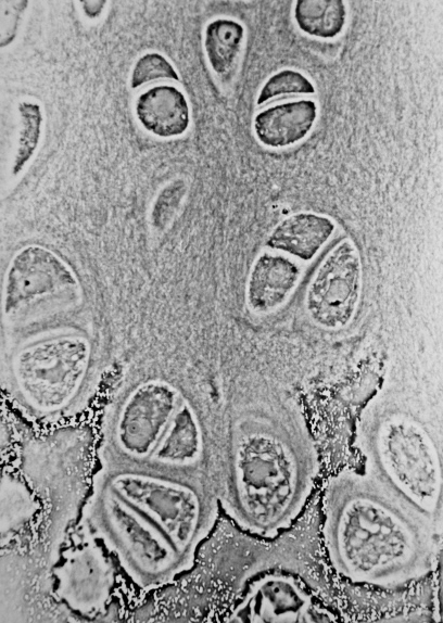
When a slice of hyaline cartilage is examined under the microscope, it is shown to consist of chondrocytes of a rounded or bluntly angular form, lying in groups of two or more in a granular, or almost homogeneous matrix. When arranged in groups of two or more, the chondrocytes have rounded, but generally straight outlines, where they are in contact with each other, and in the rest of their circumference, they are rounded.
They consist of translucent protoplasm with fine interlacing filaments and minute granules are sometimes present. Embedded in this are one or two round nuclei, having the usual intranuclear network.
The cells are contained in cavities in the matrix, called cartilage lacunae. These cavities are actually artificial gaps formed from the shrinking of the cells during the staining and setting of the tissue for examination. The inter-territorial space between the isogenous cell groups contains relatively more collagen fibers, allowing it to maintain its shape while the actual cells shrink, creating the lacunae. This constitutes the so-called 'capsule' of the space. Each lacuna is usually occupied by a single cell, but during mitosis, it may contain two, four, or even eight cells.[medical citation needed]
Hub AI
Hyaline cartilage AI simulator
(@Hyaline cartilage_simulator)
Hyaline cartilage
Hyaline cartilage is the glass-like (hyaline) and translucent cartilage found on many joint surfaces. It is also most commonly found in the ribs, nose, larynx, and trachea. Hyaline cartilage is pearl-gray in color, with a firm consistency and has a considerable amount of collagen. It contains no nerves or blood vessels, and its structure is relatively simple.
Hyaline cartilage is the most common kind of cartilage in the human body. It is primarily composed of type II collagen and proteoglycans. Hyaline cartilage is located in the trachea, nose, epiphyseal plate, sternum, and ribs.
Hyaline cartilage is covered externally by a fibrous membrane known as the perichondrium. The primary cells of cartilage are chondrocytes, which are in a matrix of fibrous tissue, proteoglycans and glycosaminoglycans.
As cartilage does not have lymph glands or blood vessels, the movements of solutes, including nutrients, occur via diffusion within the fluid compartments contiguous with adjacent tissues. Cartilage gives the structures a definite but pliable form, making them strong, but with limited mobility and flexibility. Cartilage has no nerves.
Hyaline cartilage also forms the temporary embryonic skeleton, which is gradually replaced by bone, and the skeleton of elasmobranch fish.[citation needed]
When a slice of hyaline cartilage is examined under the microscope, it is shown to consist of chondrocytes of a rounded or bluntly angular form, lying in groups of two or more in a granular, or almost homogeneous matrix. When arranged in groups of two or more, the chondrocytes have rounded, but generally straight outlines, where they are in contact with each other, and in the rest of their circumference, they are rounded.
They consist of translucent protoplasm with fine interlacing filaments and minute granules are sometimes present. Embedded in this are one or two round nuclei, having the usual intranuclear network.
The cells are contained in cavities in the matrix, called cartilage lacunae. These cavities are actually artificial gaps formed from the shrinking of the cells during the staining and setting of the tissue for examination. The inter-territorial space between the isogenous cell groups contains relatively more collagen fibers, allowing it to maintain its shape while the actual cells shrink, creating the lacunae. This constitutes the so-called 'capsule' of the space. Each lacuna is usually occupied by a single cell, but during mitosis, it may contain two, four, or even eight cells.[medical citation needed]