Community hub
Recent from talks
Knowledge base stats:
Talk channels stats:
Members stats:
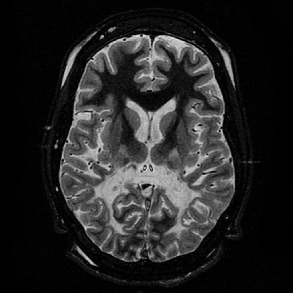
Leukodystrophy
Leukodystrophies are a group of, usually, inherited disorders, characterized by degeneration of the white matter in the brain. The word leukodystrophy comes from the Greek roots leuko, "white", dys, "abnormal" and troph, "growth". The leukodystrophies are caused by imperfect growth or development of the glial cells which produce the myelin sheath, the fatty insulating covering around nerve fibers. Leukodystrophies may be classified as hypomyelinating or demyelinating diseases, respectively, depending on whether the damage is present before birth or occurs after. While all leukodystrophies are the result of genetic mutations, other demyelinating disorders have an autoimmune, infectious, or metabolic etiology.
When damage occurs to white matter, subsequent immune responses can lead to inflammation in the central nervous system (CNS), along with the loss of myelin. The degeneration of white matter can be seen in an MRI scan and is used to diagnose leukodystrophy. Leukodystrophy is characterized by specific symptoms, including decreased motor function, muscle rigidity, and eventual degeneration of sight and hearing. While the disease is fatal, the age of onset is a key factor, as infants have a typical life expectancy of 2–8 years, while adults typically live more than a decade after onset. Treatment options are limited, although hematopoietic stem cell transplantations using bone marrow or cord blood seem to help in certain leukodystrophy types, while further research is being done.
The combined incidence of the leukodystrophies is estimated at 1 in 7,600. The majority of types involve the inheritance of an X-linked recessive, or X-linked dominant trait, while others, although involving a defective gene, are the result of spontaneous mutation rather than genetic inheritance.
Some specific symptoms vary from one type of leukodystrophy to the next, but the vast majority of symptoms are shared as the causes for the disease generally have the same effects. Symptoms are dependent on the age of onset, which is predominantly in infancy and early childhood, although the exact time of onset may be difficult to determine. Hyperirritability and hypersensitivity to the environment are common, as well as some tell-tale physical signs including muscle rigidity and a backwards-bent head. Botox therapy is often used to treat patients with spasticity. Juvenile and adult onsets display similar symptoms including a decrease or loss in hearing and vision. While children do experience optic and auditory degeneration, the course of the disease is usually too rapid, causing death relatively quickly, whereas adults may live with these conditions for many years. In children, spastic activity often precedes progressive ataxia and rapid cognitive deterioration which has been described as intellectual disability. Epilepsy is commonplace for patients of all ages. More progressed patients show weakness in deglutition, leading to spastic coughing fits due to inhaled saliva. Classic symptomatic progression of juvenile X-linked adrenoleukodystrophy is shown in the 1992 film, Lorenzo's Oil.
Course and timetable are dependent on the age of onset with infants showing a lifespan of 2–8 years, juveniles 2–10 years and adults typically 10+ years. Adults typically see an extended period of stability followed by a decline to a vegetative state and death. While treatments do exist, most are in the experimental phase and can only promise a halt in the progression of symptoms, although some gene therapies have shown some symptomatic improvement. The debilitating course of the disease has led to numerous philosophical and ethical arguments over experimental clinical trials, patients' rights and physician-assisted suicide.
While the more specific underlying causes of leukodystrophy are dependent upon the type, there are common pathophysiological patterns that can be seen amongst all types. First and foremost, leukodystrophy is a neurodegenerative disease that is always the result of both impairment and maintenance of myelin sheaths surrounding neuronal axons in the central nervous system as the result of a genetic mutation. Myelin is a fatty white substance that acts as an electrical insulator and coats axons in order to speed up impulses (i.e., action potentials) traveling down the axon. Thus, the natural result of a loss of this substance is decreased efficiency in impulse propagation. As myelin is produced by oligodendrocytes (a type of glial cell) in the central nervous system, an easy place to look for the cause is a mutation or malfunctioning of these cells and in other glial cells.[citation needed]
Inherited forms of leukodystrophy are usually the result of an autosomal recessive inheritance pattern, although dominant inheritance patterns are not unheard of, as in the case of adult-onset leukodystrophy. This means that the affected allele is carried on an autosomal, or non-sex, chromosome and is masked by the dominant, unaffected phenotype. In other words, for an individual to inherit the leukodystrophy phenotype, he or she must carry two of the recessive, mutant alleles. Krabbe disease and metachromatic leukodystrophy (MLD) are two of such type. MLD is found on human chromosome 22 at position q13.31. Another type of inherited leukodystrophy is X-linked adrenoleukodystrophy (X-ALD). As its name implies, this type of leukodystrophy is the result of a mutation found on the X-chromosome. It is also carried in a recessive pattern. The X chromosome is a sex chromosome, and since women have two "chances" of acquiring a normal X chromosome (one maternal x, one paternal x), and males only one chance (one maternal x), this disease is more likely to be seen in males than in females. The mutation resulting in adult-onset leukodystrophy is mapped at 5q23.
Although there are nearly 40 different types of leukodystrophy, many are lacking in formal and comprehensive research. Most of the research so far has been done on five types: (1) metachromatic leukodystrophy (MLD), (2) Krabbe disease, (3) X-Linked adrenoleukodystrophy (ALD), (4) Canavan disease, and (5) Alexander disease. Each type of leukodystrophy has a unique pathophysiology, but all five of these in some way affect a subset of glial cells, therefore disrupting myelin production and maintenance, and usually involve a mutation involving genes that code for enzymes necessary for the catabolism of very long chain fatty acids (VLCFAs) that are toxic to the myelin-producing cells of the central nervous system.
Hub AI
Leukodystrophy AI simulator
(@Leukodystrophy_simulator)
Leukodystrophy
Leukodystrophies are a group of, usually, inherited disorders, characterized by degeneration of the white matter in the brain. The word leukodystrophy comes from the Greek roots leuko, "white", dys, "abnormal" and troph, "growth". The leukodystrophies are caused by imperfect growth or development of the glial cells which produce the myelin sheath, the fatty insulating covering around nerve fibers. Leukodystrophies may be classified as hypomyelinating or demyelinating diseases, respectively, depending on whether the damage is present before birth or occurs after. While all leukodystrophies are the result of genetic mutations, other demyelinating disorders have an autoimmune, infectious, or metabolic etiology.
When damage occurs to white matter, subsequent immune responses can lead to inflammation in the central nervous system (CNS), along with the loss of myelin. The degeneration of white matter can be seen in an MRI scan and is used to diagnose leukodystrophy. Leukodystrophy is characterized by specific symptoms, including decreased motor function, muscle rigidity, and eventual degeneration of sight and hearing. While the disease is fatal, the age of onset is a key factor, as infants have a typical life expectancy of 2–8 years, while adults typically live more than a decade after onset. Treatment options are limited, although hematopoietic stem cell transplantations using bone marrow or cord blood seem to help in certain leukodystrophy types, while further research is being done.
The combined incidence of the leukodystrophies is estimated at 1 in 7,600. The majority of types involve the inheritance of an X-linked recessive, or X-linked dominant trait, while others, although involving a defective gene, are the result of spontaneous mutation rather than genetic inheritance.
Some specific symptoms vary from one type of leukodystrophy to the next, but the vast majority of symptoms are shared as the causes for the disease generally have the same effects. Symptoms are dependent on the age of onset, which is predominantly in infancy and early childhood, although the exact time of onset may be difficult to determine. Hyperirritability and hypersensitivity to the environment are common, as well as some tell-tale physical signs including muscle rigidity and a backwards-bent head. Botox therapy is often used to treat patients with spasticity. Juvenile and adult onsets display similar symptoms including a decrease or loss in hearing and vision. While children do experience optic and auditory degeneration, the course of the disease is usually too rapid, causing death relatively quickly, whereas adults may live with these conditions for many years. In children, spastic activity often precedes progressive ataxia and rapid cognitive deterioration which has been described as intellectual disability. Epilepsy is commonplace for patients of all ages. More progressed patients show weakness in deglutition, leading to spastic coughing fits due to inhaled saliva. Classic symptomatic progression of juvenile X-linked adrenoleukodystrophy is shown in the 1992 film, Lorenzo's Oil.
Course and timetable are dependent on the age of onset with infants showing a lifespan of 2–8 years, juveniles 2–10 years and adults typically 10+ years. Adults typically see an extended period of stability followed by a decline to a vegetative state and death. While treatments do exist, most are in the experimental phase and can only promise a halt in the progression of symptoms, although some gene therapies have shown some symptomatic improvement. The debilitating course of the disease has led to numerous philosophical and ethical arguments over experimental clinical trials, patients' rights and physician-assisted suicide.
While the more specific underlying causes of leukodystrophy are dependent upon the type, there are common pathophysiological patterns that can be seen amongst all types. First and foremost, leukodystrophy is a neurodegenerative disease that is always the result of both impairment and maintenance of myelin sheaths surrounding neuronal axons in the central nervous system as the result of a genetic mutation. Myelin is a fatty white substance that acts as an electrical insulator and coats axons in order to speed up impulses (i.e., action potentials) traveling down the axon. Thus, the natural result of a loss of this substance is decreased efficiency in impulse propagation. As myelin is produced by oligodendrocytes (a type of glial cell) in the central nervous system, an easy place to look for the cause is a mutation or malfunctioning of these cells and in other glial cells.[citation needed]
Inherited forms of leukodystrophy are usually the result of an autosomal recessive inheritance pattern, although dominant inheritance patterns are not unheard of, as in the case of adult-onset leukodystrophy. This means that the affected allele is carried on an autosomal, or non-sex, chromosome and is masked by the dominant, unaffected phenotype. In other words, for an individual to inherit the leukodystrophy phenotype, he or she must carry two of the recessive, mutant alleles. Krabbe disease and metachromatic leukodystrophy (MLD) are two of such type. MLD is found on human chromosome 22 at position q13.31. Another type of inherited leukodystrophy is X-linked adrenoleukodystrophy (X-ALD). As its name implies, this type of leukodystrophy is the result of a mutation found on the X-chromosome. It is also carried in a recessive pattern. The X chromosome is a sex chromosome, and since women have two "chances" of acquiring a normal X chromosome (one maternal x, one paternal x), and males only one chance (one maternal x), this disease is more likely to be seen in males than in females. The mutation resulting in adult-onset leukodystrophy is mapped at 5q23.
Although there are nearly 40 different types of leukodystrophy, many are lacking in formal and comprehensive research. Most of the research so far has been done on five types: (1) metachromatic leukodystrophy (MLD), (2) Krabbe disease, (3) X-Linked adrenoleukodystrophy (ALD), (4) Canavan disease, and (5) Alexander disease. Each type of leukodystrophy has a unique pathophysiology, but all five of these in some way affect a subset of glial cells, therefore disrupting myelin production and maintenance, and usually involve a mutation involving genes that code for enzymes necessary for the catabolism of very long chain fatty acids (VLCFAs) that are toxic to the myelin-producing cells of the central nervous system.