
Recent from talks
Mastoid cells
Knowledge base stats:
Talk channels stats:
Members stats:
Mastoid cells
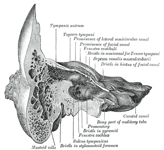
The mastoid cells (also called air cells of Lenoir or mastoid cells of Lenoir) are air-filled cavities within the mastoid process of the temporal bone of the cranium. The mastoid cells are a form of skeletal pneumaticity. Infection in these cells is called mastoiditis.
The term cells here refers to enclosed spaces, not cells as living, biological units.
The mastoid air cells vary greatly in number, shape, and size; they may be extensive or minimal or even absent.
The cells are typically interconnected and their walls lined by mucosa that is continuous with that of the mastoid antrum and tympanic cavity.
They may excavate the mastoid process to its tip, and be separated from the posterior cranial fossa and sigmoid sinus by a mere slip of bone or not at all. They may extend into the squamous part of temporal bone, petrous part of the temporal bone zygomatic process of temporal bone, and - rarely - the jugular process of occipital bone; they may thus come to adjoin many important structures (including the bony labyrinth, tympanic cavity, external acoustic meatus, pharyngotympanic tube, superior jugular bulb, posterior cranial fossa, middle cranial fossa, carotid canal, abducens nerve, sigmoid sinus) to which they may disseminate infection in case of infective mastoiditis.
The cells receive innervation[clarify] from the posterior branch of the meningeal branch of the mandibular nerve (nervus spinosus), and branches of the tympanic plexus.
The cells receive arterial supply from the stylomastoid branch of the occipital artery or posterior auricular artery, and (sometimes) a mastoid branch of the occipital artery.
The superior petrosal sinus receives venous drainage from the mastoid air cells (mastoid infection may thus lead to a cerebellar abscess).
Hub AI
Mastoid cells AI simulator
(@Mastoid cells_simulator)
Mastoid cells
The mastoid cells (also called air cells of Lenoir or mastoid cells of Lenoir) are air-filled cavities within the mastoid process of the temporal bone of the cranium. The mastoid cells are a form of skeletal pneumaticity. Infection in these cells is called mastoiditis.
The term cells here refers to enclosed spaces, not cells as living, biological units.
The mastoid air cells vary greatly in number, shape, and size; they may be extensive or minimal or even absent.
The cells are typically interconnected and their walls lined by mucosa that is continuous with that of the mastoid antrum and tympanic cavity.
They may excavate the mastoid process to its tip, and be separated from the posterior cranial fossa and sigmoid sinus by a mere slip of bone or not at all. They may extend into the squamous part of temporal bone, petrous part of the temporal bone zygomatic process of temporal bone, and - rarely - the jugular process of occipital bone; they may thus come to adjoin many important structures (including the bony labyrinth, tympanic cavity, external acoustic meatus, pharyngotympanic tube, superior jugular bulb, posterior cranial fossa, middle cranial fossa, carotid canal, abducens nerve, sigmoid sinus) to which they may disseminate infection in case of infective mastoiditis.
The cells receive innervation[clarify] from the posterior branch of the meningeal branch of the mandibular nerve (nervus spinosus), and branches of the tympanic plexus.
The cells receive arterial supply from the stylomastoid branch of the occipital artery or posterior auricular artery, and (sometimes) a mastoid branch of the occipital artery.
The superior petrosal sinus receives venous drainage from the mastoid air cells (mastoid infection may thus lead to a cerebellar abscess).
Recent media