Hubbry Logo
Sigmoid sinusSigmoid sinusMain
Open search
Sigmoid sinus
Community hub
Sigmoid sinus
logo
8 pages, 0 posts
0 subscribers
Be the first to start a discussion here.
Sigmoid sinus
Sigmoid sinus
from Wikipedia
Sigmoid Sinus
Dural veins. ("Pars sigmoid" labeled at lower right.)
Details
SourceTransverse sinus
Drains toInternal jugular vein
Identifiers
Latinsinus sigmoideus
TA98A12.3.05.108
TA24847
FMA50782
Anatomical terminology

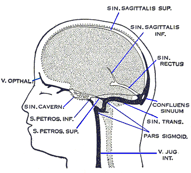
The sigmoid sinuses (sigma- or s-shaped hollow curve), also known as the pars sigmoid, are paired dural venous sinuses within the skull that receive blood from posterior transverse sinuses.

Structure

[edit]

The sigmoid sinus is a dural venous sinus situated within the dura mater. The sigmoid sinus receives blood from the transverse sinuses, which track the posterior wall of the cranial cavity, travels inferiorly along the parietal bone, temporal bone and occipital bone, and converges with the inferior petrosal sinuses to form the internal jugular vein.[1] : 795–6 

Each sigmoid sinus begins beneath the temporal bone and follows a tortuous course to the jugular foramen, at which point the sinus becomes continuous with the internal jugular vein.

Function

[edit]

The sigmoid sinus receives blood from the transverse sinuses, which receive blood from the posterior aspect of the skull. Along its course, the sigmoid sinus also receives blood from the cerebral veins, cerebellar veins, diploic veins, and emissary veins.[1] : 795 

References

[edit]
[edit]
Revisions and contributorsEdit on WikipediaRead on Wikipedia
from Grokipedia
The sigmoid sinus is a paired, S-shaped dural venous sinus situated within the of the , representing the direct continuation of the transverse sinus and primarily responsible for draining deoxygenated blood from the posterior aspects of the , including the , , and , into the . Anatomically, each sigmoid sinus begins where the transverse sinus exits the tentorium cerebelli attachment, then courses inferiorly along a groove on the inner surface of the adjacent to the mastoid process, forming its characteristic sigmoid curvature before terminating at the , where it expands into the jugular bulb. This positioning places the sinus deep within the , posteromedial to the mastoid air cells and lateral to the and . The bilateral structure exhibits variability in size and dominance, with the right sigmoid sinus often larger, influencing venous flow dynamics. The sigmoid sinus receives venous inflow primarily from the transverse sinus but also incorporates several tributaries, including the superior petrosal sinus, , , , and small veins from the inferior , , and medulla. These connections facilitate the collection of blood from the posterior and adjacent extracranial veins via emissary routes. As a valveless channel, it permits bidirectional flow, ultimately converging with the to return blood to the systemic circulation. Clinically, the sigmoid sinus's anatomical relations are critical in neurosurgical procedures, such as retrosigmoid or presigmoid approaches for posterior fossa lesions, where its variable course and proximity to the mastoid can necessitate careful management to avoid or hemorrhage. Pathologies like or may lead to or increased , highlighting its role in venous drainage integrity.

Anatomy

Location and course

The sigmoid sinus is a paired, S-shaped dural venous sinus situated in the of the base. It originates as the direct continuation of the transverse sinus at the point where the latter turns inferiorly near the posterolateral aspect of the and the free edge of the tentorium cerebelli. From its origin, the sigmoid sinus follows an initial vertical descent along the posterior aspect of the petrous before curving laterally and inferiorly in a characteristic S-shaped trajectory. This path is accommodated within a dedicated groove on the inner surface of the mastoid portion of the , positioning it immediately adjacent to the mastoid air cells. The sinus then traverses the jugular process of the as it directs toward the . Typically, the sigmoid sinus measures approximately 1 cm in diameter along its course. The sigmoid sinus concludes its intracranial course at the posterolateral aspect of the , where it dilates to form the jugular bulb before continuing extracranially as the .

Relations and tributaries

The sigmoid sinus is housed within a distinct S-shaped groove on the inner table of the mastoid portion of the and the jugular process of the , positioning it posteromedial to the mastoid air cells. This bony enclosure places the sinus posterior to the petrous apex of the and lateral to the , facilitating its role in the . In terms of soft tissue relations, the lies between the periosteal and meningeal layers of the , providing a protective dural covering. It is intimately adjacent to the and mastoid air cells, often appearing as a bluish structure beneath thin overlying bone during surgical exposure. Laterally, it is positioned relative to the and , with the cerebellar tonsils and hemisphere situated medially to its course. The major tributaries of the sigmoid sinus include the superior petrosal sinus, which joins at its superior junction with the transverse sinus. It also receives the mastoid emissary vein, which connects extracranial veins to the dural system, along with the condylar emissary vein and small veins from the , such as inferior cerebellar veins. Additionally, diploic and cerebral veins contribute to its inflow, collectively draining from the sigmoid and transverse sinus territories. Anatomically, the right sigmoid sinus is typically larger and more dominant than the left, a pattern observed in venous drainage where right-sided is less common than left-sided. This asymmetry often extends to the jugular bulb, with the right side exhibiting greater prominence due to higher flow volume.

Connections to other structures

The sigmoid sinus serves as a direct continuation of the transverse sinus, receiving venous drainage proximally at the point where the transverse sinus bends inferiorly along the petrous temporal bone. This junction marks the proximal anastomosis, facilitating the flow of blood from the posterior cranial fossa toward the base of the skull. Distally, the sigmoid sinus curves in an S-shaped path through a groove in the mastoid and jugular processes of the temporal and occipital bones, respectively, before transitioning into the internal jugular vein at the jugular foramen. Just prior to this exit, it forms the jugular bulb, a dilated segment that represents an enlargement at the confluence of the sigmoid and internal jugular systems. The walls of the sigmoid sinus, composed of the endosteal (periosteal) layer of dura adherent to the surrounding bone, particularly along the groove and at the jugular foramen, create a fixed channel that supports stable venous outflow. At the jugular foramen, the sigmoid sinus passes through the pars vascularis alongside the inferior petrosal sinus, which joins the to provide additional drainage from the . This exit pathway also accommodates IX (glossopharyngeal), X (vagus), and XI (accessory), which traverse the in close proximity to the venous structures. The indirect input from the occurs via the , integrating flows from both the sigmoid sinus and petrosal drainage before continuation into the .

Embryology and variations

Embryonic development

The sigmoid sinus forms during early embryonic development from the pro-otic sinus and segments of the primary head vein system, in conjunction with the differentiation of the dura mater's meningeal layers between weeks 6 and 8 of . This derivation involves the coalescence of venous plexuses within the surrounding the developing , where the pro-otic sinus serves as a transitional channel connecting anterior and posterior drainage pathways. Key formative stages begin with the initial appearance of the sigmoid sinus as part of a in the posterior mesencephalon around the 17- to 20-mm stage (approximately weeks 6-7), derived from the middle and posterior dural plexuses that replace portions of the primary head . By the 21-mm stage (week 8), an dorsal to the otic capsule establishes the basic course, with remodeling of the trigeminal and otic veins contributing to the transverse-sigmoid junction; this junction solidifies by week 10 (around 50 mm ), integrating drainage from the posterior fossa. These changes occur as the posterior dural plexus merges with the middle plexus, forming the sigmoid's characteristic S-shaped configuration. The development of the parallels the expansion of the and the of the , which begins around week 8 and guides the sinusal channel's caudal positioning along the dural folds. Specifically, the sigmoid portion emerges from caudal extensions of the transverse sinus, influenced by the growing otic capsule and surrounding that direct venous remodeling toward the . Asymmetry in the sigmoid sinus arises early in embryogenesis due to differential regression of embryonic veins, with the right side often persisting more prominently as a result of uneven involution and hemodynamic preferences during cranial growth.

Anatomical variations

The sigmoid sinus exhibits several anatomical variations that deviate from its typical S-shaped course along the posterior aspect of the petrous and mastoid process. or complete absence of the sinus is a recognized variant, occurring more frequently on the left side; affects approximately 10-20% of cases (often involving the contiguous transverse sinus), while aplasia is rare (less than 1%). Duplication or fenestration of the sinus wall represents another structural anomaly, though these are less prevalent and typically identified incidentally during or , contributing to irregular luminal architecture. Size variations are prominent, with the right sigmoid sinus demonstrating dominance in 60-70% of individuals, resulting in a larger cross-sectional area and correspondingly enlarged on the dominant side to accommodate increased venous outflow. The diameter of the sigmoid sinus typically ranges from 4 to 12 mm in adults, with the right side often measuring larger (average ~10 mm) compared to the left, reflecting overall in dural venous drainage patterns. Positional anomalies include high-riding configurations, where the sinus extends superiorly beyond its standard position, and lateral or anterior displacement relative to the mastoid bone, which can narrow the surgical corridor during temporal bone procedures. Sigmoid plate dehiscence, characterized by thinning or absence of the overlying bone, occurs in 5-10% of cases and is more common with advancing age. These variations often correlate with skull base asymmetry, influencing the overall pattern of cerebral venous dominance, and are best assessed using CT venography, which provides detailed visualization of sinus patency and morphology for preoperative planning.

Physiology

Venous drainage role

The sigmoid sinus functions primarily as a conduit for deoxygenated blood originating from the , receiving venous inflow mainly from the transverse sinus and channeling it toward the to facilitate cerebral venous return. This sinus collects blood from an extensive drainage territory, including indirect contributions from the inferior sagittal sinus via the transverse sinus, as well as direct tributaries from local structures such as the inferior through the inferior cerebellar veins, the via labyrinthine veins, and the posterior through the vein of Labbé (which drains into the transverse sinus). Additional inputs include the mastoid and condylar , the venous plexus of the , and small veins from the and , ensuring comprehensive drainage of the posterior fossa region. Physiologically, the sigmoid sinus is vital for maintaining efficient cerebral venous outflow, serving as a key segment in the pathway that returns blood from deeper cerebral surfaces and posterior structures to ic circulation. It operates within a low-pressure that supports steady drainage from the parenchyma and , preventing venous congestion. As part of the dural venous network, the sigmoid sinus acts as a vessel, expanding to buffer fluctuations in and thereby preserving unimpeded blood flow during variations in cerebral volume or activity. At the jugular foramen, the sigmoid sinus enlarges into the jugular bulb before continuing as the , completing its role in extracranial venous drainage.

Hemodynamic characteristics

The sigmoid sinus exhibits predominantly laminar blood flow under normal physiological conditions, with maximal systolic velocities up to 20 cm/s as measured by ultrasound in dural venous structures. This steady, streamlined flow pattern facilitates efficient drainage of venous blood from upstream structures, including the transverse sinus. However, at the junction between the transverse and sigmoid sinuses, the inherent curvature of the S-shaped pathway introduces mild flow disturbances, potentially leading to localized transitions toward turbulent or helical patterns due to geometric constraints that accelerate and redirect the flow. A modest is maintained along the sigmoid sinus, typically less than 5 mmHg from the transverse sinus to the jugular bulb in healthy individuals, which supports unidirectional drainage and minimizes risk through the sinus's tortuous morphology. This gradient arises from frictional losses and gravitational effects in the upright posture, ensuring balanced intracranial venous outflow without excessive resistance in healthy individuals. Flow regulation in the sigmoid sinus is modulated by structural features such as dural attachments that anchor the sinus walls, providing compliance to accommodate fluctuations in and prevent collapse or overdistension. Although generally lack traditional valves, chordae Willisii—thin endothelial strands within the sinus lumen—aid in directing flow and reducing stagnation, particularly in response to hemodynamic variations. The sinus demonstrates adaptability to intracranial pressure changes via its elastic dural envelope, which buffers volume shifts during activities like Valsalva maneuvers. Quantitative assessment of sigmoid sinus hemodynamics is achieved through non-invasive modalities such as Doppler ultrasound, which measures velocity profiles, and magnetic resonance venography, which visualizes flow direction and asymmetry. Asymmetry in sinus dominance significantly influences cerebral hemodynamics, with the right sigmoid sinus handling a greater volume of drainage in approximately 70% of cases, reflecting broader patterns of transverse sinus laterality.

Clinical significance

Associated pathologies

The sigmoid sinus is susceptible to , a condition known as sigmoid sinus (SST), which is often secondary to infectious processes such as or acute . This complication arises due to the sinus's anatomical proximity to the mastoid air cells, allowing direct spread of infection. Common presenting symptoms include severe headache, otalgia, fever, and , reflecting increased from venous outflow obstruction. The incidence of SST in acute is estimated at 0-3%, though it has declined with use. SST can propagate proximally to the transverse sinus or distally to the , potentially leading to extensive cerebral or septic emboli. Risk factors include hypercoagulable states, such as or , as well as , which increases blood viscosity and promotes clot formation. Diagnosis typically involves (MRI) or magnetic resonance venography (MRV), which reveal filling defects or flow voids in the affected sinus. Other pathologies associated with the sigmoid sinus include dehiscence, where thinning or absence of the overlying bone leads to pulsatile due to turbulent blood flow transmission to adjacent structures. This condition accounts for up to 25% of pulsatile cases and is often identified via temporal bone computed tomography (CT). Stenosis of the sigmoid sinus contributes to (IIH) by impeding venous drainage and elevating , frequently co-occurring with transverse sinus narrowing. Rare tumors, such as meningiomas, can compress the sigmoid sinus, causing venous hypertension and symptoms like or visual disturbances. Historically, SST has been linked to , a septic originating from oropharyngeal s that extends to the via the sigmoid sinus. Anticoagulation is commonly employed in SST management to prevent propagation, alongside addressing the underlying , though its role in septic cases remains supportive rather than definitive.

Surgical and procedural considerations

The sigmoid sinus plays a critical role in neurosurgical and otologic procedures, particularly in approaches to the posterior fossa and cerebellopontine angle. In the retrosigmoid craniotomy, commonly used for resection of acoustic neuromas (vestibular schwannomas) and other petrous region tumors, the sinus serves as a key anatomical landmark, with the bone overlying it often skeletonized to expand the surgical corridor and facilitate dural retraction. This technique involves careful drilling along the sinus edges to avoid injury while maximizing exposure. Similarly, during microvascular decompression for trigeminal neuralgia or hemifacial spasm, the dura adjacent to the sigmoid-transverse sinus junction is precisely identified and incised to access the cranial nerve root entry zones. In select cases, such as aggressive petroclival meningiomas encroaching on the sinus, ligation may be required after confirming collateral venous drainage to prevent hemodynamic compromise. Intraoperative injury to the sigmoid sinus remains a significant risk in posterior fossa surgery, with venous sinus compromise occurring in approximately 5-12% of cases due to its proximity to the operative field and variable position. Management strategies emphasize rapid hemostasis through direct compression, suturing of the dural tear, or patch grafting with autologous tissue or synthetic materials to restore patency and minimize thrombosis risk. For patients with high-flow variants or dominant sigmoid sinuses identified preoperatively, embolization may be performed to reduce intraoperative blood loss, particularly in hypervascular tumor resections involving the sinus wall. Thrombosis represents a potential postoperative complication, warranting vigilant monitoring. Preoperative imaging is essential for procedural planning, with CT angiography routinely employed to delineate the sigmoid sinus position relative to surface landmarks like the asterion, which approximates the transverse-sigmoid junction in about 87% of cases and guides burr hole placement to avoid inadvertent entry. Anatomical variations, such as anterior displacement of the sinus, heighten complication risks during by increasing the likelihood of unintended exposure or laceration, necessitating tailored surgical trajectories. In translabyrinthine approaches for removal, the sigmoid sinus is typically decompressed and retracted laterally to broaden access to the internal auditory canal, often with bone removal posterior to the sinus for optimal exposure without compromising venous outflow. Postoperative monitoring for involves to assess flow velocities and detect early occlusion, enabling prompt anticoagulation if needed.

References

Add your contribution
Related Hubs
User Avatar
No comments yet.