Hubbry Logo
Multifidus muscleMultifidus muscleMain
Open search
Multifidus muscle
Community hub
Multifidus muscle
logo
8 pages, 0 posts
0 subscribers
Be the first to start a discussion here.
Multifidus muscle
Multifidus muscle
from Wikipedia
Multifidus muscle
Deep muscles of the back. (Multifidus shaded in red.)
Sacrum, dorsal surface. (Multifidus attachment outlined in red.)
Details
OriginSacrum, erector spinae aponeurosis, PSIS, and iliac crest
InsertionSpinous process
NervePosterior branches
ActionsProvides proprioceptive feedback and input due to high muscle spindle density; Bilateral backward extension, unilateral ipsilateral side-bending and contralateral rotation.
Identifiers
Latinmusculus multifidus spinae
TA98A04.3.02.202
TA22276
FMA22827
Anatomical terms of muscle

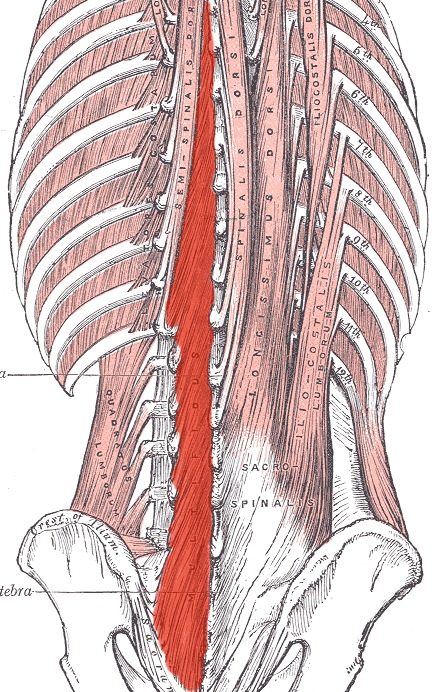
The multifidus (multifidus spinae; pl.: multifidi) muscle consists of a number of fleshy and tendinous fasciculi, which fill up the groove on either side of the spinous processes of the vertebrae, from the sacrum to the axis. While very thin, the multifidus muscle plays an important role in stabilizing the joints within the spine. The multifidus is one of the transversospinales.

Located just superficially to the spine itself, the multifidus muscle spans three joint segments and works to stabilize these joints at each level.

The stiffness and stability makes each vertebra work more effectively, and reduces the degeneration of the joint structures caused by friction from normal physical activity.

These fasciculi arise:

Each fasciculus, passing obliquely upward and medially, is inserted into the whole length of the spinous process of one of the vertebræ above.

These fasciculi vary in length: the most superficial, the longest, pass from one vertebra to the third or fourth above; those next in order run from one vertebra to the second or third above; while the deepest connect two adjacent vertebrae.

The multifidus lies deep relative to the spinal erectors, transverse abdominis, abdominal internal oblique muscle and abdominal external oblique muscle.

Atrophy and association with low back pain

[edit]

Dysfunction in the lumbar multifidus muscles is strongly associated with low back pain. The dysfunction can be caused by inhibition of pain by the spine. The dysfunction frequently persists even after the pain has disappeared. Such persistence may help explain the high recurrence rates of low back pain. Persistent lumbar multifidus dysfunction is diagnosed by atrophic replacement of the multifidus with fat, as visualized by magnetic resonance imaging or ultrasound.[1][2] One way to help recruit and strengthen the lumbar multifidus muscles is by tensing the pelvic floor muscles for a few seconds "as if stopping urination midstream".[3]

Additional images

[edit]

See also

[edit]

References

[edit]
[edit]
Revisions and contributorsEdit on WikipediaRead on Wikipedia
from Grokipedia
The multifidus muscle is a group of short, triangular-shaped deep back muscles belonging to the transversospinales group, extending along the vertebral column from the sacral region to the cervical spine, where it primarily functions to stabilize individual vertebral segments and facilitate controlled spinal movements such as extension, lateral flexion, and . Anatomically, the multifidus is organized into cervical, thoracic, and portions, with its fibers arranged in a multipennate structure that spans two to five vertebral levels. In the region, where it is most developed and thickest, the muscle originates from the mamillary processes of the , the posterior surface of the , the , and the posterior sacroiliac ; these fibers then insert into the lateral aspects and tips of the spinous processes two to five levels superiorly, often interdigitating with the supraspinous ligaments and . In the thoracic and cervical regions, origins shift to the transverse processes of the and superior articular processes of C4-C7, respectively, maintaining the pattern of ascending insertions on spinous processes. The muscle's layered architecture, consisting of up to four distinct strata in the area, allows for precise intersegmental control, with deeper layers connecting contiguous vertebrae and superficial layers extending further to support broader stability. Innervation of the multifidus arises from the medial branches of the posterior rami of the spinal nerves corresponding to each region, enabling segmental activation for fine-tuned . Blood supply varies by level: the cervical portion receives from vertebral, deep cervical, and occipital arteries; the thoracic from dorsal branches of posterior intercostal and subcostal arteries; and the from arteries and lateral sacral branches. Functionally, bilateral contraction produces spinal extension, while unilateral action causes ipsilateral lateral flexion and contralateral ; however, its primary role is proprioceptive and stabilizing, preventing excessive motion at facet joints and maintaining posture during , load transfer, and daily activities. Clinically, the multifidus is critical for lumbar spine health, as its or dysfunction—often seen in chronic low back pain, disc herniation, , or post-surgical scenarios like —can lead to segmental instability, fibrosis, and impaired , persisting for years without targeted rehabilitation. Its role in exercises underscores its importance in conservative treatments for back disorders, highlighting the need for preserving its multipennate fiber integrity to support intervertebral mobility across sagittal, frontal, and transverse planes.

Anatomy

Origin and insertion

The multifidus muscle originates from multiple sites along the posterior spine, varying by region. In the lumbar region, it arises from the mammillary processes of the (L1–L5), the , the between the sacral foramina, the posterior sacroiliac ligament, and the erector spinae aponeurosis. In the thoracic region, the origins are from the transverse processes of the (T1–T12). For the cervical region, the muscle originates from the superior articular processes of the C4–C7 vertebrae. The insertions of the multifidus occur on the lateral aspects and tips of the spinous processes of vertebrae located 2 to 4 levels superior to the origins. These attachments form oblique, overlapping bands that contribute to the muscle's segmented architecture. Regional differences in the multifidus attachments include variations in length and orientation, with the region featuring the most robust and multilayered connections, including interdigitating fascicles from the mamillary processes and superior articular processes that insert via tendinous slips to spinous processes two levels above. In contrast, thoracic and cervical attachments are generally shorter and more uniform in their transverse process or articular origins, reflecting adaptations to regional spinal dynamics.

Structure and relations

The multifidus muscle forms part of the transversospinales group of deep back muscles, alongside the semispinalis and rotatores muscles, and collectively these muscles occupy the grooves between the spinous and transverse processes of the vertebrae. It presents as a series of short, triangular-shaped muscle bundles that extend continuously from the sacrum to the axis (C2) vertebra, with regional variations in the cervical, thoracic, and lumbar segments. The lumbar portion is the most developed, exhibiting the largest cross-sectional area and greatest volume among the regions, which underscores its prominence in supporting the lower spine. The muscle is organized into layered fascicles based on their depth and segmental span, reflecting adaptations for both local and broader spinal support. The deepest layer consists of short fascicles that span one vertebral segment, originating from the superior articular processes and laminae of the below to insert on the facet capsule and lamina of the above, providing targeted segmental stability. Intermediate fascicles bridge 2-3 segments, while the superficial layer features longer fascicles extending 3-5 (or up to 6) vertebral levels, arranged in a multipennate with interdigitating fibers that enhance force transmission across multiple levels. These layers lack clear cleavage planes in some regions, allowing for integrated function, though they are delineated by thin septa. In terms of positional relations, the multifidus lies deep to the and the semispinalis group, positioned medially within the paraspinal compartment and enclosed posteriorly by the thoracodorsal . It is superficial to the rotatores and interspinales muscles, while overlying the laminae of the vertebrae and the posterior surface of the , filling the shallow grooves formed by these bony structures. Laterally, it abuts the facet joints and transverse processes, contributing to the medial boundary of the paraspinal muscles. Histologically, the multifidus is composed predominantly of type I slow-twitch oxidative fibers, comprising approximately 59% of its fiber population, which are notably larger in cross-sectional area compared to those in adjacent muscles like the erector spinae, supporting its role in sustained postural maintenance. Type II fibers, including subtypes IIa, IIax, and IIx, make up the remaining roughly 41%, enabling intermittent phasic activity. The muscle also exhibits a high of muscle spindles, particularly concentrated in the deep layers, which provide proprioceptive feedback essential for spinal position sense and control.

Function

Spinal stabilization

The multifidus muscle serves as a primary stabilizer of individual spinal segments in the region, with its deep fibers attaching directly to the laminae and mamillary processes to provide precise intersegmental control. This architecture enables the muscle to counteract excessive intervertebral translation and , particularly during dynamic activities such as walking or lifting, by generating stabilizing forces that maintain segmental alignment. Its fascicular organization, spanning two to five vertebral levels, further enhances this segmental control, allowing for targeted stabilization without broad spinal motion. In coordination with the transversus abdominis and muscles, the multifidus forms a integrated "core" system that supports lumbopelvic stability, functioning as an anatomical to distribute forces across the spine and during posture maintenance and movement. This synergistic ensures that the multifidus contributes to overall spinal , accounting for more than two-thirds of the total stiffness in the spine, which is essential for withstanding compressive loads and preventing shear forces at the intervertebral levels. The muscle's high density of muscle spindles provides substantial proprioceptive feedback, enabling fine-tuned neuromuscular adjustments to preserve neutral spine posture and natural under varying loads. This sensory input allows for rapid responses to postural perturbations, optimizing spinal alignment and minimizing deviations from the neutral zone. Additionally, by actively sharing mechanical loads, the multifidus reduces stress on passive structures like ligaments and intervertebral discs; for instance, simulations show that its absence can increase compressive forces by up to 1.82 times during forward flexion.

Movements produced

The multifidus muscle, when contracting bilaterally, primarily produces extension of the vertebral column, with a particular emphasis on the and thoracic regions to counteract flexion and restore neutral posture. This action arises from its oblique orientation spanning multiple vertebral levels, pulling the spinous processes toward the transverse processes and elevating the spinous processes. Unilateral contraction of the multifidus generates ipsilateral lateral flexion (side-bending) of the spine while producing contralateral , such as the right multifidus rotating the spine to the left. These movements result from the muscle's attachment from transverse processes to spinous processes, creating a that bends and twists individual segments. Regional variations in multifidus actions reflect its segmental architecture across the spine. In the cervical region, the multifidus aids in head and neck rotation, extension, and lateral flexion, supporting precise control during upper body turns. Conversely, the lumbar multifidus emphasizes segmental control over global motion, fine-tuning small intervertebral adjustments rather than large-scale trunk movements to maintain alignment during daily activities. The multifidus integrates with other muscles, such as the semispinalis and rotatores in the transversospinalis group, to facilitate compound movements like trunk twisting, where it contributes rotational alongside oblique abdominal muscles for coordinated spinal dynamics.

Innervation and blood supply

Innervation

The multifidus muscle is innervated by the medial branches of the dorsal (posterior) rami of the s, with each segment corresponding to the vertebral levels it spans, such as L1 through L5 for the portion. These medial branches emerge from the dorsal rami after they divide near the and travel posteriorly to supply the deep back muscles, including the multifidus. In the cervical and thoracic regions, similar segmental contributions occur from the respective levels. This segmental innervation pattern enables independent control of the muscle's fascicles, as each medial branch supplies specific bundles without interconnecting branches to adjacent segments. The deep fibers of the multifidus, which span fewer vertebral levels and attach directly to the laminae, receive particularly localized neural input from these branches, facilitating precise intersegmental adjustments. This architecture supports fine-tuned activation for spinal stability, with each fascicle responding to its dedicated nerve supply. There is no crossover innervation in the multifidus; each band or fascicle activates exclusively via the medial branch of the dorsal ramus from the same vertebral level as its insertion on the spinous process. For instance, the L3 medial branch innervates only the multifidus fascicles inserting onto the L3 spinous process, ensuring isolated segmental function without overlap from neighboring levels. This precise, non-communicating supply underscores the muscle's role in localized control mechanisms.

Blood supply

The multifidus muscle is supplied by segmental arteries that align with its vertebral levels, providing consistent vascular support for its continuous activity in spinal stabilization. This pattern of blood flow facilitates efficient oxygenation and nutrient delivery to the muscle fibers, which are essential for maintaining posture over extended periods. In the cervical region, the multifidus receives branches from the , deep cervical artery, and occipital artery, originating primarily from the subclavian and external carotid arteries. These vessels ensure robust to the upper segments, supporting the muscle's role in mobility and stability. For the thoracic region, blood supply comes from the dorsal branches of the posterior and the subcostal artery, which arise directly from the . This arrangement matches the segmental distribution of the thoracic multifidus, aiding its contribution to torso posture. In the lumbar region, the multifidus is vascularized by the arteries (branching from the ) and the lateral sacral artery (from the ). These arteries provide the primary supply to the more developed lumbar portions, critical for load-bearing and functions.

Clinical significance

Role in low back pain

The multifidus muscle undergoes significant atrophy and fatty infiltration in individuals with chronic low back pain (LBP), often manifesting unilaterally at the levels corresponding to the painful segment. This pathological change is characterized by a reduced cross-sectional area (CSA) of the muscle on the symptomatic side, with studies showing significant decreases in multifidus CSA at the affected level and adjacent segments in chronic LBP patients compared to those with acute pain. Furthermore, fatty infiltration, quantified by increased fat-to-muscle ratios on MRI, is strongly associated with LBP severity, with severe infiltration linked to higher odds of experiencing LBP (odds ratio 9.2 for lifetime LBP). These alterations persist even after pain resolution, contributing to elevated recurrence rates due to ongoing muscle dysfunction. Beyond LBP, multifidus dysfunction plays a role in other spinal conditions. In , particularly adolescent idiopathic and degenerative types, asymmetric is observed, with greater degeneration on the concave side of the curve, correlating with curve severity and contributing to spinal (r = -0.45 for CSA and ). Post-surgical scenarios, such as after , often result in significant multifidus due to and iatrogenic damage from open approaches, leading to persistent and higher reoperation rates; minimally invasive techniques preserve muscle better, reducing by up to 50%. Weakness and atrophy of the multifidus impair its role in spinal stabilization, leading to inefficient control of intervertebral shear forces and increased mechanical stress on the lumbar spine. This dysfunction exacerbates spinal instability, as the multifidus normally provides over two-thirds of the stiffness needed to maintain segmental stability during motion. Consequently, reduced multifidus function is implicated in the progression of disc degeneration, with fatty atrophy showing a positive correlation (r=0.37) to the grade of lumbar disc degeneration at levels like L4/L5 and L5/S1. Similarly, multifidus atrophy correlates with facet joint osteoarthritis, evidenced by negative associations between functional CSA and facet joint degeneration scores, independent of age or disc pathology. MRI evidence consistently demonstrates reduced multifidus CSA and elevated fatty infiltration in LBP patients, particularly those with disc herniation, where atrophy is more pronounced ipsilateral to the . At the histological level, affected multifidus segments exhibit increased fat content alongside neurogenic and myopathological changes, including fiber disorganization and , which correlate with symptom duration. These findings underscore the multifidus's central role in LBP , where muscle degeneration at painful levels perpetuates instability and degenerative cascades.

Diagnostic and therapeutic approaches

Diagnostic approaches to multifidus muscle health primarily involve non-invasive and electrophysiological techniques to evaluate morphology, composition, and function. Real-time enables measurement of the multifidus cross-sectional area (CSA) and thickness changes during contraction, providing reliable assessment of muscle size and in clinical settings, particularly for patients with (LBP). (MRI) quantifies ty infiltration within the multifidus, a key indicator of degeneration, with methods like water/ MRI allowing precise of muscle quality and its association with LBP severity. (EMG) assesses patterns, revealing delayed or reduced multifidus recruitment in individuals with chronic LBP compared to healthy controls, often through fine-wire intramuscular recordings for deep fiber analysis. Therapeutic interventions emphasize targeted exercises to enhance segmental control and restore multifidus function. Strengthening exercises such as the bird-dog (quadruped contralateral arm and ) promote high levels of multifidus , with EMG studies showing greater deep fiber recruitment during this movement than in simpler core exercises. Side planks similarly elicit substantial multifidus engagement, particularly when performed with hollowing maneuvers, as evidenced by increased muscle thickness on during these holds. Pelvic floor co-activation strategies, involving simultaneous contraction of muscles with transverse abdominis, facilitate multifidus recruitment by enhancing intra-abdominal pressure and trunk stability, supporting integrated core training protocols. Evidence from rehabilitation programs demonstrates that targeted multifidus training restores muscle size and function, with exercises leading to significant increases in CSA on in both healthy individuals and those with chronic LBP. Such interventions reduce LBP recurrence rates, as specific stabilizing exercises have been shown to lower the incidence of symptoms compared to general advice or no treatment in first-episode patients followed over 2-3 years. Post-surgical rehabilitation prioritizes early multifidus activation, with dynamic stabilization exercises improving outcomes after microdiscectomy by enhancing muscle endurance and reducing postoperative and disability. Preventive strategies incorporate core training programs to maintain multifidus integrity in at-risk populations, such as those with recurrent LBP or occupational demands involving spinal loading, where regular stabilization exercises prevent and support long-term spinal stability.

References

Add your contribution
Related Hubs
User Avatar
No comments yet.