Hubbry Logo
search
logo
1821381

Perineal nerve

logo
Community Hub0 Subscribers
Read side by side
from Wikipedia
Perineal nerve
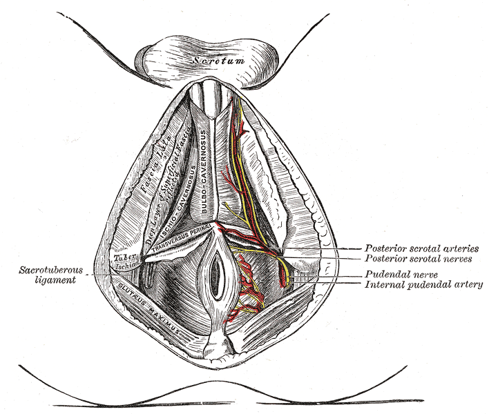
The superficial branches of the internal pudendal artery. (Perineal nerve visible but not labeled.)
Pudendal nerve, its course through the lesser sciatic foramen, and branches, including inferior anal at bottom right.
Details
FromPudendal nerve
InnervatesPerineum, scrotum or labia majora, superficial transverse perineal muscle, bulbospongiosus muscle, ischiocavernosus muscle, bulb of penis, levator ani, external anal sphincter
Identifiers
Latinnervi perineales
TA98A14.2.07.039
TA26556
FMA21866
Anatomical terms of neuroanatomy

The perineal nerve is a nerve of the pelvis. It arises from the pudendal nerve in the pudendal canal. It gives superficial branches to the skin, and a deep branch to muscles. It supplies the skin and muscles of the perineum. Its latency is tested with electrodes.

Structure

[edit]

The perineal nerve is a branch of the pudendal nerve.[1][2] It lies below the internal pudendal artery. It accompanies the perineal artery. It passes through the pudendal canal for around 2 or 3 cm.[1][2] Whilst still in the canal, it divides into superficial branches and a deep branch.[1] The superficial branches of the perineal nerve become the posterior scrotal nerves in men,[3] and the posterior labial nerves in women.[1] The deep branch of the perineal nerve (also known as the "muscular" branch) travels to the muscles of the perineum.[1] Both of these are superficial to the dorsal nerve of the penis or the dorsal nerve of the scrotum.[4]

Function

[edit]

The perineal nerve supplies the skin and muscles of the perineum.[1] The superficial branches supply sensation to the perineum, and the scrotum in men or the labia majora in women.[1] The deep branch supplies superficial transverse perineal muscle, the bulbospongiosus muscle, the ischiocavernosus muscle, the bulb of penis, levator ani, and the external anal sphincter.[1]

Clinical significance

[edit]

The latency of the perineal nerve can be measured with electrodes.[5] It is used to test nerve function.[5]

Additional images

[edit]

References

[edit]
[edit]
Revisions and contributorsEdit on WikipediaRead on Wikipedia
from Grokipedia
The perineal nerve is a mixed peripheral nerve that represents the largest terminal branch of the pudendal nerve, deriving from the ventral rami of the sacral spinal nerves S2-S4, and it provides essential motor and sensory innervation to the structures of the perineum and urogenital triangle.[1] It originates within or just after the pudendal canal in the lateral wall of the ischioanal fossa, where the pudendal nerve bifurcates, and courses anteriorly through the perineum accompanied by the perineal artery for approximately 2-3 cm before dividing into its superficial and deep branches.[2] This nerve plays a critical role in pelvic floor function, including control of micturition, defecation, and sexual responses, by innervating key muscles and providing cutaneous sensation to the posterior scrotum in males or the labia minora and vaginal vestibule in females.[3] The superficial branch of the perineal nerve is primarily sensory, supplying the skin of the urogenital triangle, including the posterior scrotal nerves in males and the posterior labial nerves in females, which transmit sensations from the perineal skin and adjacent genital areas.[1] In contrast, the deep branch is predominantly motor, innervating the muscles of the superficial and deep perineal pouches, such as the bulbospongiosus (which aids in urethral compression and erection), ischiocavernosus (supporting penile or clitoral erection), superficial transverse perineal (stabilizing the perineal body), and external urethral sphincter (facilitating urinary continence).[2] Additionally, it contributes fibers to the levator ani muscle, enhancing overall pelvic floor support.[3] Clinically, the perineal nerve is vulnerable to injury during childbirth, pelvic surgeries, or prolonged pressure, potentially leading to perineal pain, urinary incontinence, sexual dysfunction, or sensory deficits in the urogenital region.[1] Conditions like pudendal nerve entrapment syndrome, which can affect the perineal branch, may cause chronic neuropathic pain impacting up to 1 in 100,000 individuals, often managed through nerve blocks or physical therapy.[4] Its anatomical course through the pudendal canal underscores its importance in procedures like pudendal nerve blocks for labor analgesia, where targeted anesthesia alleviates perineal discomfort.[1]

Anatomy

Origin and relations

The perineal nerve originates as the second primary branch of the pudendal nerve, which derives from the ventral rami of the S2-S4 sacral spinal nerves, within the pudendal canal (also known as Alcock's canal). This canal forms a fascial tunnel on the lateral wall of the ischioanal fossa, created by a splitting of the obturator fascia covering the medial aspect of the obturator internus muscle.[1][5] The perineal nerve emerges distal to the inferior rectal nerve—the first branch of the pudendal nerve, which courses medially through the ischioanal fossa—and proximal to the dorsal nerve of the penis or clitoris, the terminal continuation of the pudendal nerve. In terms of spatial relations, it lies medial to the obturator internus muscle, which forms the lateral boundary of the canal, and inferior to the levator ani muscle of the pelvic floor. The nerve maintains close proximity to the sacrospinous and sacrotuberous ligaments, as the parent pudendal neurovascular bundle passes between these ligaments near the ischial spine before entering the canal; the perineal nerve itself is positioned lateral to the main pudendal neurovascular bundle within this confined space. Additionally, upon exiting the canal, the perineal nerve relates anteriorly to the perineal membrane and passes through the urogenital diaphragm (deep perineal pouch) to reach the superficial perineal space.[1][5] Anatomical variations in the origin of the perineal nerve include occasional multiple trunk formations from the pudendal nerve, such as double or triple trunks, observed in approximately 36.6% and 11.5% of cases, respectively, in cadaveric studies of the extrapelvic pudendal nerve. Less commonly, communications or fusions with adjacent nerves, like the sciatic nerve, occur in up to 26.9% of specimens, potentially altering the precise branching point within the pudendal canal.[5]

Course and distribution

The perineal nerve arises as the largest terminal branch of the pudendal nerve, typically within the distal portion of the pudendal canal or immediately after its exit.[2][3] It emerges inferiorly from the pudendal canal, accompanied by the perineal artery and vein, and pierces the perineal membrane to enter the superficial perineal pouch.[2][6] From there, the nerve travels transversely and slightly anteriorly across the ischioanal fossa, passing over the superficial transverse perineal muscle toward the perineal body.[2][3][7] In the superficial perineal pouch, the perineal nerve continues forward along the lateral aspect of the urogenital triangle, supplying the anterior perineal region with its general distribution pattern.[2][6] Its fibers distribute to superficial structures within the urogenital triangle, penetrating the membranous layer of the superficial perineal fascia, known as Colles' fascia, to reach underlying tissues.[3][8] The course length of the perineal nerve after exiting the pudendal canal typically measures 2-3 cm, though anatomical variations can extend this to 2-4 cm in some individuals.[2][9] A potential site for compression occurs at the inner margin of the falciform process of the sacrospinous ligament, where the nerve may become entrapped, contributing to perineal neuropathies.[10] Modern imaging techniques, such as magnetic resonance neurography at 3T, enable visualization of the perineal nerve's trajectory within the pudendal canal and its distal course, aiding in the diagnosis of entrapment syndromes.[11][12]

Branches and terminal divisions

The perineal nerve, the largest terminal branch of the pudendal nerve, divides within the superficial perineal pouch into a superficial branch and a deep branch, each with distinct distributions.[8][2] The superficial perineal nerve provides sensory innervation and typically bifurcates into two to four small twigs that supply the skin of the posterior scrotum in males or the posterior labia majora and minora in females, as well as the adjacent posterior perineal skin.[3][7] These twigs course alongside the posterior scrotal or labial branches of the internal pudendal artery, forming terminal anastomoses that enhance regional vascular-nerve coordination.[8] Sex-specific differences in the superficial branch include a broader cutaneous territory in females, extending to the vaginal vestibule and lower vaginal canal, compared to the more confined scrotal coverage in males.[3] The deep perineal nerve carries both motor and sensory fibers, piercing the perineal membrane to reach its targets. It provides motor innervation to the bulbospongiosus, ischiocavernosus, superficial transverse perineal, and external urethral sphincter muscles, supporting perineal structural integrity.[2][7][1] Sensory contributions from this branch include somatic innervation to structures within the superficial perineal space and the perineal membrane itself.[3] Additionally, in a notable anatomical variant observed in up to 60% of cases, accessory branches from the deep perineal nerve may contribute to the innervation of the pubococcygeus component of the levator ani muscle.[13] Terminal divisions of both branches occasionally exhibit rare anomalies, such as duplication of the perineal nerve trunk proximal to its bifurcation, though such variations are infrequently documented in cadaveric studies.[8]

Function

Sensory innervation

The perineal nerve primarily provides sensory innervation through its superficial branch, which transmits general somatic sensations including touch, pain, and temperature to the skin of the posterior perineum, encompassing the midline raphe and adjacent areas.[3][14] This branch arises as a terminal division of the perineal nerve within the urogenital triangle and distributes cutaneous fibers to the posterior scrotum in males or the posterior labia majora and minora in females, as well as the perineal skin with minimal overlap from the inferior rectal nerve.[1][15] Sex-specific variations in sensory coverage reflect anatomical differences, with females exhibiting supply to the labia minora, vaginal vestibule via the superficial and deep branches, and inferior portion of the vaginal canal primarily via the deep branch, while males receive innervation primarily to the posterior scrotum and perineal skin.[3][1] Anteriorly, there is sensory overlap with the ilioinguinal nerve, which supplies the mons pubis and anterior aspects of the scrotum or labia majora.[1] The deep branch contributes minor sensory input to somatic structures such as erectile tissues and the lower vaginal canal, though its primary role is motor.[3] The perineal nerve serves as the afferent limb in the perineal reflex arc, particularly for the bulbocavernosus reflex, where sensory stimuli from the perineum or genitalia trigger contraction of the bulbocavernosus muscle via sacral spinal segments S2-S4.[16] Irritation of the perineal nerve, often seen in pudendal neuralgia associated with prolonged cycling, can produce referred pain patterns radiating to the perineum, genitalia, and posterior thigh, mimicking chronic pelvic pain syndromes.[17][18]

Motor innervation

The perineal nerve, a terminal branch of the pudendal nerve arising from the ventral rami of spinal nerves S2-S4, provides somatic motor innervation primarily via its deep branch to the muscles of the superficial and deep perineal pouches. This deep branch supplies the bulbospongiosus muscle, which compresses the bulb of the penis (in males) or vestibular bulb (in females) and constricts the spongy urethra, facilitating urethral closure during ejaculation, expulsion of residual urine, and rhythmic contractions during orgasm. It also innervates the ischiocavernosus muscle, which compresses the crus of the penis or clitoris to maintain erectile rigidity by increasing intracavernosal pressure. Additionally, the deep branch targets the superficial transverse perineal muscle, which stabilizes the perineal body and central tendinous point, contributing to overall pelvic floor integrity during muscle contractions, as well as the external urethral sphincter for voluntary urinary continence, the deep transverse perineal muscle, and in females, the compressor urethrae and sphincter urethrovaginalis. The perineal nerve also contributes fibers to the levator ani muscle, enhancing pelvic floor support.[1][19][20] These motor supplies play a critical role in pelvic floor dynamics, enabling coordinated muscle actions that support micturition by aiding voluntary control of the external urethral sphincter, defecation through perineal support, and sexual function via erection maintenance and ejaculatory propulsion. The somatic motor fibers originate from motoneurons in Onuf's nucleus, a distinct cluster in the ventral horn of the sacral spinal cord (S2-S4), which exclusively innervates striated pelvic floor musculature without autonomic involvement in these efferent pathways. The perineal nerve itself contains no autonomic fibers for motor control of these skeletal muscles, distinguishing its role from visceral innervation elsewhere in the pelvis.[1][21] Electromyography (EMG) serves as a key diagnostic tool for evaluating motor innervation by the perineal nerve, recording motor unit action potentials from innervated muscles such as the bulbospongiosus, ischiocavernosus, and superficial transverse perineal. Concentric needle EMG, for instance, detects spontaneous activity like fibrillation potentials indicating denervation or polyphasic potentials suggesting reinnervation, providing insights into neuromuscular integrity and pelvic floor function. Kinesiological EMG further assesses muscle activation timing during dynamic tasks, such as sphincter coordination in urodynamic studies.[21]

Clinical significance

Injuries and associated conditions

Injuries to the perineal nerve, a terminal branch of the pudendal nerve, often arise from compression, stretching, or direct trauma, leading to pudendal neuropathy that manifests in the perineal region. One common form is pudendal nerve entrapment, also known as cyclist's syndrome, where repetitive pressure from prolonged bicycle saddle contact compresses the nerve, particularly at the pudendal canal, affecting up to 8% of long-distance cyclists with symptoms of perineal discomfort.[22] Obstetric trauma during vaginal childbirth represents another frequent injury mechanism, involving stretch damage to the pudendal nerve in approximately 38-42% of deliveries, though clinically significant neuropathy develops less often due to the nerve's anatomical vulnerability during fetal descent.[23] Associated conditions include perineal pain syndrome and pudendal neuralgia, a chronic neuropathic disorder characterized by persistent perineal pain exacerbated by sitting, with an estimated prevalence of approximately 1 in 100,000 in the general population and increased rates following abdominopelvic surgeries.[17] Pudendal neuralgia may also link to iatrogenic complications from procedures such as prostatectomy, or other pelvic surgeries involving nerve irritation.[24][25] Symptoms of perineal nerve injuries typically involve sensory disturbances such as numbness, tingling, or hyperesthesia in the perineum, genitals, and anal region, alongside motor deficits like pelvic floor muscle weakness and urinary or fecal incontinence due to impaired innervation.[26][17] Diagnosis of pudendal neuropathy relies on the Nantes criteria, which require pain in the pudendal nerve territory worsened by sitting, absence of sensory loss on objective testing, no pain at rest or nocturnally, and symptom relief upon standing.[27] Key risk factors encompass prolonged cycling, which applies sustained perineal pressure, pelvic fractures from falls or trauma that directly injure the nerve, and iatrogenic damage from perineal surgeries involving dissection near the pudendal canal.[26][25] Additionally, endometriosis has been associated with pudendal neuralgia through inflammatory lesions or scarring that entrap the nerve, exacerbating perineal symptoms in affected women.[28]

Surgical and diagnostic considerations

Diagnosis of perineal nerve disorders, often as part of pudendal neuralgia affecting its branches, relies on a combination of clinical evaluation and specialized tests. Pudendal nerve motor conduction studies, including perineal electroneuromyography (ENMG), assess nerve function but have limited sensitivity and specificity for entrapment syndromes, providing supportive rather than definitive evidence.[29] Magnetic resonance imaging (MRI) and MR neurography are key for visualizing entrapment, particularly at sites like Alcock's canal, where high-resolution 3T imaging reveals nerve swelling, signal changes, or compression by adjacent structures in pudendal branch neuropathies.[30] Diagnostic nerve blocks, such as pudendal nerve infiltration with local anesthetics, confirm the nerve's role in pain generation; a positive response, defined by temporary relief, supports the diagnosis per Nantes criteria.[31] Surgical interventions target perineal nerve compression, typically approached through pudendal nerve decompression or neurolysis. Decompression at Alcock's canal involves releasing the nerve from fascial constraints via transgluteal, transperineal, or laparoscopic routes, with neurolysis freeing adhesions along the nerve course.[32] In randomized trials, pudendal nerve decompression yields significant pain relief in 71% of patients at 12 months compared to conservative care, with long-term benefits in over 50% at 4 years and no major complications.[33] Outcomes for neuralgia cases show 70-80% improvement in pain and function post-neurolysis, particularly when performed early after failed conservative therapy.[34] Procedural considerations emphasize nerve preservation during pelvic surgeries. In episiotomy, midline incisions minimize pudendal branch injury risk compared to mediolateral approaches, which may stretch the perineal nerve.[35] Perineal prostatectomy requires careful dissection to avoid pudendal nerve traction, with intraoperative monitoring recommended to map branches and reduce postoperative neuropathy.[36] Colorectal procedures, such as low anterior resection, necessitate awareness of pudendal nerve proximity to the levator ani, using nerve-sparing techniques to prevent perineal pain or incontinence.[37] Therapeutic options beyond surgery include conservative measures for perineal nerve-related pain. Pelvic floor physical therapy targets muscle hypertonicity contributing to entrapment, improving symptoms in multidisciplinary protocols for pudendal neuralgia.[38] Medications like gabapentin, a neuropathic pain agent, provide relief in refractory cases, often combined with tricyclics or opioids for multimodal management.[39] Emerging techniques enhance precision in interventions. Ultrasound-guided pudendal nerve blocks offer real-time visualization for accurate perineal analgesia, improving safety and efficacy over landmark-based methods in diagnostic and therapeutic applications.[40] Regenerative therapies, such as platelet-rich plasma injections applied during neurolysis, promote nerve healing by reducing inflammation and supporting repair, showing promise in persistent pudendal entrapment cases.[34] As of 2025, ultrasound-guided pudendal nerve hydrodissection has emerged as a novel minimally invasive treatment to release entrapped nerves.[41]

References

User Avatar
No comments yet.