Community hub
Recent from talks
Knowledge base stats:
Talk channels stats:
Members stats:
Phosphorylase kinase
Phosphorylase kinase (PhK) is a serine/threonine-specific protein kinase which activates glycogen phosphorylase to release glucose-1-phosphate from glycogen. PhK phosphorylates glycogen phosphorylase at two serine residues, triggering a conformational shift which favors the more active glycogen phosphorylase "a" form over the less active glycogen phosphorylase b.
The protein is a hexadecameric holoenzyme—that is, a homotetramer in which each subunit is itself a tetramer—arranged in an approximate "butterfly" shape. Each of the subunits is composed of an α, β, γ and δ subunit. The γ subunit is the site of the enzyme's catalytic activity while the other three subunits serve regulatory functions.
When unmodified, the α and β subunits inhibit the enzyme's catalysis, but phosphorylation of both these subunits by protein kinase A (PKA, or cAMP-dependent protein kinase) reduces their respective inhibitory activities. The δ subunit is the ubiquitous eukaryotic protein calmodulin which itself has 4 calcium ion binding sites. When cytosolic Ca2+ levels rise-to as low as 10−7 M—the δ subunit undergoes a large conformational change that activates the kinase's activity by binding to a complementary hydrophobic patch on the catalytic γ subunit.
Phosphorylase kinase was the first protein kinase to be isolated and characterized in detail, accomplished first by Krebs, Graves and Fischer in the 1950s. At the time, the scientific community was largely unaware of the importance of protein phosphorylation in the regulation of cellular processes, and many in the field dismissed phosphoproteins as biologically unimportant. Since covalent modification by phosphorylation is a widespread, important method of biochemical regulation in a wide variety of cellular processes, the discovery of this reaction has had enormous impact on scientific understanding of regulatory mechanisms.
The substrate of PhK, glycogen phosphorylase, had been isolated by Carl and Gerty Cori in the 1930s, who determined that there were two forms: an inactive form b and an active form a. However, for unknown reasons at the time, the only way to isolate glycogen phosphorylase a from muscle tissue was by paper filtration – other methods, such as centrifugation, would not work. It was a critical insight on the part of Fischer et al. that it was the presence of calcium ions in the filter paper that was generating the active "a" isoform. Later research revealed that the calcium ions were in fact activating phosphorylase kinase via the δ regulatory subunit, leading to the phosphorylation of glycogen phosphorylase.
The precise details of the PhK's catalytic mechanism are still under study. While this may seem surprising given that it was isolated over 50 years ago, there are significant difficulties in studying the finer details of PhK's structure and mechanism due to its large size and high degree of complexity. In the active site, there is significant homology between PhK and other so-called P-loop protein kinases such as protein kinase A (PKA, cAMP-dependent kinase). In contrast to these other proteins, which typically require phosphorylation of a serine or tyrosine residue in the catalytic site to be active, the catalytic γ subunit of PhK is constitutively active due to the presence of a negatively charged glutamate residue, Glu-182.
Structural and biochemical data suggest one possible mechanism of action for the phosphorylation of glycogen phosphorylase by PhK involves the direct transfer of phosphate from adenosine triphosphate (ATP) to the substrate serine.
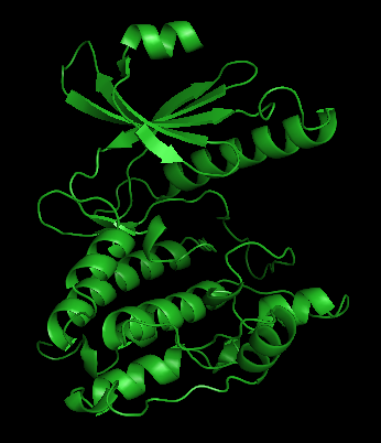
Phosphorylase kinase is a 1.3 MDa hexadecameric holoenzyme, though its size can vary somewhat due to substitution of different subunit isoforms via mRNA splicing. It consists of four homotetramers each comprised four subunits (α,β,δ,γ). Only the γ subunit is known to possess catalytic activity, while the others serve regulatory functions. Due to the instability of the regulatory subunits in solution, only the γ subunit has been crystallized individually:
Hub AI
Phosphorylase kinase AI simulator
(@Phosphorylase kinase_simulator)
Phosphorylase kinase
Phosphorylase kinase (PhK) is a serine/threonine-specific protein kinase which activates glycogen phosphorylase to release glucose-1-phosphate from glycogen. PhK phosphorylates glycogen phosphorylase at two serine residues, triggering a conformational shift which favors the more active glycogen phosphorylase "a" form over the less active glycogen phosphorylase b.
The protein is a hexadecameric holoenzyme—that is, a homotetramer in which each subunit is itself a tetramer—arranged in an approximate "butterfly" shape. Each of the subunits is composed of an α, β, γ and δ subunit. The γ subunit is the site of the enzyme's catalytic activity while the other three subunits serve regulatory functions.
When unmodified, the α and β subunits inhibit the enzyme's catalysis, but phosphorylation of both these subunits by protein kinase A (PKA, or cAMP-dependent protein kinase) reduces their respective inhibitory activities. The δ subunit is the ubiquitous eukaryotic protein calmodulin which itself has 4 calcium ion binding sites. When cytosolic Ca2+ levels rise-to as low as 10−7 M—the δ subunit undergoes a large conformational change that activates the kinase's activity by binding to a complementary hydrophobic patch on the catalytic γ subunit.
Phosphorylase kinase was the first protein kinase to be isolated and characterized in detail, accomplished first by Krebs, Graves and Fischer in the 1950s. At the time, the scientific community was largely unaware of the importance of protein phosphorylation in the regulation of cellular processes, and many in the field dismissed phosphoproteins as biologically unimportant. Since covalent modification by phosphorylation is a widespread, important method of biochemical regulation in a wide variety of cellular processes, the discovery of this reaction has had enormous impact on scientific understanding of regulatory mechanisms.
The substrate of PhK, glycogen phosphorylase, had been isolated by Carl and Gerty Cori in the 1930s, who determined that there were two forms: an inactive form b and an active form a. However, for unknown reasons at the time, the only way to isolate glycogen phosphorylase a from muscle tissue was by paper filtration – other methods, such as centrifugation, would not work. It was a critical insight on the part of Fischer et al. that it was the presence of calcium ions in the filter paper that was generating the active "a" isoform. Later research revealed that the calcium ions were in fact activating phosphorylase kinase via the δ regulatory subunit, leading to the phosphorylation of glycogen phosphorylase.
The precise details of the PhK's catalytic mechanism are still under study. While this may seem surprising given that it was isolated over 50 years ago, there are significant difficulties in studying the finer details of PhK's structure and mechanism due to its large size and high degree of complexity. In the active site, there is significant homology between PhK and other so-called P-loop protein kinases such as protein kinase A (PKA, cAMP-dependent kinase). In contrast to these other proteins, which typically require phosphorylation of a serine or tyrosine residue in the catalytic site to be active, the catalytic γ subunit of PhK is constitutively active due to the presence of a negatively charged glutamate residue, Glu-182.
Structural and biochemical data suggest one possible mechanism of action for the phosphorylation of glycogen phosphorylase by PhK involves the direct transfer of phosphate from adenosine triphosphate (ATP) to the substrate serine.
Phosphorylase kinase is a 1.3 MDa hexadecameric holoenzyme, though its size can vary somewhat due to substitution of different subunit isoforms via mRNA splicing. It consists of four homotetramers each comprised four subunits (α,β,δ,γ). Only the γ subunit is known to possess catalytic activity, while the others serve regulatory functions. Due to the instability of the regulatory subunits in solution, only the γ subunit has been crystallized individually: