Recent from talks
Radiology
Knowledge base stats:
Talk channels stats:
Members stats:
Radiology
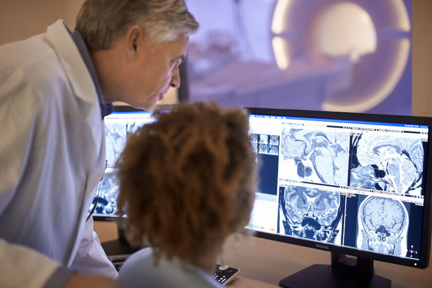
Radiology (/ˌreɪdiˈɒlədʒi/ RAY-dee-AHL-ə-jee) is the medical specialty that uses medical imaging to diagnose diseases and guide treatment within the bodies of humans and other animals. It began with radiography (which is why its name has a root referring to radiation), but today it includes all imaging modalities. This includes technologies that use no ionizing electromagnetic radiation, such as ultrasonography and magnetic resonance imaging (MRI), as well as others that do use radiation, such as computed tomography (CT), fluoroscopy, and nuclear medicine including positron emission tomography (PET). Interventional radiology is the performance of usually minimally invasive medical procedures with the guidance of imaging technologies such as those mentioned above.
The modern practice of radiology involves a team of several different healthcare professionals. A radiologist, who is a medical doctor with specialized post-graduate training, interprets medical images, communicates these findings to other physicians through reports or verbal communication, and uses imaging to perform minimally invasive medical procedures The nurse is involved in the care of patients before and after imaging or procedures, including administration of medications, monitoring of vital signs and monitoring of sedated patients. The radiographer, also known as a radiologic technologist in countries such as the United States and Canada, is a specialized healthcare professional who performs radiographic procedures and radiation therapy for the diagnosis and treatment of diseases such as cancer. The images produced through radiographic procedures are used for interpretation by radiologists, and depending on their education, training, and the regulations of the country in which they practice, radiographers in some regions also have an extended role in image interpretation and reporting.
Radiographs (originally called roentgenographs, named after the discoverer of X-rays, Wilhelm Conrad Röntgen) are produced by transmitting X-rays through a patient. The X-rays are projected through the body onto a detector; an image is formed based on which rays pass through (and are detected) versus those that are absorbed or scattered in the patient (and thus are not detected). Röntgen discovered X-rays on November 8, 1895, and received the first Nobel Prize in Physics in 1901 for this discovery. In film-screen radiography, an X-ray tube generates a beam of X-rays, which is aimed at the patient. The X-rays that pass through the patient are filtered through a device called a grid or X-ray filter, to reduce scatter, and strike an undeveloped film, which is held tightly to a screen of light-emitting phosphors in a light-tight cassette. The film is then developed chemically and an image appears on the film. Film-screen radiography is being replaced by phosphor plate radiography but more recently by digital radiography (DR) and the EOS imaging. In the two latest systems, the X-rays strike sensors that converts the signals generated into digital information, which is transmitted and converted into an image displayed on a computer screen. In digital radiography the sensors shape a plate, but in the EOS system, which is a slot-scanning system, a linear sensor vertically scans the patient.[citation needed]
Plain radiography was one of the earliest imaging modalities used in clinical medicine and remained the most widely used for several decades. Due to its broad availability, speed, and relatively low cost, it continues to be a common first-line tool in radiologic evaluation. Despite advances in CT, MRI, and other imaging techniques, there are many conditions in which traditional radiographs remain helpful in diagnosis. These include arthritis, pneumonia, bone tumors, fractures, congenital skeletal anomalies, and certain types of kidney stones.
Mammography and DXA are two applications of low energy projectional radiography, used for the evaluation of breast cancer and osteoporosis, respectively.
Fluoroscopy and angiography are special applications of X-ray imaging, in which a fluorescent screen and image intensifier tube is connected to a closed-circuit television system. This allows real-time imaging of structures in motion or augmented with a radiocontrast agent. Radiocontrast agents are usually administered by swallowing or injecting into the body of the patient to delineate anatomy and functioning of the blood vessels, the genitourinary system, or the gastrointestinal tract (GI tract). Two radiocontrast agents are presently in common use. Barium sulfate (BaSO4) is given orally or rectally for evaluation of the GI tract. Iodine, in multiple proprietary forms, is given by oral, rectal, vaginal, intra-arterial or intravenous routes. These radiocontrast agents strongly absorb or scatter X-rays, and in conjunction with the real-time imaging, allow demonstration of dynamic processes, such as peristalsis in the digestive tract or blood flow in arteries and veins. Iodine contrast may also be concentrated in abnormal areas more or less than in normal tissues and make abnormalities (tumors, cysts, inflammation) more conspicuous. Additionally, in specific circumstances, air can be used as a contrast agent for the gastrointestinal system and carbon dioxide can be used as a contrast agent in the venous system; in these cases, the contrast agent attenuates the X-ray radiation less than the surrounding tissues.
CT imaging uses X-rays in conjunction with computing algorithms to image the body. In CT, an X-ray tube opposite an X-ray detector (or detectors) in a ring-shaped apparatus rotate around a patient, producing a computer-generated cross-sectional image (tomogram). CT is acquired in the axial plane, with coronal and sagittal images produced by computer reconstruction. Radiocontrast agents are often used with CT for enhanced delineation of anatomy. Although radiographs provide higher spatial resolution, CT can detect more subtle variations in attenuation of X-rays (higher contrast resolution). CT exposes the patient to significantly more ionizing radiation than a radiograph.[citation needed]
Spiral multidetector CT uses 16, 64, 254 or more detectors during continuous motion of the patient through the radiation beam to obtain fine detail images in a short exam time. With rapid administration of intravenous contrast during the CT scan, these fine detail images can be reconstructed into three-dimensional (3D) images of carotid, cerebral, coronary or other arteries.[citation needed]
Hub AI
Radiology AI simulator
(@Radiology_simulator)
Radiology
Radiology (/ˌreɪdiˈɒlədʒi/ RAY-dee-AHL-ə-jee) is the medical specialty that uses medical imaging to diagnose diseases and guide treatment within the bodies of humans and other animals. It began with radiography (which is why its name has a root referring to radiation), but today it includes all imaging modalities. This includes technologies that use no ionizing electromagnetic radiation, such as ultrasonography and magnetic resonance imaging (MRI), as well as others that do use radiation, such as computed tomography (CT), fluoroscopy, and nuclear medicine including positron emission tomography (PET). Interventional radiology is the performance of usually minimally invasive medical procedures with the guidance of imaging technologies such as those mentioned above.
The modern practice of radiology involves a team of several different healthcare professionals. A radiologist, who is a medical doctor with specialized post-graduate training, interprets medical images, communicates these findings to other physicians through reports or verbal communication, and uses imaging to perform minimally invasive medical procedures The nurse is involved in the care of patients before and after imaging or procedures, including administration of medications, monitoring of vital signs and monitoring of sedated patients. The radiographer, also known as a radiologic technologist in countries such as the United States and Canada, is a specialized healthcare professional who performs radiographic procedures and radiation therapy for the diagnosis and treatment of diseases such as cancer. The images produced through radiographic procedures are used for interpretation by radiologists, and depending on their education, training, and the regulations of the country in which they practice, radiographers in some regions also have an extended role in image interpretation and reporting.
Radiographs (originally called roentgenographs, named after the discoverer of X-rays, Wilhelm Conrad Röntgen) are produced by transmitting X-rays through a patient. The X-rays are projected through the body onto a detector; an image is formed based on which rays pass through (and are detected) versus those that are absorbed or scattered in the patient (and thus are not detected). Röntgen discovered X-rays on November 8, 1895, and received the first Nobel Prize in Physics in 1901 for this discovery. In film-screen radiography, an X-ray tube generates a beam of X-rays, which is aimed at the patient. The X-rays that pass through the patient are filtered through a device called a grid or X-ray filter, to reduce scatter, and strike an undeveloped film, which is held tightly to a screen of light-emitting phosphors in a light-tight cassette. The film is then developed chemically and an image appears on the film. Film-screen radiography is being replaced by phosphor plate radiography but more recently by digital radiography (DR) and the EOS imaging. In the two latest systems, the X-rays strike sensors that converts the signals generated into digital information, which is transmitted and converted into an image displayed on a computer screen. In digital radiography the sensors shape a plate, but in the EOS system, which is a slot-scanning system, a linear sensor vertically scans the patient.[citation needed]
Plain radiography was one of the earliest imaging modalities used in clinical medicine and remained the most widely used for several decades. Due to its broad availability, speed, and relatively low cost, it continues to be a common first-line tool in radiologic evaluation. Despite advances in CT, MRI, and other imaging techniques, there are many conditions in which traditional radiographs remain helpful in diagnosis. These include arthritis, pneumonia, bone tumors, fractures, congenital skeletal anomalies, and certain types of kidney stones.
Mammography and DXA are two applications of low energy projectional radiography, used for the evaluation of breast cancer and osteoporosis, respectively.
Fluoroscopy and angiography are special applications of X-ray imaging, in which a fluorescent screen and image intensifier tube is connected to a closed-circuit television system. This allows real-time imaging of structures in motion or augmented with a radiocontrast agent. Radiocontrast agents are usually administered by swallowing or injecting into the body of the patient to delineate anatomy and functioning of the blood vessels, the genitourinary system, or the gastrointestinal tract (GI tract). Two radiocontrast agents are presently in common use. Barium sulfate (BaSO4) is given orally or rectally for evaluation of the GI tract. Iodine, in multiple proprietary forms, is given by oral, rectal, vaginal, intra-arterial or intravenous routes. These radiocontrast agents strongly absorb or scatter X-rays, and in conjunction with the real-time imaging, allow demonstration of dynamic processes, such as peristalsis in the digestive tract or blood flow in arteries and veins. Iodine contrast may also be concentrated in abnormal areas more or less than in normal tissues and make abnormalities (tumors, cysts, inflammation) more conspicuous. Additionally, in specific circumstances, air can be used as a contrast agent for the gastrointestinal system and carbon dioxide can be used as a contrast agent in the venous system; in these cases, the contrast agent attenuates the X-ray radiation less than the surrounding tissues.
CT imaging uses X-rays in conjunction with computing algorithms to image the body. In CT, an X-ray tube opposite an X-ray detector (or detectors) in a ring-shaped apparatus rotate around a patient, producing a computer-generated cross-sectional image (tomogram). CT is acquired in the axial plane, with coronal and sagittal images produced by computer reconstruction. Radiocontrast agents are often used with CT for enhanced delineation of anatomy. Although radiographs provide higher spatial resolution, CT can detect more subtle variations in attenuation of X-rays (higher contrast resolution). CT exposes the patient to significantly more ionizing radiation than a radiograph.[citation needed]
Spiral multidetector CT uses 16, 64, 254 or more detectors during continuous motion of the patient through the radiation beam to obtain fine detail images in a short exam time. With rapid administration of intravenous contrast during the CT scan, these fine detail images can be reconstructed into three-dimensional (3D) images of carotid, cerebral, coronary or other arteries.[citation needed]
Recent media