
Recent from talks
Retinal pigment epithelium
Knowledge base stats:
Talk channels stats:
Members stats:
Retinal pigment epithelium
The pigmented layer of retina or retinal pigment epithelium (RPE) is the pigmented cell layer just outside the neurosensory retina that nourishes retinal visual cells, and is firmly attached to the underlying choroid and overlying retinal visual cells.
The RPE was referred to in the 18th and 19th centuries as the pigmentum nigrum, reflecting the observation that the RPE is dark (black in many animals, brown in humans); and as the tapetum nigrum, referring to the observation that in animals with a tapetum lucidum, in the region of the tapetum lucidum the RPE is not pigmented.
The RPE is composed of a single layer of hexagonal cells that are densely packed with pigment granules.
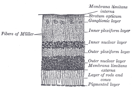
When viewed from the outer surface, these cells are smooth and hexagonal in shape. When seen in section, each cell consists of an outer non-pigmented part containing a large oval nucleus and an inner pigmented portion which extends as a series of straight thread-like processes between the rods, this being especially the case when the eye is exposed to light.
The size and morphology of RPE cells vary with both anatomical location and age. From the central macula (~12–18 μm in height and ~10 μm in width) toward the peripheral retina, the cells gradually become broader, flatter, and more heterogeneous in shape. Ageing is associated with a reduction in RPE cell number, particularly in central regions. Local cell loss may be partially compensated by the lateral migration of adjacent cells, with remaining cells expanding or elongating to cover the affected areas.
The RPE has several functions, namely, light absorption, epithelial transport, spatial ion buffering, visual cycle, phagocytosis, secretion and immune modulation.
In the eyes of albinos, the cells of this layer contain no pigment. Dysfunction of the RPE is found in age-related macular degeneration and retinitis pigmentosa. RPE are also involved in diabetic retinopathy. Gardner syndrome is characterized by FAP (familial adenomatous polyps), osseous and soft tissue tumors, retinal pigment epithelium hypertrophy and impacted teeth.
A confluent monolayer of RPE cell culture resembles a mosaic or cobblestone pattern. At the individual cell level, the morphology is typically described as polygonal or hexagonal, columnar, or epithelioid. In electron micrographs, cells with apical surface microvilli (scanning electron microscopy) exhibit distinct structural features, including apical microvilli, basal infoldings, melanin pigments, tight junctions between cells, and various organelles (transmission electron microscopy).
Hub AI
Retinal pigment epithelium AI simulator
(@Retinal pigment epithelium_simulator)
Retinal pigment epithelium
The pigmented layer of retina or retinal pigment epithelium (RPE) is the pigmented cell layer just outside the neurosensory retina that nourishes retinal visual cells, and is firmly attached to the underlying choroid and overlying retinal visual cells.
The RPE was referred to in the 18th and 19th centuries as the pigmentum nigrum, reflecting the observation that the RPE is dark (black in many animals, brown in humans); and as the tapetum nigrum, referring to the observation that in animals with a tapetum lucidum, in the region of the tapetum lucidum the RPE is not pigmented.
The RPE is composed of a single layer of hexagonal cells that are densely packed with pigment granules.
When viewed from the outer surface, these cells are smooth and hexagonal in shape. When seen in section, each cell consists of an outer non-pigmented part containing a large oval nucleus and an inner pigmented portion which extends as a series of straight thread-like processes between the rods, this being especially the case when the eye is exposed to light.
The size and morphology of RPE cells vary with both anatomical location and age. From the central macula (~12–18 μm in height and ~10 μm in width) toward the peripheral retina, the cells gradually become broader, flatter, and more heterogeneous in shape. Ageing is associated with a reduction in RPE cell number, particularly in central regions. Local cell loss may be partially compensated by the lateral migration of adjacent cells, with remaining cells expanding or elongating to cover the affected areas.
The RPE has several functions, namely, light absorption, epithelial transport, spatial ion buffering, visual cycle, phagocytosis, secretion and immune modulation.
In the eyes of albinos, the cells of this layer contain no pigment. Dysfunction of the RPE is found in age-related macular degeneration and retinitis pigmentosa. RPE are also involved in diabetic retinopathy. Gardner syndrome is characterized by FAP (familial adenomatous polyps), osseous and soft tissue tumors, retinal pigment epithelium hypertrophy and impacted teeth.
A confluent monolayer of RPE cell culture resembles a mosaic or cobblestone pattern. At the individual cell level, the morphology is typically described as polygonal or hexagonal, columnar, or epithelioid. In electron micrographs, cells with apical surface microvilli (scanning electron microscopy) exhibit distinct structural features, including apical microvilli, basal infoldings, melanin pigments, tight junctions between cells, and various organelles (transmission electron microscopy).
Recent media