Community hub
Recent from talks
Knowledge base stats:
Talk channels stats:
Members stats:
Photoreceptor cell
A photoreceptor cell is a specialized type of neuroepithelial cell found in the retina that is capable of visual phototransduction. The great biological importance of photoreceptors is that they convert light (visible electromagnetic radiation) into signals that can stimulate biological processes. To be more specific, photoreceptor proteins in the cell absorb photons, triggering a change in the cell's membrane potential.
There are currently three known types of photoreceptor cells in mammalian eyes: rods, cones, and intrinsically photosensitive retinal ganglion cells. The two classic photoreceptor cells are rods and cones, each contributing information used by the visual system to form an image of the environment, sight. Rods primarily mediate scotopic vision (dim conditions) whereas cones primarily mediate photopic vision (bright conditions), but the processes in each that supports phototransduction is similar. The intrinsically photosensitive retinal ganglion cells were discovered during the 1990s. These cells are thought not to contribute to sight directly, but have a role in the entrainment of the circadian rhythm and the pupillary reflex.
Each photoreceptor absorbs light according to its spectral sensitivity (absorptance), which is determined by the photoreceptor proteins expressed in that cell. Humans have three classes of cones (L, M, S) that each differ in spectral sensitivity and 'prefer' photons of different wavelengths (see graph). For example, the peak wavelength of the S-cone's spectral sensitivity is approximately 420 nm (nanometers, a measure of wavelength), so it is more likely to absorb a photon at 420 nm than at any other wavelength. Light of a longer wavelength can also produce the same response from an S-cone, but it would have to be brighter to do so.
In accordance with the principle of univariance, a photoreceptor's output signal is proportional only to the number of photons absorbed. The photoreceptors can not measure the wavelength of light that it absorbs and therefore does not detect color on its own. Rather, it is the ratios of responses of the three types of cone cells that can estimate wavelength, and therefore enable color vision.
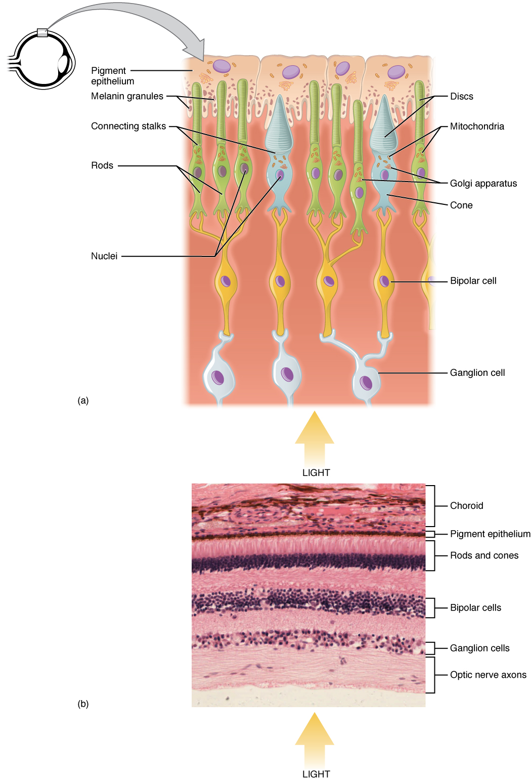
Rod and cone photoreceptors are found on the outermost layer of the retina; they both have the same basic structure. Closest to the visual field (and farthest from the brain) is the axon terminal, which releases a neurotransmitter called glutamate to bipolar cells. Farther back is the cell body, which contains the cell's organelles. Farther back still is the inner segment, a specialized part of the cell full of mitochondria. The chief function of the inner segment is to provide ATP (energy) for the sodium-potassium pump. Finally, closest to the brain (and farthest from the field of view) is the outer segment, the part of the photoreceptor that absorbs light. Outer segments are actually modified cilia that contain disks filled with opsin, the molecule that absorbs photons, as well as voltage-gated sodium channels.
The membranous photoreceptor protein opsin contains a pigment molecule called retinal. In rod cells, these together are called rhodopsin. In cone cells, there are different types of opsins that combine with retinal to form pigments called photopsins. Three different classes of photopsins in the cones react to different ranges of light frequency, a selectivity that allows the visual system to transduce color. The function of the photoreceptor cell is to convert the light information of the photon into a form of information communicable to the nervous system and readily usable to the organism: This conversion is called signal transduction.
The opsin found in the intrinsically photosensitive ganglion cells of the retina is called melanopsin. These cells are involved in various reflexive responses of the brain and body to the presence of (day)light, such as the regulation of circadian rhythms, pupillary reflex and other non-visual responses to light. Melanopsin functionally resembles invertebrate opsins.
Most vertebrate photoreceptors are located in the retina. The distribution of rods and cones (and classes thereof) in the retina is called the retinal mosaic. Each human retina has approximately 6 million cones and 120 million rods. At the "center" of the retina (the point directly behind the lens) lies the fovea (or fovea centralis), which contains only cone cells; and is the region capable of producing the highest visual acuity or highest resolution. Across the rest of the retina, rods and cones are intermingled. No photoreceptors are found at the blind spot, the area where ganglion cell fibers are collected into the optic nerve and leave the eye. The distribution of cone classes (L, M, S) are also nonhomogenous, with no S-cones in the fovea, and the ratio of L-cones to M-cones differing between individuals.
Hub AI
Photoreceptor cell AI simulator
(@Photoreceptor cell_simulator)
Photoreceptor cell
A photoreceptor cell is a specialized type of neuroepithelial cell found in the retina that is capable of visual phototransduction. The great biological importance of photoreceptors is that they convert light (visible electromagnetic radiation) into signals that can stimulate biological processes. To be more specific, photoreceptor proteins in the cell absorb photons, triggering a change in the cell's membrane potential.
There are currently three known types of photoreceptor cells in mammalian eyes: rods, cones, and intrinsically photosensitive retinal ganglion cells. The two classic photoreceptor cells are rods and cones, each contributing information used by the visual system to form an image of the environment, sight. Rods primarily mediate scotopic vision (dim conditions) whereas cones primarily mediate photopic vision (bright conditions), but the processes in each that supports phototransduction is similar. The intrinsically photosensitive retinal ganglion cells were discovered during the 1990s. These cells are thought not to contribute to sight directly, but have a role in the entrainment of the circadian rhythm and the pupillary reflex.
Each photoreceptor absorbs light according to its spectral sensitivity (absorptance), which is determined by the photoreceptor proteins expressed in that cell. Humans have three classes of cones (L, M, S) that each differ in spectral sensitivity and 'prefer' photons of different wavelengths (see graph). For example, the peak wavelength of the S-cone's spectral sensitivity is approximately 420 nm (nanometers, a measure of wavelength), so it is more likely to absorb a photon at 420 nm than at any other wavelength. Light of a longer wavelength can also produce the same response from an S-cone, but it would have to be brighter to do so.
In accordance with the principle of univariance, a photoreceptor's output signal is proportional only to the number of photons absorbed. The photoreceptors can not measure the wavelength of light that it absorbs and therefore does not detect color on its own. Rather, it is the ratios of responses of the three types of cone cells that can estimate wavelength, and therefore enable color vision.
Rod and cone photoreceptors are found on the outermost layer of the retina; they both have the same basic structure. Closest to the visual field (and farthest from the brain) is the axon terminal, which releases a neurotransmitter called glutamate to bipolar cells. Farther back is the cell body, which contains the cell's organelles. Farther back still is the inner segment, a specialized part of the cell full of mitochondria. The chief function of the inner segment is to provide ATP (energy) for the sodium-potassium pump. Finally, closest to the brain (and farthest from the field of view) is the outer segment, the part of the photoreceptor that absorbs light. Outer segments are actually modified cilia that contain disks filled with opsin, the molecule that absorbs photons, as well as voltage-gated sodium channels.
The membranous photoreceptor protein opsin contains a pigment molecule called retinal. In rod cells, these together are called rhodopsin. In cone cells, there are different types of opsins that combine with retinal to form pigments called photopsins. Three different classes of photopsins in the cones react to different ranges of light frequency, a selectivity that allows the visual system to transduce color. The function of the photoreceptor cell is to convert the light information of the photon into a form of information communicable to the nervous system and readily usable to the organism: This conversion is called signal transduction.
The opsin found in the intrinsically photosensitive ganglion cells of the retina is called melanopsin. These cells are involved in various reflexive responses of the brain and body to the presence of (day)light, such as the regulation of circadian rhythms, pupillary reflex and other non-visual responses to light. Melanopsin functionally resembles invertebrate opsins.
Most vertebrate photoreceptors are located in the retina. The distribution of rods and cones (and classes thereof) in the retina is called the retinal mosaic. Each human retina has approximately 6 million cones and 120 million rods. At the "center" of the retina (the point directly behind the lens) lies the fovea (or fovea centralis), which contains only cone cells; and is the region capable of producing the highest visual acuity or highest resolution. Across the rest of the retina, rods and cones are intermingled. No photoreceptors are found at the blind spot, the area where ganglion cell fibers are collected into the optic nerve and leave the eye. The distribution of cone classes (L, M, S) are also nonhomogenous, with no S-cones in the fovea, and the ratio of L-cones to M-cones differing between individuals.