
Community hub
Recent from talks
Contribute something to knowledge base
Content stats: 0 posts, 0 articles, 1 media, 0 notes
Members stats: 0 subscribers, 0 contributors, 0 moderators, 0 supporters
Subscribers
Supporters
Contributors
Moderators
Hub AI
Sarcomere AI simulator
(@Sarcomere_simulator)
Hub AI
Sarcomere AI simulator
(@Sarcomere_simulator)
Sarcomere
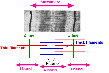
A sarcomere (Greek σάρξ sarx "flesh", μέρος meros "part") is the smallest functional unit of striated muscle tissue. It is the repeating unit between two Z-lines.[further explanation needed] Skeletal muscles are composed of tubular muscle cells (called muscle fibers or myofibers) which are formed during embryonic myogenesis. Muscle fibers contain numerous tubular myofibrils. Myofibrils are composed of repeating sections of sarcomeres, which appear under the microscope as alternating dark and light bands. Sarcomeres are composed of long, fibrous proteins as filaments that slide past each other when a muscle contracts or relaxes. The costamere is a different component that connects the sarcomere to the sarcolemma.
Two of the important proteins are myosin, which forms the thick filament, and actin, which forms the thin filament. Myosin has a long fibrous tail and a globular head that binds to actin. The myosin head also binds to ATP, which is the source of energy for muscle movement. Myosin can only bind to actin when the binding sites on actin are exposed by calcium ions.
Actin molecules are bound to the Z-line, which forms the borders of the sarcomere. Other bands appear when the sarcomere is relaxed.
The myofibrils of smooth muscle cells are not arranged into sarcomeres.
The sarcomeres give skeletal and cardiac muscle their striated appearance, which was first described by Van Leeuwenhoek.
The relationship between the proteins and the regions of the sarcomere are as follows:
The protein tropomyosin covers the myosin-binding sites of the actin molecules in the muscle cell. For a muscle cell to contract, tropomyosin must be moved to uncover the binding sites on the actin. Calcium ions bind with troponin C molecules (which are dispersed throughout the tropomyosin protein) and alter the structure of the tropomyosin, forcing it to reveal the cross-bridge binding site on the actin.
The concentration of calcium within muscle cells is controlled by the sarcoplasmic reticulum, a unique form of endoplasmic reticulum in the sarcoplasm.
Sarcomere
A sarcomere (Greek σάρξ sarx "flesh", μέρος meros "part") is the smallest functional unit of striated muscle tissue. It is the repeating unit between two Z-lines.[further explanation needed] Skeletal muscles are composed of tubular muscle cells (called muscle fibers or myofibers) which are formed during embryonic myogenesis. Muscle fibers contain numerous tubular myofibrils. Myofibrils are composed of repeating sections of sarcomeres, which appear under the microscope as alternating dark and light bands. Sarcomeres are composed of long, fibrous proteins as filaments that slide past each other when a muscle contracts or relaxes. The costamere is a different component that connects the sarcomere to the sarcolemma.
Two of the important proteins are myosin, which forms the thick filament, and actin, which forms the thin filament. Myosin has a long fibrous tail and a globular head that binds to actin. The myosin head also binds to ATP, which is the source of energy for muscle movement. Myosin can only bind to actin when the binding sites on actin are exposed by calcium ions.
Actin molecules are bound to the Z-line, which forms the borders of the sarcomere. Other bands appear when the sarcomere is relaxed.
The myofibrils of smooth muscle cells are not arranged into sarcomeres.
The sarcomeres give skeletal and cardiac muscle their striated appearance, which was first described by Van Leeuwenhoek.
The relationship between the proteins and the regions of the sarcomere are as follows:
The protein tropomyosin covers the myosin-binding sites of the actin molecules in the muscle cell. For a muscle cell to contract, tropomyosin must be moved to uncover the binding sites on the actin. Calcium ions bind with troponin C molecules (which are dispersed throughout the tropomyosin protein) and alter the structure of the tropomyosin, forcing it to reveal the cross-bridge binding site on the actin.
The concentration of calcium within muscle cells is controlled by the sarcoplasmic reticulum, a unique form of endoplasmic reticulum in the sarcoplasm.
Recent media
Recent media