



Community hub
Recent from talks
Knowledge base stats:
Talk channels stats:
Members stats:
Superior vena cava syndrome
Superior vena cava syndrome (SVCS) is a group of symptoms caused by obstruction of the superior vena cava ("SVC"), a short, wide vessel carrying circulating blood into the heart. The majority of cases are caused by malignant tumors within the mediastinum, most commonly lung cancer and non-Hodgkin's lymphoma, directly compressing or invading the SVC wall. Non-malignant causes are increasing in prevalence due to expanding use of intravascular devices (such as permanent central venous catheters and leads for pacemakers and defibrillators), which can result in thrombosis. Other non-malignant causes include benign mediastinal tumors, aortic aneurysm, infections, and fibrosing mediastinitis.
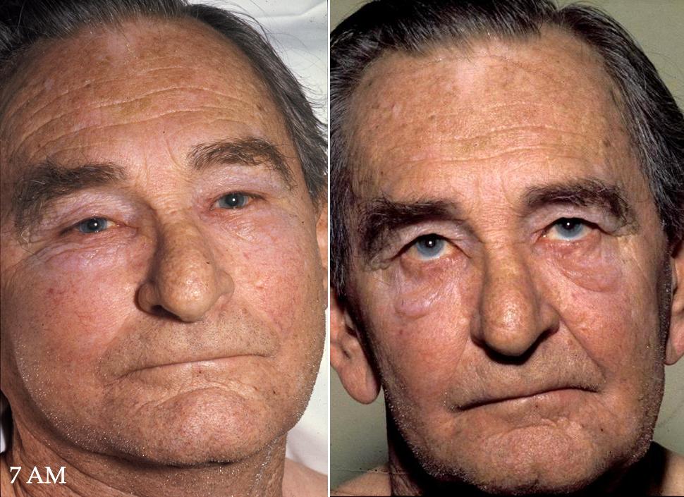
Characteristic features are edema (swelling due to excess fluid) of the face and arms and development of swollen collateral veins on the front of the chest wall. Shortness of breath and coughing are quite common symptoms; difficulty swallowing is reported in 11% of cases, headache in 6% and stridor (a high-pitched wheeze) in 4%. The symptoms are rarely life-threatening, though edema of the epiglottis can make breathing difficult, edema of the brain can cause reduced alertness, and in less than 5% of cases of SVCO, severe neurological symptoms or airway compromise are reported. Resolution of superior vena cava syndrome is directly related to the treatment of the underlying compression.
Shortness of breath is the most common symptom, followed by face or arm swelling.
Following are frequent symptoms:
Superior vena cava syndrome usually presents more gradually with an increase in symptoms over time as malignancies increase in size or invasiveness.
Over 80% of cases are caused by malignant tumors compressing the superior vena cava. Lung cancer, most commonly squamous cell carcinoma, comprises 75–80% of these cases and non-Hodgkin lymphoma, most commonly diffuse large B-cell lymphoma, comprises 10–15%. Rare malignant causes include Hodgkin's lymphoma, metastatic cancers, leukemia, leiomyosarcoma of the mediastinal vessels, and plasmocytoma. Syphilis and tuberculosis have also been known to cause superior vena cava syndrome. SVCS can be caused by invasion or compression by a pathological process or by a deep vein thrombosis in the vein itself, although this latter is less common (approximately 35% due to the use of intravascular devices).
The main techniques of diagnosing SVCS are with chest X-rays (CXR), CT scans, transbronchial needle aspiration at bronchoscopy and mediastinoscopy. CXRs often provide the ability to show mediastinal widening and may show the presenting primary cause of SVCS. However, 16% of people with SVC syndrome have a normal chest X-ray. CT scans should be contrast enhanced and be taken on the neck, chest, lower abdomen, and pelvis. They may also show the underlying cause and the extent to which the disease has progressed.
Several methods of treatment are available, mainly consisting of careful drug therapy and surgery. Glucocorticoids (such as prednisone or methylprednisolone) decrease the inflammatory response to tumor invasion and edema surrounding the tumor. Glucocorticoids are most helpful if the tumor is steroid-responsive, such as lymphomas. In addition, diuretics (such as furosemide) are used to reduce venous return to the heart which relieves the increased pressure.
Hub AI
Superior vena cava syndrome AI simulator
(@Superior vena cava syndrome_simulator)
Superior vena cava syndrome
Superior vena cava syndrome (SVCS) is a group of symptoms caused by obstruction of the superior vena cava ("SVC"), a short, wide vessel carrying circulating blood into the heart. The majority of cases are caused by malignant tumors within the mediastinum, most commonly lung cancer and non-Hodgkin's lymphoma, directly compressing or invading the SVC wall. Non-malignant causes are increasing in prevalence due to expanding use of intravascular devices (such as permanent central venous catheters and leads for pacemakers and defibrillators), which can result in thrombosis. Other non-malignant causes include benign mediastinal tumors, aortic aneurysm, infections, and fibrosing mediastinitis.
Characteristic features are edema (swelling due to excess fluid) of the face and arms and development of swollen collateral veins on the front of the chest wall. Shortness of breath and coughing are quite common symptoms; difficulty swallowing is reported in 11% of cases, headache in 6% and stridor (a high-pitched wheeze) in 4%. The symptoms are rarely life-threatening, though edema of the epiglottis can make breathing difficult, edema of the brain can cause reduced alertness, and in less than 5% of cases of SVCO, severe neurological symptoms or airway compromise are reported. Resolution of superior vena cava syndrome is directly related to the treatment of the underlying compression.
Shortness of breath is the most common symptom, followed by face or arm swelling.
Following are frequent symptoms:
Superior vena cava syndrome usually presents more gradually with an increase in symptoms over time as malignancies increase in size or invasiveness.
Over 80% of cases are caused by malignant tumors compressing the superior vena cava. Lung cancer, most commonly squamous cell carcinoma, comprises 75–80% of these cases and non-Hodgkin lymphoma, most commonly diffuse large B-cell lymphoma, comprises 10–15%. Rare malignant causes include Hodgkin's lymphoma, metastatic cancers, leukemia, leiomyosarcoma of the mediastinal vessels, and plasmocytoma. Syphilis and tuberculosis have also been known to cause superior vena cava syndrome. SVCS can be caused by invasion or compression by a pathological process or by a deep vein thrombosis in the vein itself, although this latter is less common (approximately 35% due to the use of intravascular devices).
The main techniques of diagnosing SVCS are with chest X-rays (CXR), CT scans, transbronchial needle aspiration at bronchoscopy and mediastinoscopy. CXRs often provide the ability to show mediastinal widening and may show the presenting primary cause of SVCS. However, 16% of people with SVC syndrome have a normal chest X-ray. CT scans should be contrast enhanced and be taken on the neck, chest, lower abdomen, and pelvis. They may also show the underlying cause and the extent to which the disease has progressed.
Several methods of treatment are available, mainly consisting of careful drug therapy and surgery. Glucocorticoids (such as prednisone or methylprednisolone) decrease the inflammatory response to tumor invasion and edema surrounding the tumor. Glucocorticoids are most helpful if the tumor is steroid-responsive, such as lymphomas. In addition, diuretics (such as furosemide) are used to reduce venous return to the heart which relieves the increased pressure.