Recent from talks
Thymus
Knowledge base stats:
Talk channels stats:
Members stats:
Thymus
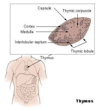
The thymus (pl.: thymuses or thymi) is a specialized primary lymphoid organ of the immune system. Within the thymus, T cells mature. T cells are critical to the adaptive immune system, where the body adapts to specific foreign invaders. The thymus is located in the upper front part of the chest, in the anterior superior mediastinum, behind the sternum, and in front of the heart. It is made up of two lobes, each consisting of a central medulla and an outer cortex, surrounded by a capsule.
The thymus is made up of immature T cells called thymocytes, as well as lining cells called epithelial cells which help the thymocytes develop. T cells that successfully develop react appropriately with MHC immune receptors of the body (called positive selection) and not against proteins of the body (called negative selection). The thymus is the largest and most active during the neonatal and pre-adolescent periods. By the early teens, the thymus begins to decrease in size and activity and the tissue of the thymus is gradually replaced by fatty tissue. Nevertheless, some T cell development continues throughout adult life.
Abnormalities of the thymus can result in a decreased number of T cells and autoimmune diseases such as autoimmune polyendocrine syndrome type 1 and myasthenia gravis. These are often associated with cancer of the tissue of the thymus, called thymoma, or tissues arising from immature lymphocytes such as T cells, called lymphoma. Removal of the thymus is called a thymectomy. Although the thymus has been identified as a part of the body since the time of the Ancient Greeks, it is only since the 1960s that the function of the thymus in the immune system has become clearer.
The thymus is an organ that sits behind the sternum in the upper front part of the chest, stretching upwards towards the neck. In children, the thymus is pinkish-gray, soft, and lobulated on its surfaces. At birth, it is about 4–6 cm long, 2.5–5 cm wide, and about 1 cm thick. It increases in size until puberty, where it may have a size of about 40–50 g, following which it decreases in size in a process known as involution.
The thymus is located in the anterior mediastinum. It is made up of two lobes that meet in the upper midline, and stretch from below the thyroid in the neck to as low as the cartilage of the fourth rib. The lobes are covered by a capsule. The thymus lies behind the sternum, rests on the pericardium, and is separated from the aortic arch and great vessels by a layer of fascia. The left brachiocephalic vein may even be embedded within the thymus. In the neck, it lies on the front and sides of the trachea, behind the sternohyoid and sternothyroid muscles.
The thymus consists of two lobes, merged in the middle, surrounded by a capsule that extends with blood vessels into the interior. The lobes consist of an outer cortex rich with cells and an inner less dense medulla. The lobes are divided into smaller lobules 0.5-2 mm diameter, between which extrude radiating insertions from the capsule along septa.
The cortex is mainly made up of thymocytes and epithelial cells. The thymocytes, immature T cells, are supported by a network of the finely-branched epithelial reticular cells, which is continuous with a similar network in the medulla. This network forms an adventitia to the blood vessels, which enter the cortex via septa near the junction with the medulla. Other cells are also present in the thymus, including macrophages, dendritic cells, and a small amount of B cells, neutrophils and eosinophils.
In the medulla, the network of epithelial cells is coarser than in the cortex, and the lymphoid cells are relatively fewer in number. Concentric, nest-like bodies called Hassall's corpuscles (also called thymic corpuscles) are formed by aggregations of the medullary epithelial cells. These are concentric, layered whorls of epithelial cells that increase in number throughout life. They are the remains of the epithelial tubes, which grow out from the third pharyngeal pouches of the embryo to form the thymus.
Hub AI
Thymus AI simulator
(@Thymus_simulator)
Thymus
The thymus (pl.: thymuses or thymi) is a specialized primary lymphoid organ of the immune system. Within the thymus, T cells mature. T cells are critical to the adaptive immune system, where the body adapts to specific foreign invaders. The thymus is located in the upper front part of the chest, in the anterior superior mediastinum, behind the sternum, and in front of the heart. It is made up of two lobes, each consisting of a central medulla and an outer cortex, surrounded by a capsule.
The thymus is made up of immature T cells called thymocytes, as well as lining cells called epithelial cells which help the thymocytes develop. T cells that successfully develop react appropriately with MHC immune receptors of the body (called positive selection) and not against proteins of the body (called negative selection). The thymus is the largest and most active during the neonatal and pre-adolescent periods. By the early teens, the thymus begins to decrease in size and activity and the tissue of the thymus is gradually replaced by fatty tissue. Nevertheless, some T cell development continues throughout adult life.
Abnormalities of the thymus can result in a decreased number of T cells and autoimmune diseases such as autoimmune polyendocrine syndrome type 1 and myasthenia gravis. These are often associated with cancer of the tissue of the thymus, called thymoma, or tissues arising from immature lymphocytes such as T cells, called lymphoma. Removal of the thymus is called a thymectomy. Although the thymus has been identified as a part of the body since the time of the Ancient Greeks, it is only since the 1960s that the function of the thymus in the immune system has become clearer.
The thymus is an organ that sits behind the sternum in the upper front part of the chest, stretching upwards towards the neck. In children, the thymus is pinkish-gray, soft, and lobulated on its surfaces. At birth, it is about 4–6 cm long, 2.5–5 cm wide, and about 1 cm thick. It increases in size until puberty, where it may have a size of about 40–50 g, following which it decreases in size in a process known as involution.
The thymus is located in the anterior mediastinum. It is made up of two lobes that meet in the upper midline, and stretch from below the thyroid in the neck to as low as the cartilage of the fourth rib. The lobes are covered by a capsule. The thymus lies behind the sternum, rests on the pericardium, and is separated from the aortic arch and great vessels by a layer of fascia. The left brachiocephalic vein may even be embedded within the thymus. In the neck, it lies on the front and sides of the trachea, behind the sternohyoid and sternothyroid muscles.
The thymus consists of two lobes, merged in the middle, surrounded by a capsule that extends with blood vessels into the interior. The lobes consist of an outer cortex rich with cells and an inner less dense medulla. The lobes are divided into smaller lobules 0.5-2 mm diameter, between which extrude radiating insertions from the capsule along septa.
The cortex is mainly made up of thymocytes and epithelial cells. The thymocytes, immature T cells, are supported by a network of the finely-branched epithelial reticular cells, which is continuous with a similar network in the medulla. This network forms an adventitia to the blood vessels, which enter the cortex via septa near the junction with the medulla. Other cells are also present in the thymus, including macrophages, dendritic cells, and a small amount of B cells, neutrophils and eosinophils.
In the medulla, the network of epithelial cells is coarser than in the cortex, and the lymphoid cells are relatively fewer in number. Concentric, nest-like bodies called Hassall's corpuscles (also called thymic corpuscles) are formed by aggregations of the medullary epithelial cells. These are concentric, layered whorls of epithelial cells that increase in number throughout life. They are the remains of the epithelial tubes, which grow out from the third pharyngeal pouches of the embryo to form the thymus.
Recent media