Hubbry Logo
Alanine scanningAlanine scanningMain
Open search
Alanine scanning
Community hub
Alanine scanning
logo
8 pages, 0 posts
0 subscribers
Be the first to start a discussion here.
Alanine scanning
Alanine scanning
from Wikipedia
Example of alanine scanning. The native protein (top row) and each possible point mutation to alanine is considered.

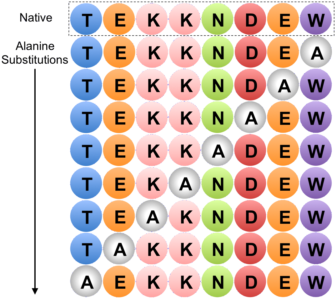
In molecular biology, alanine scanning is a site-directed mutagenesis technique used to determine the contribution of a specific residue to the stability or function of a given protein.[1] Alanine is used because of its non-bulky, chemically inert, methyl functional group that nevertheless mimics the secondary structure preferences that many of the other amino acids possess. Sometimes bulky amino acids such as valine or leucine are used in cases where conservation of the size of mutated residues is needed.

This technique can also be used to determine whether the side chain of a specific residue plays a significant role in bioactivity. This is usually accomplished by site-directed mutagenesis or randomly by creating a PCR library. Furthermore, computational methods to estimate thermodynamic parameters based on theoretical alanine substitutions have been developed.

This technique is rapid, because many side chains are analyzed simultaneously and the need for protein purification and biophysical analysis is circumvented.[2] The technology is very mature at this point and is widely used in biochemical fields. The data can be tested by IR, NMR Spectroscopy, mathematical methods, bioassays, etc.[3][4][5]

One good example of alanine scanning is the examination of the role of charged residues on the surface of proteins.[6] In a systematic study on the roles of conserved charged residues on the surface of epithelial sodium channel (ENaC), alanine scanning was used to reveal the importance of charged residues for the process of transport of the proteins to the cell surface.[6]

Applications

[edit]

Alanine scanning was used to determine simultaneously the functional contributions of 19 side chains buried at the interface between human growth hormone and the extracellular domain of its receptor.[2] Each amino acid in the side chains was substituted by alanine. Then shotgun scanning method which combines the concepts of alanine scanning mutagenesis and binomial mutagenesis with phage display technology was used.

Another critical application of alanine scanning is to determine the influence of individual residues on structure and activity in the prototypic cyclotide kalata B1.[3] Cyclotides display a wide range of pharmaceutically important bioactivities, but their natural function is in plant defense as insecticidal agents. On the structure of cyclotides kalata B1, all 23 non-cysteine residues were successively substituted with alanine. The data were tested by NMR Spectroscopy.

In addition, alanine scanning is also used to determine which functional motif of Cry4Aa has the mosquitocidal activity.[4] Cry4Aa was produced by Bacillus thuringiensis. It is a dipteran-specific toxin and it plays an important role in how to produce a bioinsecticide to control mosquitoes. So, it is very essential to determine which functional motif of Cry4Aa contributes to this activity. In this study, several Cry4Aa mutants were made by replacing the residues of potential receptor binding site, loops 1, 2, and 3 in domain II with alanine. A bioassay Culex pipiens was followed to test the activities.

Alanine-World model

[edit]

The alanine scanning method takes advantage of the fact that most canonical amino acids can be exchanged with Ala by point mutations, while the secondary structure of mutated protein remains intact, as Ala mimics the secondary structure preferences of the majority of the encoded or canonical amino acids. This is predicted by the Alanine-World model.

References

[edit]

Further reading

[edit]
Revisions and contributorsEdit on WikipediaRead on Wikipedia
from Grokipedia
Alanine scanning mutagenesis is a site-directed mutagenesis technique employed in protein engineering and structural biology to systematically replace individual amino acid residues in a protein with alanine, thereby evaluating the specific contributions of each side chain to the protein's function, stability, binding interactions, or enzymatic activity. Alanine is selected for these substitutions due to its compact, non-polar methyl side chain, which minimally disrupts the protein's backbone conformation or secondary structure while effectively neutralizing larger, potentially interactive side chains beyond the β-carbon. This method enables precise mapping of functional epitopes and hotspots—residues that disproportionately influence molecular recognition or catalysis—without introducing confounding steric or electrostatic effects. The technique was pioneered in 1989 by Brian C. Cunningham and James A. Wells, who applied it to dissect the binding interface between human growth hormone (hGH) and its receptor, generating 62 mutants to identify a cluster of 12 key residues responsible for high-affinity interactions. Their work demonstrated that substitutions could reduce binding affinity by over fourfold for critical residues while preserving overall , as verified by reactivity with monoclonal antibodies. Since its introduction, scanning has evolved into a foundational tool, with thousands of mutations analyzed across dozens of protein interfaces, revealing that binding energies are often concentrated in a few conserved hotspots rather than distributed evenly. In typical implementations, oligonucleotide-directed creates a of single-point alanine variants, whose effects are quantified via biophysical assays such as for binding kinetics, for stability, or enzymatic turnover rates. The approach has broad applications, including the design of therapeutic proteins and antibodies by optimizing interfaces, the identification of druggable sites in protein-protein interactions (e.g., in viral proteins like gp120), and the elucidation of catalytic mechanisms in enzymes. Despite challenges like conformational plasticity that may require complementary computational modeling, alanine scanning remains invaluable for its simplicity, reliability, and ability to guide rational protein modifications.

Fundamentals

Definition and purpose

Alanine scanning is a technique that systematically replaces selected residues in a protein with to assess their specific contributions to protein stability, folding, binding affinity, or enzymatic activity. This method allows researchers to probe the functional roles of individual side chains by generating a series of single-point mutants and evaluating alterations in protein behavior. Introduced in studies of protein-ligand interactions, it has become a cornerstone for dissecting structure-function relationships in proteins. The primary purpose of alanine scanning is to identify "hotspot" residues that are critical for protein function, as evidenced by significant changes in measurable properties following substitution. By comparing the wild-type protein to alanine variants, it quantifies the energetic impact of side chains, such as through shifts in binding free energy (ΔΔG, typically in kcal/mol) derived from affinity measurements. This approach highlights residues involved in key interactions, enabling the mapping of functional epitopes or active sites without introducing confounding effects from larger substitutions. Alanine scanning isolates side-chain effects by leveraging alanine's minimal side chain—a methyl group—which minimally perturbs the polypeptide backbone geometry and secondary structure, preserving overall protein folding. For example, in ligand binding studies, it has revealed essential residues at the human growth hormone (hGH)-receptor interface, where alanine mutations reduced binding affinity by over fourfold for a cluster of hydrophobic and charged side chains, underscoring their role in stabilizing the complex. Similarly, applications to enzymatic activity, such as in Pseudomonas aeruginosa elastase, have pinpointed residues vital for substrate recognition and catalysis by showing substantial activity losses upon substitution.

Biochemical rationale for alanine substitution

Alanine is selected as the substitute residue in scanning mutagenesis due to its unique chemical properties that minimize perturbations to the protein's overall structure and function while allowing precise assessment of individual side-chain contributions. Possessing the smallest chiral side chain—a simple methyl group (-CH₃)—alanine is non-polar and hydrophobic, introducing minimal steric hindrance and avoiding the introduction of charges or reactive groups that could alter interactions such as hydrogen bonding or electrostatics. This enables the isolation of the wild-type residue's specific role by effectively "deleting" larger side chains beyond the β-carbon without significantly disrupting the polypeptide backbone geometry. In comparison to wild-type residues, alanine substitution effectively removes bulkier side chains, such as the long, charged chain of or the aromatic ring of , thereby revealing their involvement in key interactions. For instance, replacing a residue eliminates potential electrostatic attractions or hydrogen bonds, while substituting disrupts van der Waals contacts or hydrophobic packing, allowing researchers to quantify the energetic cost of these losses. This approach highlights the functional importance of specific side chains in maintaining protein stability, binding affinity, or catalytic activity, as the small size of ensures that observed changes are primarily attributable to the removed interactions rather than compensatory structural shifts. From a structural perspective, alanine preserves secondary structure elements like α-helices and β-sheets better than other small residues, owing to its high helix-forming propensity, which is among the strongest of all . This propensity arises from the methyl group's ability to stabilize helical conformations through favorable van der Waals interactions without excessive rigidity or flexibility. In contrast, , with its side chain, promotes greater backbone conformational and flexibility, potentially destabilizing local structures and complicating interpretation of mutational effects. Thus, alanine substitutions maintain the native , as evidenced by retained immunoreactivity with conformation-sensitive antibodies in early studies. Quantitatively, alanine substitutions at key residues typically result in changes in folding free energy (ΔΔG) ranging from 0.5 to 3 kcal/mol, reflecting the moderate energetic contributions of individual side chains to overall stability or binding. For non-critical positions, ΔΔG values are often near zero, indicating negligible impact, while hotspots at interfaces may exceed 2 kcal/mol, underscoring their disproportionate role in protein function. These values, derived from binding affinity measurements (where ΔΔG = -RT ln(K_d^{mut}/K_d^{wt})), provide a direct gauge of side-chain importance without confounding effects from alanine's inert .

Historical development

Origins in site-directed mutagenesis

Alanine scanning mutagenesis originated within the framework of site-directed mutagenesis techniques developed in the 1970s and 1980s, which enabled precise alterations to DNA sequences to study protein function. Early methods, such as the oligonucleotide-directed approach introduced by Hutchison and colleagues in 1978, utilized mismatched synthetic primers on single-stranded DNA templates to introduce specific base changes, allowing targeted amino acid substitutions in proteins. This technique laid the groundwork for systematic protein engineering by providing a means to generate mutants with minimal disruption to the overall sequence. The concept of alanine scanning specifically emerged as a refined application of these methods, focusing on replacing amino acid side chains with alanine to assess their contributions to protein interactions and stability. It was first formally described in 1989 by Cunningham and Wells, who applied it to map the binding interface of human growth hormone (hGH) with its receptor, identifying key residues through 62 alanine mutations that revealed side-chain roles in affinity modulation. This approach capitalized on alanine's small, non-reactive methyl side chain, which minimizes structural perturbations while isolating the effects of larger residues. Preceding this milestone, influential work by Alan Fersht in the used to quantify the energetic contributions of s and other interactions in proteins, such as in tyrosyl-tRNA synthetase, establishing quantitative frameworks for interpreting mutant phenotypes. Fersht's studies on barnase further advanced these ideas by employing mutagenesis to dissect intramolecular strengths and protein stability during the early 1990s. Similarly, Greg Winter's application of to in the , including reshaping rodent antibodies for human therapy by grafting hypervariable regions, demonstrated the power of targeted substitutions for functional analysis. By the early 1990s, advancements in PCR-based facilitated the shift from individual substitutions to comprehensive alanine scanning libraries, enabling high-throughput generation of mutant variants across protein interfaces. This evolution expanded alanine scanning's utility beyond isolated studies, such as the hGH-receptor example, to broader and interaction profiling.

Key studies and advancements

In the , alanine scanning was applied to dissect the receptor-binding interface of insulin, where systematic alanine substitutions in the A and B chains revealed critical residues such as TyrA19, which caused a 1,000-fold decrease in affinity, and GlyB8, leading to a 33-fold reduction, thereby mapping key side-chain contributions to binding. Concurrently, alanine-stretch scanning emerged as an efficient method to replace contiguous stretches of residues with alanines, enabling rapid assessment of multi-residue regions' roles in and function without exhaustive single-point mutations. Advancements in the early introduced combinatorial alanine scanning, which generates libraries of alanine-substituted proteins via to simultaneously evaluate multiple residues' impacts on function, stability, and shape, significantly accelerating hotspot identification compared to sequential . During the and , high-throughput adaptations integrated surface display with alanine scanning for and variant screening, allowing parallel assessment of thousands of mutants. Recent developments up to 2025 have coupled scanning with computational tools like Rosetta's Flex ddG protocol, which predicts ΔΔG values for mutants using ensemble-based modeling to forecast stability changes and guide experimental design, enhancing efficiency in . Whole-protein scans, such as the comprehensive of 431 residues in human liver , demonstrated that a large proportion of the protein contributes to allosteric mechanisms, with many mutations subtly modulating by fructose-1,6-bisphosphate. These innovations have enabled detailed mapping of over 100 protein interfaces and amassed more than 5,000 citations in the literature by 2025, underscoring scanning's enduring impact on .

Methodology

Generating alanine mutants

Generating alanine mutants primarily involves techniques to introduce specific substitutions where target are replaced by , typically using codons such as GCT or GCC to minimize changes in DNA sequence context. This approach allows precise alteration of protein residues while preserving the overall . For single-site , the QuikChange method, developed by Stratagene in the , is widely used; it employs (PCR) with complementary primers incorporating the alanine codon, amplifying the entire template in a single reaction. The reaction mixture includes high-fidelity (e.g., KOD or Pfu), dNTPs, and the template, cycled at 95–98°C for denaturation, an annealing temperature matching the primers (often 55–68°C), and extension at 68–72°C for 1–3 minutes per kb, followed by DpnI digestion to remove parental methylated DNA. The resulting nicked s are transformed into competent E. coli cells, yielding colonies with the mutant . For multiple alanine substitutions, overlap extension PCR (OE-PCR) facilitates the introduction of several changes by generating overlapping fragments with the desired mutations, which are then assembled in a second PCR round using outer primers. This method is particularly useful for constructing sequential alanine scans across protein domains, requiring 40–50 cycles total and allowing up to six base changes per reaction, though efficiency decreases with proximity of sites. Alternatively, a two-fragment PCR approach amplifies the vector backbone and mutagenic insert separately, followed by for seamless ligation, enabling high-throughput generation of alanine libraries with success rates of 70–85% across hundreds of mutations. Library construction for comprehensive alanine scanning often employs cassette mutagenesis, where synthetic oligonucleotides containing clusters of alanine codons replace targeted DNA segments via ligation into a gapped plasmid, avoiding redundancy by selecting only non-alanine and non-glycine residues for substitution. Error-prone PCR can complement this by introducing low-level random mutations alongside targeted alanine changes to create diverse scanning libraries, though it requires subsequent screening to isolate alanine-specific variants. These libraries are cloned into expression vectors such as pET series for bacterial hosts like E. coli, which utilize T7 promoters for inducible high-level expression. Codon optimization of the mutant sequences, adjusting synonymous codons to match the host's tRNA abundance (e.g., increasing for E. coli), enhances translation efficiency and protein yield without altering the sequence. Verification of alanine mutants typically involves of DNA extracted from transformed colonies, confirming the alanine codon at the intended position with efficiencies of 80–90% for single-site changes using QuikChange. For libraries, next-generation sequencing may assess coverage, but individual clones are routinely sequenced to ensure fidelity before expression.

Characterization and analysis

Once alanine mutants have been generated, they must be expressed and purified to assess their properties. Heterologous expression systems such as Escherichia coli, yeast, and mammalian cells are commonly employed for producing these variants, allowing for high-yield production of recombinant proteins. Affinity tags, particularly polyhistidine (), facilitate purification via immobilized metal affinity chromatography (), enabling isolation of mutants from cellular debris with high specificity. Yields of alanine mutants are typically comparable to wild-type proteins when substitutions do not disrupt folding, though certain mutants may exhibit reduced expression levels due to instability, often necessitating optimization of induction conditions or co-expression of chaperones. Functional impacts of alanine substitutions are evaluated through binding affinity assays, which quantify changes in interaction strength. (SPR) is widely used to measure dissociation constants (Kd) in real-time, allowing comparison of and wild-type binding kinetics to ligands or partners. Similarly, (ITC) provides thermodynamic parameters such as and contributions to binding, offering insights into the energetic role of substituted residues. The change in binding free energy (ΔΔGbind) is calculated from Kd ratios using ΔΔGbind = RT ln(Kd,mut/Kd,wt), where R is the and T is temperature in ; values exceeding 2 kcal/mol typically indicate critical "hotspot" residues contributing significantly to affinity. Structural effects are probed using spectroscopic and high-resolution methods to detect alterations in folding and conformation. (CD) spectroscopy assesses secondary structure integrity and thermal stability by monitoring ellipticity changes, with melting temperature (Tm) shifts revealing stabilization or destabilization upon substitution. For instance, a decrease in Tm by several degrees Celsius signals reduced folding stability. and (NMR) spectroscopy provide atomic-level details of local structural perturbations, such as side-chain rearrangements or helix distortions induced by alanine replacement. Thermal denaturation experiments, often coupled with CD or fluorescence, further quantify folding free energy changes (ΔΔGfolding) via two-state transition models, highlighting residues essential for thermodynamic stability. Interpreting alanine scanning data involves analyzing energetic contributions across the protein surface. For residues acting independently, effects on binding or stability are often additive, permitting of individual ΔΔG values to predict multi-mutant outcomes without synergistic interference. Hotspots—residues with large ΔΔG impacts—frequently cluster in contiguous regions, such as interfaces or functional motifs, reflecting cooperative networks that amplify overall protein function. This clustering pattern aids in mapping critical domains, guiding further engineering efforts.

Applications

Protein-protein interaction mapping

Alanine scanning plays a pivotal role in mapping protein-protein interactions (PPIs) by systematically replacing interface residues with to quantify their contributions to binding affinity, thereby identifying "hotspots"—specific that disproportionately drive association. These hotspots typically comprise a small subset of the interface residues, often fewer than 10-20 out of dozens in contact, yet they account for the majority of the binding free energy, enabling focused insights into molecular recognition. This approach has become a cornerstone in PPI research due to its ability to dissect energetic landscapes at atomic resolution without disrupting overall . In the human growth hormone (hGH)-receptor complex, a landmark application of alanine scanning revealed a compact cluster of hydrophobic residues as the dominant energetic hotspot. Mutating these residues, particularly the two central side chains (Trp-86 and Trp-169), resulted in affinity losses exceeding 100-fold, with the hotspot region contributing over 75% of the total binding free energy despite involving only a fraction of the ~30 contacting residues. Similarly, in -antigen interfaces, such as the HyHEL-10 bound to hen egg lysozyme, mutagenesis identified 6 residues that account for most of the interaction energy. The standard protocol involves alanine substitution of one binding partner while assaying interactions with the wild-type counterpart, often using techniques like or to measure affinity changes. For more complex interfaces, combinatorial shotgun alanine scanning generates libraries where multiple residues vary between wild-type and alanine simultaneously, allowing parallel evaluation of both partners and detection of additive or effects across the interface. By comparing binding free energy changes (ΔΔG), alanine scanning differentiates interaction types: polar residues like engaged in hydrogen bonds typically exhibit large penalties (>2 kcal/mol upon ), indicating specific electrostatic contributions, whereas hydrophobic residues often reveal nonpolar desolvation effects with more variable impacts. Hotspots are conventionally defined as those with ΔΔG ≥ 2 kcal/mol, a threshold reflecting significant energetic roles. This delineation aids in classifying interfaces as "flat" or "protruding" based on hotspot clustering. A key case study from the early applied alanine scanning to the vascular endothelial growth factor (VEGF)-kinase insert domain receptor (KDR/VEGFR-2) interaction, mapping critical residues in KDR's extracellular domain 3, such as aspartates and glutamates involved in contacts affecting VEGF binding. These findings elucidated the binding and guided rational design of inhibitors by targeting epitope disruption.

Enzyme function and stability studies

Alanine scanning has been instrumental in elucidating the roles of specific residues in by systematically replacing them with and measuring changes in kinetic parameters such as k_cat and K_m. For instance, in serine proteases like , substitution of the catalytic (His57) with alanine (H57A) greatly reduces k_cat while K_m remains relatively unaffected, distinguishing from substrate binding. Similar alanine substitutions in other enzymes have identified key residues involved in catalytic mechanisms. In stability studies, alanine scanning quantifies the energetic contributions of side chains to by determining changes in the free energy of unfolding (ΔΔG). Buried hydrophobic core residues, such as , make substantial contributions; for example, the L33A in the β-sheet core of T4 destabilizes the protein by 3.6 kcal/mol, illustrating the packing efficiency of larger aliphatic side chains in maintaining structural integrity. Multiple such mutations across the core often yield additive effects, with typical destabilizations of 3-5 kcal/mol per substitution, underscoring the cumulative role of hydrophobic interactions in folding thermodynamics. Alanine scanning has also probed helix propensity, revealing that non-alanine residues like enhance α-helix stability through side-chain interactions, as seen in variants where alanine replacements reduce helical content and overall ΔG by 1-2 kcal/mol per residue in model peptides and proteins. Seminal studies on barnase ribonuclease by Alan Fersht in the 1980s and 1990s employed alanine scanning to dissect interactions during folding and . By comparing ΔΔG values for unfolding and energies, φ-value showed that core residues form early in the (high φ-values), contributing substantially to native interactions and stabilizing the rate-limiting step, while surface residues interact later (low φ-values). This approach quantified how specific side chains modulate the folding barrier, providing insights into enzymatic efficiency. In therapeutic design, alanine scanning has aided in engineering proteins for pharmaceutical applications by identifying residues that enhance stability. Broader applications include engineering thermostable enzymes, where alanine scanning identifies destabilizing residues for targeted reinforcement. Additionally, the technique aids in predicting deleterious mutations in diseases like , where certain variants reduce stability and impair function.

Evolutionary perspectives

Alanine-World model

The Alanine-World model proposes that alanine functioned as the primordial scaffold in the evolution of , serving as the foundational α-amino acid during the transition from prebiotic chemistry to early biological systems. Introduced in 2019 by Vladimir Kubyshkin and Nediljko Budisa, the model posits alanine's central role due to its chemical simplicity and ease of synthesis in prebiotic environments, particularly through the reaction of with (HCN), which yields alanine as a dominant product under plausible conditions. This hypothesis builds on earlier evolution schemes, emphasizing alanine's structural versatility in forming stable backbones without the complexity of side-chain interactions. Central to the model are the dynamics of early formation and diversification. It envisions primordial peptides as predominantly alanine-rich α-helices, which provided a robust, amphipathic framework for nascent protein functions such as and . Subsequent are theorized to have emerged as derivatives of through biosynthetic modifications; for instance, arises via of alanine's methyl group, while others like serine and incorporate functional groups from central metabolic pathways. This evolutionary progression accounts for alanine's consistent 8% abundance in modern protein sequences, reflecting its retention as a "core" residue amid the expansion of the amino acid repertoire to 20 standard types. Supporting evidence draws from prebiotic chemistry simulations, which demonstrate alanine's selective dominance in abiotic pools—often comprising up to 50% of synthesized products—due to its non-chiral and resistance to degradation compared to more complex residues. Additionally, the structure of the reinforces the model, with alanine's codons forming the GCN family (where N denotes any ), a sector that clusters related and aligns with the physicochemical properties favoring alanine-rich helical structures in early proteins. These patterns suggest a phased code development, starting with alanine-centric assignments that later accommodated derivatives. The model yields testable predictions for , advocating experiments to recreate "alanine-world" proteomes by engineering minimal genomes with expanded alanine usage or alternative codes centered on alanine derivatives. Such approaches could validate the by assessing the foldability and functionality of alanine-enriched peptides, potentially revealing how this enabled the of modern proteomes.

Implications for prebiotic chemistry

Alanine's prominence in prebiotic chemistry is underscored by its facile synthesis in experiments simulating conditions, such as the Miller-Urey experiment conducted in 1953, where alanine emerged as one of the most abundant produced from spark discharge in a containing , , , and . This non-polar residue, with its simple methyl side chain, represents the minimal structural unit capable of forming amphiphilic peptides that could self-assemble into protocell-like structures, providing hydrophobic cores essential for encapsulating primitive biochemical reactions in a prebiotic milieu. Such properties position alanine as a foundational building block bridging abiotic synthesis to biotic polymerization. In the evolution of the , alanine's codons—GCU, GCC, GCA, and GCG—are posited as early assignments within a GNC-based primordial code, reflecting alanine's metabolic accessibility from pyruvate via in ancient biochemical networks. The expansion to the full 20-amino-acid code likely proceeded through biosynthetic and coding reassignments, with derivatives like serine emerging via codon reallocation from alanine's GCX set to UCX, enabling diversification while retaining scaffolds for structural integrity. This sequential recruitment aligns with the tricarboxylic acid cycle's role in generating precursors, underscoring alanine's centrality in transitioning from a limited prebiotic repertoire to complex proteomes. Synthetic biology leverages the Alanine-World framework to engineer minimal proteins dominated by alanine, using techniques like alanine scanning to probe and construct helix-stabilized folds that mimic putative early polypeptides. In , expanded genetic codes prioritize alanine-based scaffolds for orthogonal systems, facilitating the incorporation of non-canonical while maintaining fold stability, as demonstrated in recoded microbial strains for enhanced and novel functions. The model predicts a toward α-helix-dominated early protein folds due to alanine's high helix propensity, testable through simulations of prebiotic assemblies. However, it faces challenges from 's greater prebiotic abundance and simplicity, as lacks a and was also prominent in Miller-Urey yields, potentially competing as a primordial residue. Alanine's advantage lies in its , established via enantioselective of achiral pyruvate to L-alanine, which may have driven homochiral polymerization in prebiotic environments over 's achirality.

Advantages and limitations

Strengths of the technique

Alanine scanning mutagenesis offers simplicity and specificity due to the minimal structural perturbation introduced by substituting side chains with alanine's small, non-reactive , which preserves the protein's backbone conformation and overall folding while allowing precise mapping of individual residue contributions to function. This approach enables high-resolution identification of critical residues, with high success rates in producing soluble and functional mutants in many studies, as alanine substitutions rarely disrupt protein stability or expression. For instance, in studies of protein interfaces, this minimal perturbation facilitates the isolation of "hotspot" residues without confounding effects from larger structural changes. The technique's versatility stems from its applicability to virtually any expressible protein, regardless of size or complexity, and its scalability to high-throughput formats using standard tools and automation. Researchers can generate libraries of 100 or more mutants efficiently via robotic systems for and screening, making it suitable for systematic analysis across diverse protein classes, from enzymes to receptors. This broad adaptability has supported its integration into combinatorial strategies, enhancing its utility in mapping interaction networks. Alanine scanning provides quantitative insights into protein energetics through measurements of changes in free energy (ΔΔG), which reveal the additive contributions of side chains to binding affinity or stability, guiding rational efforts. For example, in therapeutic , such as human growth hormone variants, alanine scanning has identified hotspots, guiding subsequent mutations that lead to substantially improved affinity, such as a 400-fold increase for an affinity-matured variant. These ΔΔG values offer a thermodynamic framework for predicting and optimizing protein interactions, as demonstrated in where energetic hotspots correlate directly with functional outcomes. Furthermore, the method is cost-effective, relying on routine protocols that are faster and less resource-intensive than alternatives like deletion or multi-residue substitutions, enabling systematic scanning without specialized equipment. This efficiency has made alanine scanning a staple in workflows, particularly for iterative optimization in .

Potential drawbacks and alternatives

Alanine scanning mutagenesis has several inherent limitations that can affect its applicability and interpretation. One key drawback is that it cannot directly assess the functional role of native residues, as substituting them with results in no change, precluding evaluation of at those positions. Additionally, the technique may overlook subtle biophysical effects, such as changes in conformational , which contribute to binding or stability but are not captured by the simple side-chain truncation to . A significant portion of generated mutants can also exhibit reduced , complicating downstream expression and analysis, as observed in various protein systems where insoluble fractions predominate for certain substitutions. Furthermore, scanning often fails to account for interactions between residues, potentially underestimating the of protein interfaces or active sites. Overinterpretation of results poses another risk, as the method assumes additive effects from individual mutations, yet epistasis—non-additive interactions where the impact of one depends on others—is prevalent in proteins, leading to misleading conclusions about independent contributions. This assumption can amplify errors, particularly in regions with coupled residues. Moreover, the approach is inherently limited to proteins that can be successfully expressed and purified, excluding analysis of those prone to misfolding or aggregation upon . Several alternatives address these shortcomings by offering complementary or expanded strategies. Serine scanning serves as a useful substitute for polar or charged residues, where alanine's hydrophobicity might disrupt function differently; serine's functional neutrality in many contexts allows probing without the same burial penalties. Deep mutational scanning, which assays all 20 at each position using high-throughput sequencing, provides a more comprehensive view of variant effects, overcoming alanine scanning's restriction to one substitution type and enabling detection of at scale. Computational methods, such as FoldX for predicting changes in folding free energy (ΔΔG), offer rapid alanine scanning equivalents, predicting hotspot contributions without experimental mutant production and allowing integration of entropic factors. Alanine scanning remains best suited for initial identification of functional hotspots in protein interfaces, where its simplicity efficiently highlights critical residues. For robust validation, it is often combined with structural techniques like to confirm mutational impacts on atomic-level interactions.

References

Add your contribution
Related Hubs
User Avatar
No comments yet.