Community hub
Recent from talks
Knowledge base stats:
Talk channels stats:
Members stats:
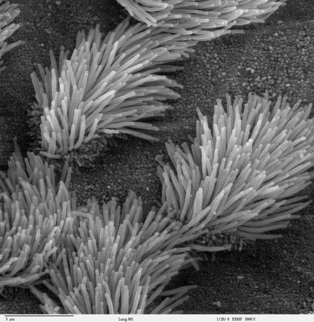
Cilium
The cilium (pl.: cilia; from Latin cilium 'eyelash'; in Medieval Latin and in anatomy, cilium) is a short hair-like membrane protrusion from many types of eukaryotic cell. (Cilia are absent in bacteria and archaea.) The cilium has the shape of a slender threadlike projection that extends from the surface of the much larger cell body. Eukaryotic flagella found on sperm cells and many protozoans have a similar structure to motile cilia that enables swimming through liquids; they are longer than cilia and have a different undulating motion.
There are two major classes of cilia: motile and non-motile cilia, each with two subtypes, giving four types in all. A cell will typically have one primary cilium or many motile cilia. The structure of the cilium core, called the axoneme, determines the cilium class. Most motile cilia have a central pair of single microtubules surrounded by nine pairs of double microtubules called a 9+2 axoneme. Most non-motile cilia have a 9+0 axoneme that lacks the central pair of microtubules. Also lacking are the associated components that enable motility including the outer and inner dynein arms, and radial spokes. Some motile cilia lack the central pair, and some non-motile cilia have the central pair, hence the four types.
Most non-motile cilia, termed primary cilia or sensory cilia, serve solely as sensory organelles. Most vertebrate cell types possess a single non-motile primary cilium, which functions as a cellular antenna. Olfactory neurons possess a great many non-motile cilia. Non-motile cilia that have a central pair of microtubules are the kinocilia present on hair cells.
Motile cilia are found in large numbers on respiratory epithelial cells – around 200 cilia per cell, where they function in mucociliary clearance, and also have mechanosensory and chemosensory functions. Motile cilia on ependymal cells move the cerebrospinal fluid through the ventricular system of the brain. Motile cilia are also present in the oviducts (fallopian tubes) of female (therian) mammals, where they function in moving egg cells from the ovary to the uterus. Motile cilia that lack the central pair of microtubules are found in the cells of the embryonic primitive node; termed nodal cells, these nodal cilia are responsible for the left-right asymmetry of bilaterians.
A cilium is assembled and built from a basal body on the cell surface. From the basal body, the ciliary rootlet forms ahead of the transition plate and transition zone where the earlier microtubule triplets change to the microtubule doublets of the axoneme.
The foundation of the cilium is the basal body, a term applied to the mother centriole when it is associated with a cilium. Mammalian basal bodies consist of a barrel of nine triplet microtubules, subdistal appendages and nine strut-like structures, known as distal appendages, which attach the basal body to the membrane at the base of the cilium. Two of each of the basal body's triplet microtubules extend during growth of the axoneme to become the doublet microtubules.
The ciliary rootlet is a cytoskeleton-like structure that originates from the basal body at the proximal end of a cilium. Rootlets are typically 80-100 nm in diameter and contain cross striae distributed at regular intervals of approximately 55-70 nm. A prominent component of the rootlet is rootletin a coiled coil rootlet protein coded for by the CROCC gene.
To achieve its distinct composition, the proximal-most region of the cilium consists of a transition zone, also known as the ciliary gate, that controls the entry and exit of proteins to and from the cilium. At the transition zone, Y-shaped structures connect the ciliary membrane to the underlying axoneme. Control of selective entry into cilia may involve a sieve-like function of transition zone. Inherited defects in components of the transition zone cause ciliopathies, such as Joubert syndrome. Transition zone structure and function is conserved across diverse organisms, including vertebrates, Caenorhabditis elegans, Drosophila melanogaster and Chlamydomonas reinhardtii. In mammals, disruption of the transition zone reduces the ciliary abundance of membrane-associated ciliary proteins, such as those involved in Hedgehog signal transduction, compromising Hedgehog-dependent embryonic development of digit number and central nervous system patterning.
Hub AI
Cilium AI simulator
(@Cilium_simulator)
Cilium
The cilium (pl.: cilia; from Latin cilium 'eyelash'; in Medieval Latin and in anatomy, cilium) is a short hair-like membrane protrusion from many types of eukaryotic cell. (Cilia are absent in bacteria and archaea.) The cilium has the shape of a slender threadlike projection that extends from the surface of the much larger cell body. Eukaryotic flagella found on sperm cells and many protozoans have a similar structure to motile cilia that enables swimming through liquids; they are longer than cilia and have a different undulating motion.
There are two major classes of cilia: motile and non-motile cilia, each with two subtypes, giving four types in all. A cell will typically have one primary cilium or many motile cilia. The structure of the cilium core, called the axoneme, determines the cilium class. Most motile cilia have a central pair of single microtubules surrounded by nine pairs of double microtubules called a 9+2 axoneme. Most non-motile cilia have a 9+0 axoneme that lacks the central pair of microtubules. Also lacking are the associated components that enable motility including the outer and inner dynein arms, and radial spokes. Some motile cilia lack the central pair, and some non-motile cilia have the central pair, hence the four types.
Most non-motile cilia, termed primary cilia or sensory cilia, serve solely as sensory organelles. Most vertebrate cell types possess a single non-motile primary cilium, which functions as a cellular antenna. Olfactory neurons possess a great many non-motile cilia. Non-motile cilia that have a central pair of microtubules are the kinocilia present on hair cells.
Motile cilia are found in large numbers on respiratory epithelial cells – around 200 cilia per cell, where they function in mucociliary clearance, and also have mechanosensory and chemosensory functions. Motile cilia on ependymal cells move the cerebrospinal fluid through the ventricular system of the brain. Motile cilia are also present in the oviducts (fallopian tubes) of female (therian) mammals, where they function in moving egg cells from the ovary to the uterus. Motile cilia that lack the central pair of microtubules are found in the cells of the embryonic primitive node; termed nodal cells, these nodal cilia are responsible for the left-right asymmetry of bilaterians.
A cilium is assembled and built from a basal body on the cell surface. From the basal body, the ciliary rootlet forms ahead of the transition plate and transition zone where the earlier microtubule triplets change to the microtubule doublets of the axoneme.
The foundation of the cilium is the basal body, a term applied to the mother centriole when it is associated with a cilium. Mammalian basal bodies consist of a barrel of nine triplet microtubules, subdistal appendages and nine strut-like structures, known as distal appendages, which attach the basal body to the membrane at the base of the cilium. Two of each of the basal body's triplet microtubules extend during growth of the axoneme to become the doublet microtubules.
The ciliary rootlet is a cytoskeleton-like structure that originates from the basal body at the proximal end of a cilium. Rootlets are typically 80-100 nm in diameter and contain cross striae distributed at regular intervals of approximately 55-70 nm. A prominent component of the rootlet is rootletin a coiled coil rootlet protein coded for by the CROCC gene.
To achieve its distinct composition, the proximal-most region of the cilium consists of a transition zone, also known as the ciliary gate, that controls the entry and exit of proteins to and from the cilium. At the transition zone, Y-shaped structures connect the ciliary membrane to the underlying axoneme. Control of selective entry into cilia may involve a sieve-like function of transition zone. Inherited defects in components of the transition zone cause ciliopathies, such as Joubert syndrome. Transition zone structure and function is conserved across diverse organisms, including vertebrates, Caenorhabditis elegans, Drosophila melanogaster and Chlamydomonas reinhardtii. In mammals, disruption of the transition zone reduces the ciliary abundance of membrane-associated ciliary proteins, such as those involved in Hedgehog signal transduction, compromising Hedgehog-dependent embryonic development of digit number and central nervous system patterning.