Hubbry Logo
Clue cellClue cellMain
Open search
Clue cell
Community hub
Clue cell
logo
8 pages, 0 posts
0 subscribers
Be the first to start a discussion here.
Clue cell
Clue cell
from Wikipedia
Micrograph of a clue cell (center), covered in bacteria, as compared to an unremarkable squamous cell at bottom left, and a neutrophil at bottom center. Pap stain
Phase contrast microscopy of clue cells in a vaginal swab

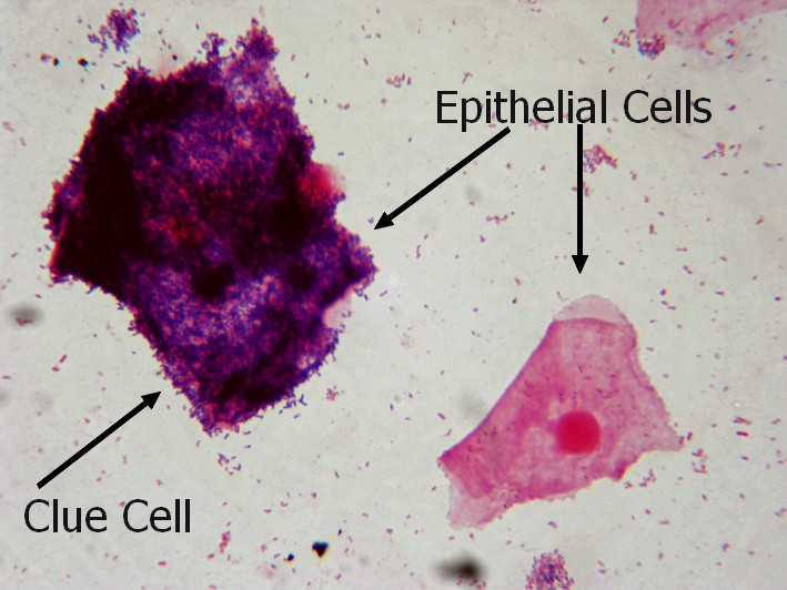
Clue cells are epithelial cells of the vagina that get their distinctive stippled appearance by being covered with bacteria. The etymology behind the term "clue" cell derives from the original research article from Gardner and Dukes describing the characteristic cells. The name was chosen for its brevity in describing the sine qua non of bacterial vaginosis.[1]

They are a medical sign of bacterial vaginosis, particularly that caused by Gardnerella vaginalis,[2] a group of Gram-variable bacteria. This bacterial infection is characterized by thin gray vaginal discharge, and an increase in vaginal pH from around 4.5 to over 5.5.


Bacteria obscuring the surfaces of vaginal epithelial cells, giving them a stippled appearance

References

[edit]
[edit]
Revisions and contributorsEdit on WikipediaRead on Wikipedia
from Grokipedia
Clue cells are squamous epithelial cells from the vaginal mucosa that exhibit a distinctive stippled or granular appearance under microscopic examination due to heavy adherence of , primarily serving as a hallmark diagnostic feature of (BV). First described in 1955 by Gardner and Dukes as a specific indicator of what was then termed "Haemophilus vaginalis" —now recognized as BV caused by polymicrobial overgrowth—these cells derive their name from providing a critical "clue" to the underlying infection. The adherent , most commonly Gardnerella vaginalis and other anaerobic species such as and Atopobium, form biofilms on the cell surface, obscuring the normal sharp borders and creating a fuzzy or beaded outline visible in wet mounts or Gram-stained smears. In clinical practice, the presence of clue cells is evaluated using Amsel's criteria for diagnosis, where their detection in more than 20% of epithelial cells in a microscopic , alongside other signs like elevated vaginal (>4.5), thin homogeneous discharge, and a positive whiff test (fishy upon KOH addition), confirms the condition with high specificity. associated with clue cells increases risks of complications including , , and enhanced susceptibility to sexually transmitted infections like , underscoring the importance of accurate identification. Recent studies using advanced techniques like have distinguished true clue cells (with Gardnerella biofilms) from pseudo-clue cells (involving non-pathogenic bacteria like Lactobacillus iners), revealing variability in morphotypes across populations.

Etymology and history

Origin of the term

The term "clue cell" originates from the English word "clue," evoking the idea of a detective's hint or lead, to underscore the cells' value as a pivotal diagnostic indicator in identifying . This nomenclature highlights their role in providing clinicians with essential evidence for without suggesting a direct causal relationship between the observed cellular changes and the infection. The designation was introduced in the seminal 1955 study by Herman L. Gardner and Charles D. Dukes, who first described these cells in the context of what they termed Haemophilus vaginalis vaginitis—a condition previously classified as nonspecific vaginitis. In their research, published in the American Journal of Obstetrics and Gynecology, Gardner and Dukes emphasized the microscopic appearance of vaginal epithelial cells obscured by adherent bacteria as a reliable "clue" to guide therapeutic decisions. By framing the cells this way, the term avoids overattribution of while stressing their practical utility in clinical settings, a convention that has persisted in despite subsequent reclassifications of the associated from Haemophilus vaginalis to in 1980.

Discovery and early descriptions

Clue cells were first described in 1955 by Herman L. Gardner and Charles D. Dukes in their seminal paper on what they termed "Haemophilus vaginalis ," a specific previously classified as non-specific . In their study of vaginal smears from women with symptomatic , Gardner and Dukes observed vaginal epithelial cells densely coated with small coccobacilli, which they identified as Haemophilus vaginalis (later reclassified as in 1980), distinguishing this condition from more commonly assumed causes like yeast infections or . These coated cells, named clue cells for their diagnostic value, were noted in nearly all cases of the they attributed to this bacterium. Throughout the 1950s and 1960s, subsequent research built on these observations, confirming the presence of similar bacterial-adherent epithelial cells in vaginal discharges from women exhibiting symptoms such as malodorous discharge and elevated , further solidifying their role as a hallmark of non-specific . By the 1970s, studies expanded on the , revealing a complex overgrowth of anaerobes alongside Gardnerella, challenging the initial view of it as a single-pathogen . The understanding evolved significantly in the 1980s, with the term "" adopted in 1983 to replace "non-specific " and emphasize its polymicrobial nature, distinguishing it from other vaginitides; this shift was supported by diagnostic criteria including the presence of clue cells.

Definition and characteristics

Medical definition

A clue cell is a vaginal squamous epithelial cell characterized by extensive adherence of , primarily along with anaerobic species such as spp., Atopobium vaginae, and Mobiluncus spp., that obscure the cell borders. This feature is defined by a granular or stippled coating of adherent that renders the cell borders indistinct, setting it apart from unaffected epithelial cells. Recent studies distinguish true clue cells, involving biofilms of BV-associated like Gardnerella, from pseudo-clue cells coated by non-pathogenic species such as Lactobacillus iners. Clue cells do not constitute a unique but instead reflect a morphological change arising from overgrowth of microbes within the vaginal .

Microscopic appearance

Clue cells are observed under wet mount , typically at 400× , as vaginal squamous epithelial cells with irregular, shaggy, or fuzzy borders resulting from the adherence of numerous coccobacilli. The adherent create a fine granular or stippled layer across the cell surface, obscuring the cell borders and giving the cell a smudged or indistinct appearance compared to normal epithelial cells, which have clear contents and sharp borders. These cells are generally round to polygonal in shape and measure approximately 20–50 μm in diameter, though their form is distorted by the bacterial coating. Under , clue cells reveal gram-variable rods or coccobacilli, predominantly Gardnerella , densely attached to the epithelial surface, further emphasizing the loss of distinct cellular outlines.

Diagnostic role

Role in bacterial vaginosis diagnosis

Clue cells play a central role in the clinical of (BV) as one of the four Amsel criteria, established in 1983 for non-invasive office-based assessment. According to these criteria, a of BV is confirmed if at least three of the following are present: homogeneous thin , vaginal greater than 4.5, a positive whiff test (fishy odor upon addition of 10% ), and the presence of clue cells on wet-mount . Specifically, clue cells are considered positive when they constitute 20% or more of the vaginal epithelial cells examined, indicating heavy adherence of that obscures cell borders. In the Amsel framework, the detection of clue cells is highly indicative of BV due to their association with overgrowth of anaerobic bacteria, such as , forming a polymicrobial on epithelial surfaces. This criterion alone is not diagnostic but contributes significantly when combined with others, enhancing overall accuracy. Studies evaluating Amsel criteria against the gold-standard report the full set achieving sensitivity of 37% to 70% and specificity of 94% to 99%. Clue cells also factor into the Nugent scoring system, a Gram stain-based method that quantifies bacterial morphotypes to grade vaginal microbiota (score 0–3: normal; 4–6: intermediate; 7–10: BV). While clue cells are not a separate scored category, their presence correlates strongly with elevated counts of Gardnerella and Bacteroides-like coccobacilli, which contribute 3–4 points when comprising 5 or more per , shifting intermediate flora toward a BV diagnosis (total score 7–10). This morphological shift reflects the bacterial coating characteristic of clue cells, aiding in confirming BV when Lactobacillus dominance is reduced. The diagnostic utility of clue cells varies across studies, with reported sensitivity ranging from 46% to 98% and specificity from 94% to 100% when compared to Nugent scoring as the reference. For instance, one evaluation found clue cells to have 98.2% sensitivity and 94.3% specificity, marking them as the most reliable single indicator among Amsel components. However, clue cells are not for , as they can occasionally appear in other vaginal conditions or states, necessitating integration with additional criteria for accurate .

Detection methods

The primary method for detecting clue cells involves preparing a wet mount from . A sample of is collected using a swab and mixed with one or two drops of 0.9% normal saline solution on a glass slide, which is then covered with a coverslip. The preparation is examined immediately under a using low power (10×) for initial screening and high dry power (400×) or to identify clue cells, defined as vaginal epithelial cells with borders obscured by adherent . This technique allows for direct visualization and is integrated into diagnostic criteria such as Amsel's, where the presence of clue cells contributes to the overall assessment. An alternative approach uses Gram staining of a vaginal smear to detect clue cells alongside bacterial morphotypes. The specimen is air-dried, heat-fixed on a slide, stained with , decolorized, and counterstained with , then examined at 1000× . Clue cells appear as epithelial cells heavily coated with Gram-variable coccobacilli, such as those from Gardnerella , and this method supports quantitative scoring systems like Nugent's for evaluation. Potassium hydroxide (KOH) preparations should be avoided for clue cell detection, as the alkaline solution lyses epithelial cells and obscures bacterial adherence. Emerging molecular methods, such as (PCR) assays targeting DNA and other BV-associated , offer sensitive detection of microbial components but do not directly visualize clue cells. These nucleic acid amplification tests (NAATs), including multiplex PCR panels, achieve sensitivities of 84.8%–97% and specificities of 77%–100% compared to traditional , yet direct microscopic identification of clue cells remains the gold standard for confirming their characteristic appearance in . Recent 2025 studies have highlighted additional point-of-care options like the BVBlue test, achieving high accuracy in rapid BV , and models for automated Nugent scoring with improved reproducibility.

Clinical significance

Association with bacterial vaginosis

Clue cells serve as a hallmark indicator of vaginal in (BV), characterized by a marked reduction in the dominance of species, typically falling below 10^6 CFU/mL, which allows for the overgrowth of anaerobic such as and species. This shift disrupts the normal acidic vaginal environment maintained by -produced and , promoting the proliferation of BV-associated microbes that adhere to and form biofilms on vaginal epithelial cells. In the pathophysiology of BV, clue cells arise from the adherence of these anaerobes to squamous epithelial cells, facilitated by bacterial sialidase enzymes produced primarily by and other BV pathogens. These sialidases cleave terminal residues from protective glycoproteins on the epithelial surface, exposing underlying receptors that enhance bacterial attachment and formation, thereby obscuring the cell's borders under . The resulting biofilms create a polymicrobial community that resists clearance and perpetuates the . Clinically, the presence of clue cells correlates with characteristic BV symptoms, including a thin, grayish-white vaginal discharge and a fishy odor upon alkalinization (positive amine or whiff test), reflecting the production of volatile amines like putrescine and cadaverine by anaerobic metabolism. In diagnostic contexts, clue cells exceeding 20% of observed vaginal epithelial cells on wet mount microscopy strongly support a BV diagnosis under Amsel's criteria. BV associated with clue cells carries significant complications, including an elevated risk of due to ascending and inflammation, through endometrial and tubal involvement, and increased susceptibility to acquisition owing to disrupted mucosal barriers and altered immune responses. Recurrence is common, affecting 30-50% of treated cases within the first year, often linked to persistent biofilms and incomplete restoration of dominance.

Implications in other conditions

The presence of clue cells in urine sediment generally signifies concurrent (BV) rather than a primary (UTI), as these cells represent vaginal epithelial elements coated with and other BV-associated that have contaminated or ascended into the urinary tract. While sample from vaginal sources is common, studies using suprapubic aspiration or catheterization confirm their occurrence in bladder , indicating potential microbial dysbiosis extending beyond the and increasing UTI susceptibility by 2.2- to 13.7-fold in affected women. Pseudo-clue cells, which resemble true clue cells under , arise in conditions mimicking but lack the characteristic adherent Gardnerella biofilms. In cytolytic vaginosis, an overgrowth of lactobacilli leads to epithelial cell fragmentation and adherence of these larger rod-shaped bacteria, producing a stippled appearance on wet mount; however, they are differentiated by an acidic vaginal (3.5–4.5), absence of clue cell border obliteration, intact epithelial borders, and no Gardnerella or . may similarly present with epithelial cells coated by inflammatory aerobic bacteria (e.g., or group B streptococci), featuring larger bacilli and preserved cell margins, in contrast to the fine coccobacilli that obscure borders in genuine clue cells associated with . Clue cells occasionally appear in rare scenarios beyond typical , such as postmenopausal vaginal or post-hysterectomy states, where deficiency promotes epithelial thinning and , elevating risk (prevalence 2%–57%) particularly in sexually active women on replacement therapy; this can lead to complications like vaginal cuff infections. Nonetheless, their diagnostic utility diminishes in women, where low predictive value results in false positives up to 20%, often from transient microbial shifts, pseudo-clue cell misidentification, or non-pathologic variations.

References

Add your contribution
Related Hubs
User Avatar
No comments yet.