
Community hub
Recent from talks
Contribute something to knowledge base
Content stats: 0 posts, 0 articles, 1 media, 0 notes
Members stats: 0 subscribers, 0 contributors, 0 moderators, 0 supporters
Subscribers
Supporters
Contributors
Moderators
Hub AI
Cotton wool spots AI simulator
(@Cotton wool spots_simulator)
Hub AI
Cotton wool spots AI simulator
(@Cotton wool spots_simulator)
Cotton wool spots
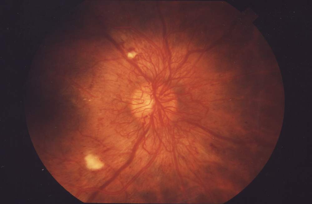
Cotton wool spots are opaque fluffy white patches on the retina of the eye that are considered an abnormal finding during a funduscopic exam (also called an ophthalmoscopic exam). Cotton wool spots are typically a sign of another disease state, most common of which is diabetic retinopathy. The irregularly shaped white patches are a result of ischemia, or reduced blood flow and oxygen, in the retinal nerve fiber layer, which is located in the distribution of the capillaries of the superficial layer of the retina. These areas with reduced blood flow reflect the obstruction of axoplasmic flow due to mechanical or vascular causes and the consequential accumulation as a result of decreased axonal transport. This reduced axonal transport can then cause swelling or bulging on the surface layer of the retina, increasing the potential for nerve fiber damage.
The presence of cotton wool spots may resolve independently over time, typically in 4–12 weeks, or may depend on the underlying disease causing the condition. Diagnosis and treatment of the underlying disease state may be beneficial in the treatment and management of cotton wool spots.
Cotton wool spots present as opaque fluffy white spots, sometimes with feather-like edges, on the retina when seen on a funduscopic exam. These opaque white areas are typically asymptomatic, but may cause some vision loss due to the underlying disease state. Cotton wool spots are a retinal condition that when present, frequently act as a significant indicator, or sign, of a variety of underlying disease states. These fluffy white spots are often accompanied by additional abnormal funduscopic findings such as intraretinal bleeds, hard exudates, and atypical small blood vessels, including obstructed or dilated blood vessels and microaneurysms. These associated findings are common due to the underlying diseases causing these manifestations seen in the eyes. Because of the use of cotton wool spots more as an indicator of other systemic diseases, Mcleod had proposed that the taxonomy of the terms 'cotton wool spot' should be changed to something else to help better understand its true meaning.
Cotton wool spots are commonly caused by changes to the retina secondary to diabetes, hypertension, or blockages to blood vessels to the retina, such as central retinal vein occlusion. While at one point believed to have been the result of nerve damage in the retina, interruptions to axoplasmic flow to these nerves may result from organelle accumulation in the axons. This is further purported by the presence of cytoid bodies in ganglion axon cells, where the accumulation of white blood cells, more specifically, eosinophils, mitochondria, and other cellular material lead to swelling of the ganglion axon cells which then lead to cotton wool spots.
Referred to as a form of retinal myopathies, cotton wools spots are commonly found to be a secondary manifestation to diseases like diabetes, hypertension, and AIDS. Cotton wool spots have become one of the hallmarks of pre-proliferative diabetic retinopathy, a condition caused by damaged blood vessels in the retina due to high blood sugar. Abundant cotton wool spots are also found in hypertensive retinopathy, including malignant hypertension, where the white patches are a result of a microvascular infarct, or a sudden occlusion of the arteriole. One study observed the presence of cotton wool spots in people with acute leukemia, referring to the increase in white blood cells as a probable cause. Although cotton wool spots are a secondary manifestation to leukemia, they have been found to be a poor prognostic sign for survival in acute leukemia.
More recently, prolonged travel in space has been found to be a probable cause of cotton wool spots in people who experience ocular changes after coming back from space. Although infrequent, conditions like HIV Retinopathy and Purtscher's retinopathy can also lead to the appearance of cotton wool spots. Although they are associated with multiple systemic diseases, it is possible for there to be an unknown etiology that would require further investigation. Cotton wool spots have different clinical characteristics when present as a result of HIV rather than in systemic diseases, like diabetes or hypertension, due to the viral complexes that cause structural changes to the retinal microvasculature.
Cotton wool spots are also associated with giant cell arteritis (GCA), a type of inflammation of the lining of the arteries, as this may reduce blood flow to the eyes. In a study of 123 subjects with vision loss due to early stage giant cell arteritis, one-third of subjects had cotton wool spots present in their eyes. Furthermore, because giant cell arteritis can lead to loss of vision and is associated with cotton wool spots, it is recommended to investigate for giant cell arteritis if cotton wool spots are found.
Another condition in which cotton wool spots are found is central retinal vein occlusion as a result of reduced blood flow from retinal arteriole obstruction.
Cotton wool spots
Cotton wool spots are opaque fluffy white patches on the retina of the eye that are considered an abnormal finding during a funduscopic exam (also called an ophthalmoscopic exam). Cotton wool spots are typically a sign of another disease state, most common of which is diabetic retinopathy. The irregularly shaped white patches are a result of ischemia, or reduced blood flow and oxygen, in the retinal nerve fiber layer, which is located in the distribution of the capillaries of the superficial layer of the retina. These areas with reduced blood flow reflect the obstruction of axoplasmic flow due to mechanical or vascular causes and the consequential accumulation as a result of decreased axonal transport. This reduced axonal transport can then cause swelling or bulging on the surface layer of the retina, increasing the potential for nerve fiber damage.
The presence of cotton wool spots may resolve independently over time, typically in 4–12 weeks, or may depend on the underlying disease causing the condition. Diagnosis and treatment of the underlying disease state may be beneficial in the treatment and management of cotton wool spots.
Cotton wool spots present as opaque fluffy white spots, sometimes with feather-like edges, on the retina when seen on a funduscopic exam. These opaque white areas are typically asymptomatic, but may cause some vision loss due to the underlying disease state. Cotton wool spots are a retinal condition that when present, frequently act as a significant indicator, or sign, of a variety of underlying disease states. These fluffy white spots are often accompanied by additional abnormal funduscopic findings such as intraretinal bleeds, hard exudates, and atypical small blood vessels, including obstructed or dilated blood vessels and microaneurysms. These associated findings are common due to the underlying diseases causing these manifestations seen in the eyes. Because of the use of cotton wool spots more as an indicator of other systemic diseases, Mcleod had proposed that the taxonomy of the terms 'cotton wool spot' should be changed to something else to help better understand its true meaning.
Cotton wool spots are commonly caused by changes to the retina secondary to diabetes, hypertension, or blockages to blood vessels to the retina, such as central retinal vein occlusion. While at one point believed to have been the result of nerve damage in the retina, interruptions to axoplasmic flow to these nerves may result from organelle accumulation in the axons. This is further purported by the presence of cytoid bodies in ganglion axon cells, where the accumulation of white blood cells, more specifically, eosinophils, mitochondria, and other cellular material lead to swelling of the ganglion axon cells which then lead to cotton wool spots.
Referred to as a form of retinal myopathies, cotton wools spots are commonly found to be a secondary manifestation to diseases like diabetes, hypertension, and AIDS. Cotton wool spots have become one of the hallmarks of pre-proliferative diabetic retinopathy, a condition caused by damaged blood vessels in the retina due to high blood sugar. Abundant cotton wool spots are also found in hypertensive retinopathy, including malignant hypertension, where the white patches are a result of a microvascular infarct, or a sudden occlusion of the arteriole. One study observed the presence of cotton wool spots in people with acute leukemia, referring to the increase in white blood cells as a probable cause. Although cotton wool spots are a secondary manifestation to leukemia, they have been found to be a poor prognostic sign for survival in acute leukemia.
More recently, prolonged travel in space has been found to be a probable cause of cotton wool spots in people who experience ocular changes after coming back from space. Although infrequent, conditions like HIV Retinopathy and Purtscher's retinopathy can also lead to the appearance of cotton wool spots. Although they are associated with multiple systemic diseases, it is possible for there to be an unknown etiology that would require further investigation. Cotton wool spots have different clinical characteristics when present as a result of HIV rather than in systemic diseases, like diabetes or hypertension, due to the viral complexes that cause structural changes to the retinal microvasculature.
Cotton wool spots are also associated with giant cell arteritis (GCA), a type of inflammation of the lining of the arteries, as this may reduce blood flow to the eyes. In a study of 123 subjects with vision loss due to early stage giant cell arteritis, one-third of subjects had cotton wool spots present in their eyes. Furthermore, because giant cell arteritis can lead to loss of vision and is associated with cotton wool spots, it is recommended to investigate for giant cell arteritis if cotton wool spots are found.
Another condition in which cotton wool spots are found is central retinal vein occlusion as a result of reduced blood flow from retinal arteriole obstruction.
Recent media
Recent media