Hubbry Logo
External intercostal musclesExternal intercostal musclesMain
Open search
External intercostal muscles
Community hub
External intercostal muscles
logo
8 pages, 0 posts
0 subscribers
Be the first to start a discussion here.
External intercostal muscles
from Wikipedia
External intercostal muscles
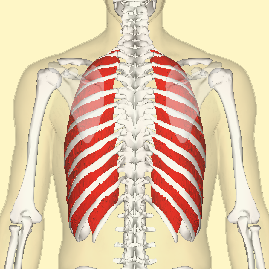
Position of the external intercostal muscles (shown in red) seen from the back.
Details
OriginLower border of ribs
InsertionUpper border of rib below
ArteryIntercostal arteries
NerveIntercostal nerves
ActionsInhalation
AntagonistIntercostales interni muscles
Identifiers
Latinmusculi intercostales externi
TA98A04.4.01.012
TA22311
FMA74084
Anatomical terms of muscle

The external intercostal muscles or external intercostals (intercostales externi) are eleven in number on both sides.

Structure

[edit]
A cutout of the thoracic wall showing the three layers of intercostal muscle - from the left wall.

The muscles extend from the tubercles of the ribs behind, to the cartilages of the ribs in front, where they end in thin membranes, the external intercostal membranes, which are continued forward to the sternum. These muscles work in unison when inhalation occurs. The internal intercostal muscles relax while the external muscles contract causing the expansion of the chest cavity and an influx of air into the lungs.

Each arises from the lower border of a rib, and is inserted into the upper border of the rib below. In the two lower spaces they extend to the ends of the cartilages, and in the upper two or three spaces they do not quite reach the ends of the ribs.

They are thicker than the internal intercostals, and fibers are directed obliquely downward and laterally on the back of the thorax, and downward, forward, and medially on the front.

Variations

[edit]

Continuation with the external oblique or serratus anterior: A supracostalis muscle, from the anterior end of the first rib down to the second, third or fourth ribs occasionally occurs.

Additional images

[edit]

See also

[edit]

References

[edit]
Revisions and contributorsEdit on WikipediaRead on Wikipedia
from Grokipedia
The external intercostal muscles are a group of skeletal muscles located in the superficial layer of the , consisting of eleven pairs that span the intercostal spaces between the . These muscles originate from the inferior border of each (from 1 to 11) and insert onto the superior border of the immediately below, with fibers running obliquely downward and forward; posteriorly, they extend from the to the costochondral junction anteriorly, where they transition into the external intercostal membrane. Their primary function is to assist in respiration by contracting during inspiration to elevate the , which increases the transverse and anteroposterior diameters of the through a "bucket-handle" motion, thereby expanding the lungs for . Innervated by the (anterior rami of thoracic spinal nerves T1 to T11), these muscles receive sensory and motor input that coordinates their role in . Blood supply is provided by the posterior intercostal arteries (from the supreme intercostal artery for T1-T2 or the for T3-T11) and anterior intercostal arteries (branches of the for upper spaces or musculophrenic artery for lower ones). As part of the intrinsic thoracic musculature, the external intercostals work in concert with the diaphragm and internal intercostals to facilitate efficient ventilation, and their dysfunction can impair respiratory mechanics.

Anatomy

Structure

The external intercostal muscles form the outermost layer of the three intercostal muscle groups in the thoracic wall, consisting of skeletal muscle fibers that lie superficial to the internal and innermost intercostal muscles. These muscles are separated from the deeper layers by thin fascia, positioning them as the most superficial component of the intercostal musculature. They span the 11 intercostal spaces between the 12 pairs of , occupying the regions from the first to the 11th intercostal space. The muscle fibers exhibit an oblique orientation, running downward and forward in a direction that is perpendicular to the fibers of the internal . In terms of thickness and appearance, the external intercostal muscles present as thin, flattened bands beneath the skin and .

Attachments

The external intercostal muscles originate from the inferior border of each , near the costal groove. They insert onto the upper border of the immediately below, where the fibers blend into the of that . These muscles occupy the 11 intercostal spaces between 1 and 12. Posteriorly, they extend to the of the , adjacent to the costotransverse ligaments, while anteriorly they reach the costochondral junction near the edge of the , transitioning into the external intercostal membrane. In terms of spatial relationships, the external intercostal muscles lie superficial to the internal intercostal muscles, with the intercostal running in the costal groove deep to the internal layer.

Function

Respiratory Role

The external intercostal muscles primarily function during the inspiratory phase of by contracting to elevate the , which expands the and facilitates lung inflation. This elevation increases both the anteroposterior and transverse diameters of the , allowing for greater air intake. The mechanism involves upward and outward movement of the at their articulations with the vertebrae and . Specifically, contraction produces a "pump-handle" motion in the anterior , enhancing the anteroposterior dimension, and a "bucket-handle" motion in the lateral , widening the transverse dimension. In forced or deep inspiration, the external intercostal muscles serve as accessory inspiratory muscles, augmenting the diaphragm's action to achieve larger thoracic expansion and increased volume, particularly during elevated ventilatory demands. Their activity is minimal during quiet but intensifies with higher respiratory efforts. During quiet expiration, the external intercostal muscles are inactive, relying instead on the of the s and chest wall to reduce thoracic volume and expel air passively.

Biomechanics

The external intercostal muscles contribute to thoracic expansion by contracting to rotate the ribs around their costovertebral axes, elevating the ribs in a bucket-handle motion that increases the transverse and anteroposterior diameters of the rib cage. This rotation, primarily in the rostral and dorsal interspaces, drives inspiratory expansion of the thoracic cavity, with the mechanical advantage diminishing caudally. During deep inspiration, this action significantly augments thoracic volume to facilitate greater air intake, working in coordination with the diaphragm to enhance overall ventilatory efficiency. The interact antagonistically with the internal intercostal muscles to enable coordinated cycles. While external intercostals primarily elevate the during inspiration, the internal interosseous intercostals depress them during expiration, with their opposing fiber orientations ensuring reciprocal activation for efficient thoracic volume changes. This opposition is regionally selective, as external intercostals exhibit inspiratory effects in dorsal regions, contrasting with the expiratory effect of the interosseous internal intercostals in dorsal regions and the inspiratory effect of the parasternal internal intercostals in ventral areas, thus optimizing respiratory mechanics without interference. Beyond expansion, the external intercostals play a critical role in stabilizing the chest wall by stiffening the intercostal spaces against inward deformation during inspiration. This stabilization counters the negative intrathoracic pressure that could otherwise cause paradoxical inward motion of the , maintaining structural integrity and efficient airflow. Postural influences modulate external intercostal activation, with greater recruitment observed in positions due to gravitational effects that displace abdominal contents cephalad, reducing diaphragmatic efficiency and increasing demands on thoracic stabilization. In this posture, the increased reliance on intercostals helps sustain expansion and prevent collapse, thereby adapting to body orientation.

Innervation and Vascular Supply

Nerve Supply

The external intercostal muscles are innervated by the , which are the ventral rami of the thoracic spinal nerves from T1 to T11. These nerves provide the primary somatic motor innervation to the muscles, enabling their contraction during inspiration. Each segment of the external intercostal muscle is supplied by the corresponding intercostal nerve, which runs along the inferior border of the in the costal groove, accompanied by the intercostal vessels and protected by the internal intercostal muscle. The muscular branches of these nerves (particularly from T3 to T6) directly innervate the external intercostals, ensuring segmental control that aligns with the rib levels. The intercostal nerves carry somatic motor fibers originating from the anterior divisions of the thoracic spinal nerves, which synapse with motor neurons in the anterior horn of the spinal cord to drive muscle contraction. These nerves also include sensory components, with afferent fibers providing proprioceptive feedback from muscle spindles and Golgi tendon organs in the external intercostals, as well as general somatic sensation to the overlying thoracic wall. Central control of the external intercostal muscles occurs through descending pathways from the medullary respiratory centers in the , specifically the dorsal respiratory group (DRG) in the nucleus tractus solitarius and the ventral respiratory group (VRG) in the and related areas. Inspiratory neurons in the DRG and VRG generate rhythmic signals that excite external intercostal motor neurons via bulbospinal tracts in the , coordinating their activity with the diaphragm during quiet . This integration allows for automatic regulation of respiratory rhythm based on metabolic demands.

Blood Supply

The external intercostal muscles receive their arterial blood supply primarily from the posterior , which course within the intercostal spaces as part of the . The first and second posterior intercostal arteries originate from the supreme intercostal artery, a branch of the costocervical trunk arising from the , while the third through eleventh posterior intercostal arteries branch directly from the descending . These arteries provide segmental supply to the posterior aspects of the intercostal spaces, including the external intercostal muscles. Additional arterial supply to the external intercostal muscles comes from the anterior intercostal arteries, which are derived from the (for intercostal spaces 1–6) and its continuation, the musculophrenic artery (for spaces 7–9). These anterior branches vascularize the anterior portions of the intercostal spaces and contribute to the muscular perfusion. Venous drainage from the external intercostal muscles follows the arterial pattern via the posterior intercostal veins, which on the right drain into the and on the left into the hemiazygos and accessory hemiazygos veins, ultimately converging toward the . Anterior venous drainage occurs through tributaries of the internal thoracic vein. Lymphatic drainage of the external intercostal muscles is collected by vessels that empty into the intercostal lymph nodes located near the heads of the and the parasternal (internal thoracic) lymph nodes along the . From these nodes, flows ultimately to the on the left side and the right lymphatic duct on the right, returning to the venous system at the junction of the internal jugular and subclavian veins.

Variations and Clinical Aspects

Anatomical Variations

The external intercostal muscles exhibit several common anatomical variations, particularly at the superior and inferior extremes of the thoracic cage. In the first , the external intercostal muscle is often reduced in thickness or replaced anteriorly by a membranous rather than distinct muscle fibers, facilitating greater mobility near the and first . Similarly, in the lower intercostal spaces, fibers of the external intercostal muscles frequently fuse or continue seamlessly with the external oblique muscle of the , blending the thoracic and abdominal musculature. Asymmetrical fiber thickness between left and right sides has been noted in some individuals, potentially influenced by habitual posture or respiratory patterns, though this is typically subclinical. These variations are generally bilateral and do not significantly alter overall respiratory mechanics. Rare anomalies include supernumerary slips or accessory muscles derived from the external intercostal layer. For instance, the musculus supracostalis anterior is an uncommon supernumerary muscle originating from the first rib and inserting onto the fourth rib, spanning multiple intercostal spaces and occasionally connecting to the serratus anterior; it is innervated by branches of the and represents an aberrant extension of external intercostal development. Such anomalies are documented in isolated cadaveric cases and may arise from incomplete segmentation during myotomal differentiation. Incomplete development of external intercostal muscles can also occur in congenital chest wall syndromes, though specific involvement is variable and often accompanies broader thoracic hypoplasia. The external intercostal muscles derive from the myotomes of thoracic somites during weeks 4–8 of gestation. Errors in somite segmentation or myotomal migration can lead to fusion, absence, or supernumerary formations, as the myoblasts migrate to form the segmented intercostal layers between developing ribs from the sclerotome.

Clinical Significance

Intercostal muscle strain involving the external intercostal muscles commonly occurs in athletes or following trauma, such as sudden twisting, heavy lifting, or direct impact to the chest wall, resulting in overstretching or tearing of the muscle fibers. This injury typically presents with sharp, localized pain that intensifies during inspiration, coughing, or twisting movements, often accompanied by tenderness, swelling, and limited chest expansion. Diagnosis is primarily based on a thorough physical examination, including palpation along the intercostal spaces and assessment of pain provocation during respiration, with imaging such as X-rays to exclude rib fractures and MRI or CT scans to confirm soft tissue tears if symptoms persist. Weakness or of the external intercostal muscles plays a critical role in respiratory distress, particularly in neuromuscular disorders like Guillain-Barré syndrome, where demyelination of leads to impaired forced inspiration and reduced , potentially necessitating . In such cases, the loss of external intercostal function contributes to restrictive lung mechanics and , exacerbating overall when combined with diaphragmatic involvement. During surgical procedures like or intercostal drain placement, the external intercostal muscles are directly accessed via incisions through the intercostal spaces to reach the , which carries risks such as postoperative formation due to vascular disruption in the . Proper technique, including blunt dissection to separate muscle fibers, minimizes these complications, but can lead to , , or impaired healing if not managed promptly. Diagnostic imaging and electrophysiology are essential for evaluating external intercostal muscle ; MRI and CT scans effectively visualize tears, , or in the muscle layers, providing detailed contrast to guide treatment. (EMG) assesses innervation integrity by detecting abnormal electrical activity in the , aiding in the of neuropathic involvement or weakness.

References

Add your contribution
Related Hubs
User Avatar
No comments yet.