
Community hub
Recent from talks
Contribute something to knowledge base
Content stats: 0 posts, 0 articles, 1 media, 0 notes
Members stats: 0 subscribers, 0 contributors, 0 moderators, 0 supporters
Subscribers
Supporters
Contributors
Moderators
Hub AI
Group C nerve fiber AI simulator
(@Group C nerve fiber_simulator)
Hub AI
Group C nerve fiber AI simulator
(@Group C nerve fiber_simulator)
Group C nerve fiber
Group C nerve fibers are one of three classes of nerve fiber in the central nervous system (CNS) and peripheral nervous system (PNS). The Group C fibers are unmyelinated and have a small diameter and low conduction velocity, whereas Groups A and B are myelinated. Group C fibers include postganglionic fibers in the autonomic nervous system (ANS), and nerve fibers at the dorsal roots (IV fiber). These fibers carry sensory information.
Damage or injury to nerve fibers causes neuropathic pain. Capsaicin activates C fibre vanilloid receptors, providing the burning sensation associated with chili peppers.
C fibers are one class of nerve fiber found in the nerves of the somatic sensory system. They are afferent fibers, conveying input signals from the periphery to the central nervous system.
C fibers are unmyelinated unlike most other fibers in the nervous system. This lack of myelination is the cause of their slow conduction velocity, which is on the order of no more than 2 m/s. C fibers are on average 0.2–1.5 μm in diameter.
C fiber axons are grouped together into what is known as Remak bundles. These occur when a non-myelinating Schwann cell bundles the axons close together by surrounding them. The Schwann cell keeps them from touching each other by squeezing its cytoplasm between the axons. The condition of Remak bundles varies with age. The number of C fiber axons in each Remak bundle varies with location. For example, in a rat model, large bundles of greater than 20 axons are found exiting the L5 dorsal root ganglion, while smaller bundles of average 3 axons are found in distal nerve segments. Multiple neurons contribute axons to the Remak bundle with an average ratio of about 2 axons contributed per bundle. The cross sectional area of a Remak bundle is proportional to the number of axons found inside it. Remak bundles in the distal peripheral nerve are clustered with other Remak bundles. The Remak Schwann cells have been shown to be electrochemically responsive to action potentials of the axons contained within them.
In experiments where nerve injury is caused but nearby C fibers remain intact, increased spontaneous activity in the C fibers is observed. This phenomenon supports the theory that damaged nerve fibers may release factors that alter the function of neighboring undamaged fibers. Study of Remak bundles has important implications in nerve regeneration after sustaining injury. Currently, recovery of distal C fiber function takes months and may still only regain incomplete function. This may result in abnormal sensory function or neuropathic pain. Remak bundles are thought to release certain trophic factors that promote the regeneration of the damaged axons.
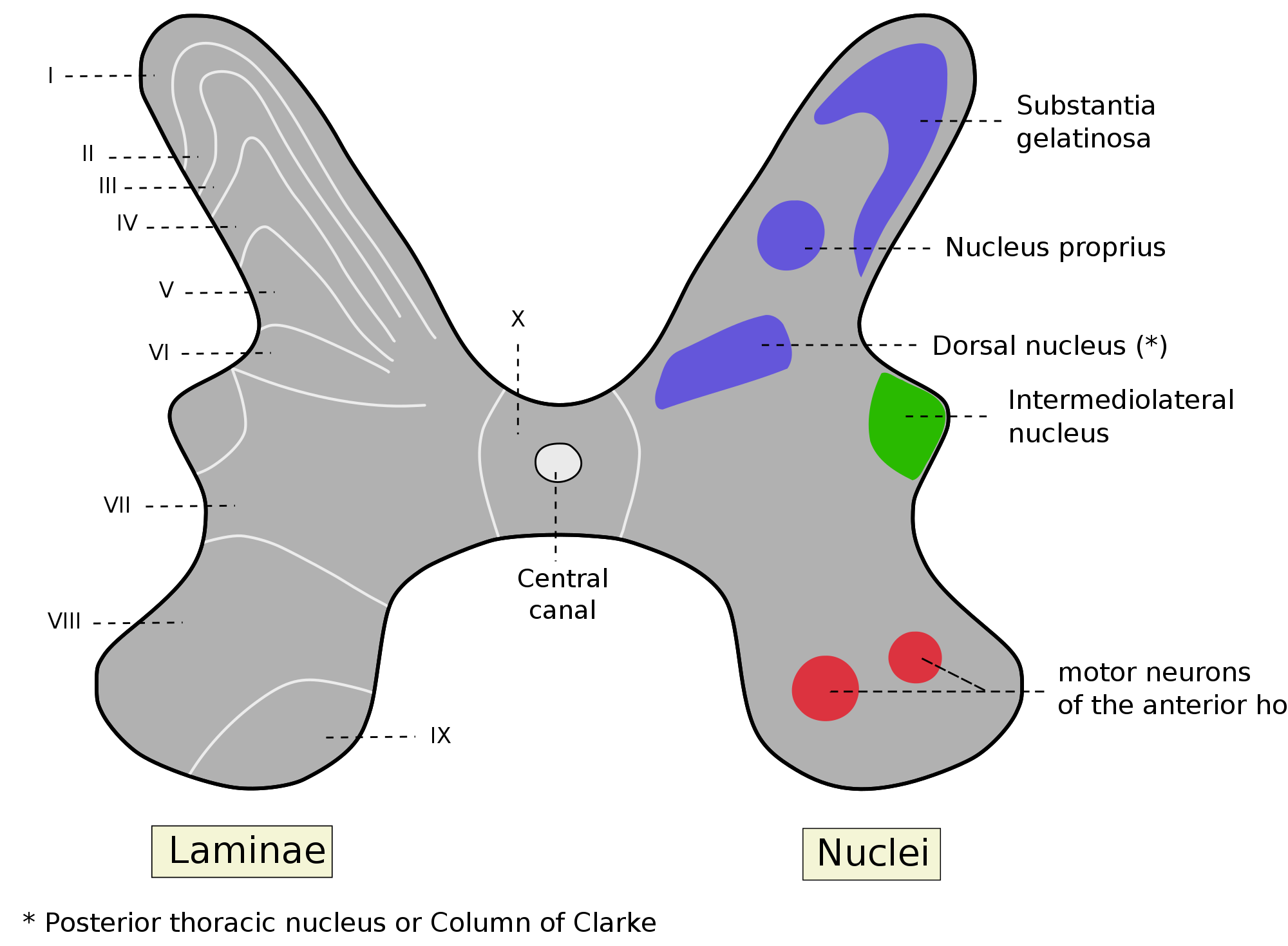
C fibers synapse to second-order projection neurons in the spinal cord at the upper laminae of the dorsal horn in the substantia gelatinosa. The second-order projection neurons are of the wide dynamic range (WDR) type, which receive input from both nociceptive terminals as well as myelinated A-type fibers. There are three types of second order projection neurons in the spinothalamic tract: wide dynamic range (WDR), high threshold (HT), and low threshold (LT). These classifications are based on their responses to mechanical stimuli. The second-order neurons ascend to the brain stem and thalamus in the ventrolateral, or anterolateral, quadrant of the contralateral half of the spinal cord, forming the spinothalamic tract. The spinothalamic tract is the main pathway associated with pain and temperature perception, which immediately crosses the spinal cord laterally. This crossover feature is clinically important because it allows for identification of the location of injury.
Because of their higher conduction velocity owing to strong myelination and different activation conditions, Aδ fibers are broadly responsible for the sensation of a quick shallow pain that is specific on one area, termed as first pain. They respond to a weaker intensity of stimulus. C fibers respond to stimuli which have stronger intensities and are the ones to account for the slow, lasting and spread out second pain. These fibers are virtually unmyelinated and their conduction velocity is, as a result, much slower which is why they presumably conduct a slower sensation of pain.
Group C nerve fiber
Group C nerve fibers are one of three classes of nerve fiber in the central nervous system (CNS) and peripheral nervous system (PNS). The Group C fibers are unmyelinated and have a small diameter and low conduction velocity, whereas Groups A and B are myelinated. Group C fibers include postganglionic fibers in the autonomic nervous system (ANS), and nerve fibers at the dorsal roots (IV fiber). These fibers carry sensory information.
Damage or injury to nerve fibers causes neuropathic pain. Capsaicin activates C fibre vanilloid receptors, providing the burning sensation associated with chili peppers.
C fibers are one class of nerve fiber found in the nerves of the somatic sensory system. They are afferent fibers, conveying input signals from the periphery to the central nervous system.
C fibers are unmyelinated unlike most other fibers in the nervous system. This lack of myelination is the cause of their slow conduction velocity, which is on the order of no more than 2 m/s. C fibers are on average 0.2–1.5 μm in diameter.
C fiber axons are grouped together into what is known as Remak bundles. These occur when a non-myelinating Schwann cell bundles the axons close together by surrounding them. The Schwann cell keeps them from touching each other by squeezing its cytoplasm between the axons. The condition of Remak bundles varies with age. The number of C fiber axons in each Remak bundle varies with location. For example, in a rat model, large bundles of greater than 20 axons are found exiting the L5 dorsal root ganglion, while smaller bundles of average 3 axons are found in distal nerve segments. Multiple neurons contribute axons to the Remak bundle with an average ratio of about 2 axons contributed per bundle. The cross sectional area of a Remak bundle is proportional to the number of axons found inside it. Remak bundles in the distal peripheral nerve are clustered with other Remak bundles. The Remak Schwann cells have been shown to be electrochemically responsive to action potentials of the axons contained within them.
In experiments where nerve injury is caused but nearby C fibers remain intact, increased spontaneous activity in the C fibers is observed. This phenomenon supports the theory that damaged nerve fibers may release factors that alter the function of neighboring undamaged fibers. Study of Remak bundles has important implications in nerve regeneration after sustaining injury. Currently, recovery of distal C fiber function takes months and may still only regain incomplete function. This may result in abnormal sensory function or neuropathic pain. Remak bundles are thought to release certain trophic factors that promote the regeneration of the damaged axons.
C fibers synapse to second-order projection neurons in the spinal cord at the upper laminae of the dorsal horn in the substantia gelatinosa. The second-order projection neurons are of the wide dynamic range (WDR) type, which receive input from both nociceptive terminals as well as myelinated A-type fibers. There are three types of second order projection neurons in the spinothalamic tract: wide dynamic range (WDR), high threshold (HT), and low threshold (LT). These classifications are based on their responses to mechanical stimuli. The second-order neurons ascend to the brain stem and thalamus in the ventrolateral, or anterolateral, quadrant of the contralateral half of the spinal cord, forming the spinothalamic tract. The spinothalamic tract is the main pathway associated with pain and temperature perception, which immediately crosses the spinal cord laterally. This crossover feature is clinically important because it allows for identification of the location of injury.
Because of their higher conduction velocity owing to strong myelination and different activation conditions, Aδ fibers are broadly responsible for the sensation of a quick shallow pain that is specific on one area, termed as first pain. They respond to a weaker intensity of stimulus. C fibers respond to stimuli which have stronger intensities and are the ones to account for the slow, lasting and spread out second pain. These fibers are virtually unmyelinated and their conduction velocity is, as a result, much slower which is why they presumably conduct a slower sensation of pain.
Recent media
Recent media