
Community hub
Recent from talks
Contribute something to knowledge base
Content stats: 0 posts, 0 articles, 1 media, 0 notes
Members stats: 0 subscribers, 0 contributors, 0 moderators, 0 supporters
Subscribers
Supporters
Contributors
Moderators
Hub AI
Lateral geniculate nucleus AI simulator
(@Lateral geniculate nucleus_simulator)
Hub AI
Lateral geniculate nucleus AI simulator
(@Lateral geniculate nucleus_simulator)
Lateral geniculate nucleus
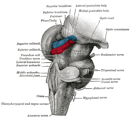
In neuroanatomy, the lateral geniculate nucleus (LGN; also called the lateral geniculate body or lateral geniculate complex) is a structure in the thalamus and a key component of the mammalian visual pathway. It is a small, ovoid, ventral projection of the thalamus where the thalamus connects with the optic nerve. There are two LGNs, one on the left and another on the right side of the thalamus. In humans, both LGNs have six layers of neurons (grey matter) alternating with optic fibers (white matter).
The LGN receives information directly from the ascending retinal ganglion cells via the optic tract and from the reticular activating system. Neurons of the LGN send their axons through the optic radiation, a direct pathway to the primary visual cortex. In addition, the LGN receives many strong feedback connections from the primary visual cortex. In humans as well as other mammals, the two strongest pathways linking the eye to the brain are those projecting to the dorsal part of the LGN in the thalamus, and to the superior colliculus.
Both the left and right hemispheres of the brain have a lateral geniculate nucleus, named after its resemblance to a bent knee (genu is Latin for "knee"). In humans as well as in many other primates, the LGN has layers of magnocellular cells and parvocellular cells that are interleaved with layers of koniocellular cells.
In humans the LGN is normally described as having six distinctive layers. The inner two layers, (1 and 2) are magnocellular layers, while the outer four layers, (3, 4, 5 and 6), are parvocellular layers. An additional set of neurons, known as the koniocellular layers, are found ventral to each of the magnocellular and parvocellular layers. This layering is variable between primate species, and extra leafleting is variable within species.
The average volume of each LGN in an adult human is about 118mm. (This is the same volume as a 4.9mm-sided cube.) A study of 24 hemispheres from 15 normal individuals with average age 59 years at autopsy found variation from about 91 to 157mm. The same study found that in each LGN, the magnocellular layers comprised about 28mm in total, and the parvocellular layers comprised about 90mm in total.

*Size describes the cell body and dendritic tree, though also can describe the receptive field
The magnocellular, parvocellular, and koniocellular layers of the LGN correspond with the similarly named types of retinal ganglion cells. Retinal P ganglion cells send axons to a parvocellular layer, M ganglion cells send axons to a magnocellular layer, and K ganglion cells send axons to a koniocellular layer.
Koniocellular cells are functionally and neurochemically distinct from M and P cells and provide a third channel to the visual cortex. They project their axons between the layers of the lateral geniculate nucleus where M and P cells project. Their role in visual perception is presently unclear; however, the koniocellular system has been linked with the integration of somatosensory system-proprioceptive information with visual perception[citation needed], and it may also be involved in color perception.
Lateral geniculate nucleus
In neuroanatomy, the lateral geniculate nucleus (LGN; also called the lateral geniculate body or lateral geniculate complex) is a structure in the thalamus and a key component of the mammalian visual pathway. It is a small, ovoid, ventral projection of the thalamus where the thalamus connects with the optic nerve. There are two LGNs, one on the left and another on the right side of the thalamus. In humans, both LGNs have six layers of neurons (grey matter) alternating with optic fibers (white matter).
The LGN receives information directly from the ascending retinal ganglion cells via the optic tract and from the reticular activating system. Neurons of the LGN send their axons through the optic radiation, a direct pathway to the primary visual cortex. In addition, the LGN receives many strong feedback connections from the primary visual cortex. In humans as well as other mammals, the two strongest pathways linking the eye to the brain are those projecting to the dorsal part of the LGN in the thalamus, and to the superior colliculus.
Both the left and right hemispheres of the brain have a lateral geniculate nucleus, named after its resemblance to a bent knee (genu is Latin for "knee"). In humans as well as in many other primates, the LGN has layers of magnocellular cells and parvocellular cells that are interleaved with layers of koniocellular cells.
In humans the LGN is normally described as having six distinctive layers. The inner two layers, (1 and 2) are magnocellular layers, while the outer four layers, (3, 4, 5 and 6), are parvocellular layers. An additional set of neurons, known as the koniocellular layers, are found ventral to each of the magnocellular and parvocellular layers. This layering is variable between primate species, and extra leafleting is variable within species.
The average volume of each LGN in an adult human is about 118mm. (This is the same volume as a 4.9mm-sided cube.) A study of 24 hemispheres from 15 normal individuals with average age 59 years at autopsy found variation from about 91 to 157mm. The same study found that in each LGN, the magnocellular layers comprised about 28mm in total, and the parvocellular layers comprised about 90mm in total.
*Size describes the cell body and dendritic tree, though also can describe the receptive field
The magnocellular, parvocellular, and koniocellular layers of the LGN correspond with the similarly named types of retinal ganglion cells. Retinal P ganglion cells send axons to a parvocellular layer, M ganglion cells send axons to a magnocellular layer, and K ganglion cells send axons to a koniocellular layer.
Koniocellular cells are functionally and neurochemically distinct from M and P cells and provide a third channel to the visual cortex. They project their axons between the layers of the lateral geniculate nucleus where M and P cells project. Their role in visual perception is presently unclear; however, the koniocellular system has been linked with the integration of somatosensory system-proprioceptive information with visual perception[citation needed], and it may also be involved in color perception.
Recent media
Recent media