Hubbry Logo
RepressorRepressorMain
Open search
Repressor
Community hub
Repressor
logo
8 pages, 0 posts
0 subscribers
Be the first to start a discussion here.
Contribute something
Repressor
Repressor
from Wikipedia
The lac operon: 1: RNA Polymerase, 2: lac repressor, 3: Promoter, 4: Operator, 5: Lactose, 6: lacZ, 7: lacY, 8: lacA. Top: The gene is essentially turned off. There is no lactose to inhibit the repressor, so the repressor binds to the operator, which obstructs the RNA polymerase from binding to the promoter and making lactase. Bottom: The gene is turned on. Lactose is inhibiting the repressor, allowing the RNA polymerase to bind with the promoter, and express the genes, which synthesize lactase. Eventually, the lactase will digest all of the lactose, until there is none to bind to the repressor. The repressor will then bind to the operator, stopping the manufacture of lactase.

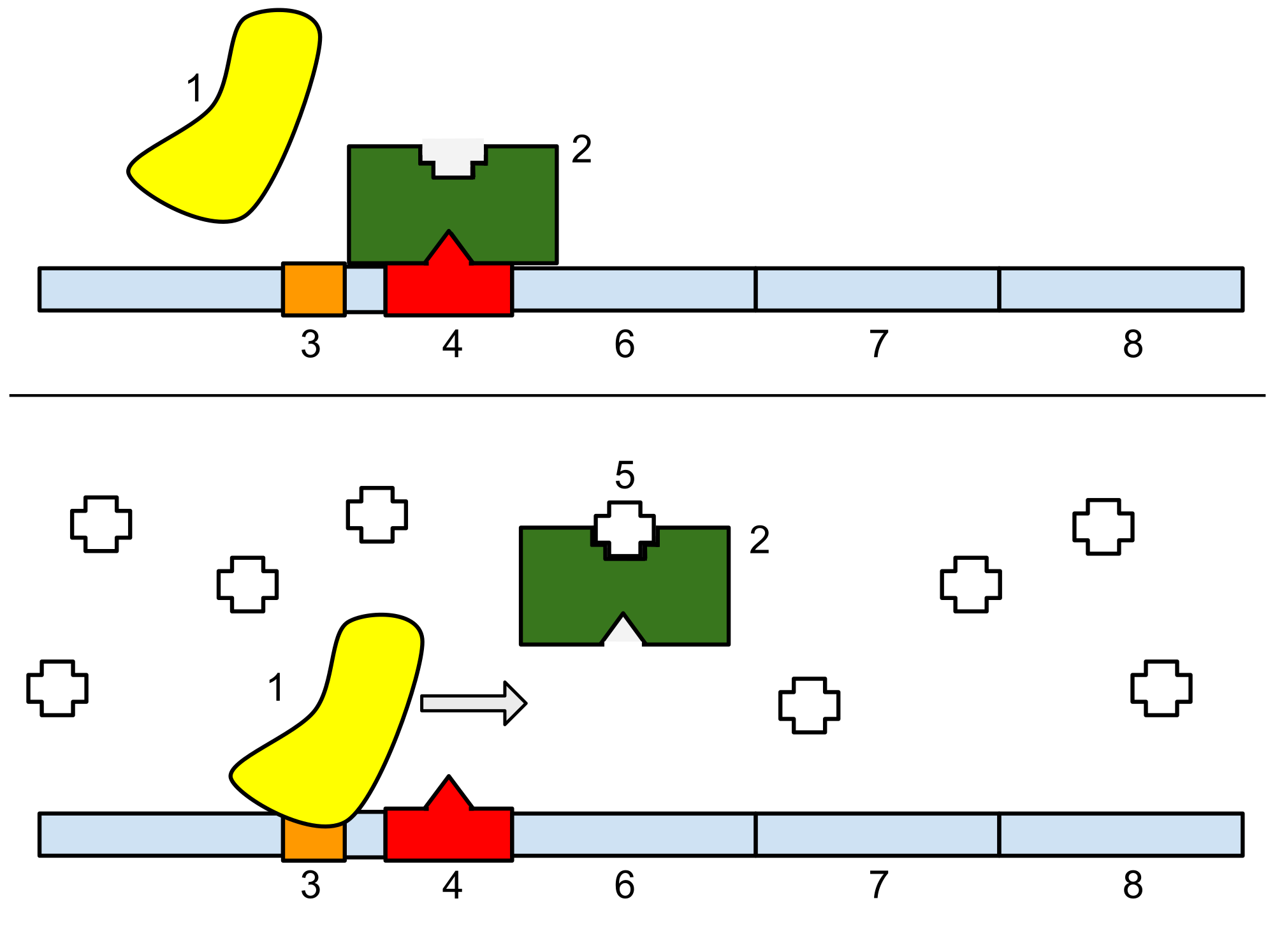
In molecular genetics, a repressor is a DNA- or RNA-binding protein that inhibits the expression of one or more genes by binding to the operator or associated silencers. A DNA-binding repressor blocks the attachment of RNA polymerase to the promoter, thus preventing transcription of the genes into messenger RNA. An RNA-binding repressor binds to the mRNA and prevents translation of the mRNA into protein. This blocking or reducing of expression is called repression.

Function

[edit]

If an inducer, a molecule that initiates the gene expression, is present, then it can interact with the repressor protein and detach it from the operator. RNA polymerase then can transcribe the message (expressing the gene). A co-repressor is a molecule that can bind to the repressor and make it bind to the operator tightly, which decreases transcription.

A repressor that binds with a co-repressor is termed an aporepressor or inactive repressor. One type of aporepressor is the trp repressor, an important metabolic protein in bacteria. The above mechanism of repression is a type of a feedback mechanism because it only allows transcription to occur if a certain condition is present: the presence of specific inducer(s). In contrast, an active repressor binds directly to an operator to repress gene expression.

While repressors are more commonly found in prokaryotes, they are rare in eukaryotes. Furthermore, most known eukaryotic repressors are found in simple organisms (e.g., yeast), and act by interacting directly with activators.[1] This contrasts prokaryotic repressors which can also alter DNA or RNA structure.

Within the eukaryotic genome are regions of DNA known as silencers. These are DNA sequences that bind to repressors to partially or fully repress a gene. Silencers can be located several bases upstream or downstream from the actual promoter of the gene. Repressors can also have two binding sites: one for the silencer region and one for the promoter. This causes chromosome looping, allowing the promoter region and the silencer region to come in proximity of each other.

Examples of Repressors

[edit]

lac operon repressor

[edit]

The lacZYA operon houses genes encoding proteins needed for lactose breakdown.[2] The lacI gene codes for a protein called "the repressor" or "the lac repressor", which functions to repressor of the lac operon.[2] The gene lacI is situated immediately upstream of lacZYA but is transcribed from a lacI promoter.[2] The lacI gene synthesizes LacI repressor protein. The LacI repressor protein represses lacZYA by binding to the operator sequence lacO.[2]

The lac repressor is constitutively expressed and usually bound to the operator region of the promoter, which interferes with the ability of RNA polymerase (RNAP) to begin transcription of the lac operon.[2] In the presence of the inducer allolactose, the repressor changes conformation, reduces its DNA binding strength and dissociates from the operator DNA sequence in the promoter region of the lac operong. RNAP is then able to bind to the promoter and begin transcription of the lacZYA gene.[2]

met operon repressor

[edit]

An example of a repressor protein is the methionine repressor MetJ. MetJ interacts with DNA bases via a ribbon-helix-helix (RHH) motif.[3] MetJ is a homodimer consisting of two monomers, which each provides a beta ribbon and an alpha helix. Together, the beta ribbons of each monomer come together to form an antiparallel beta-sheet which binds to the DNA operator ("Met box") in its major groove. Once bound, the MetJ dimer interacts with another MetJ dimer bound to the complementary strand of the operator via its alpha helices. AdoMet binds to a pocket in MetJ that does not overlap the site of DNA binding.

The Met box has the DNA sequence AGACGTCT, a palindrome (it shows dyad symmetry) allowing the same sequence to be recognized on either strand of the DNA. The junction between C and G in the middle of the Met box contains a pyrimidine-purine step that becomes positively supercoiled forming a kink in the phosphodiester backbone. This is how the protein checks for the recognition site as it allows the DNA duplex to follow the shape of the protein. In other words, recognition happens through indirect readout of the structural parameters of the DNA, rather than via specific base sequence recognition.

Each MetJ dimer contains two binding sites for the cofactor S-Adenosyl methionine (SAM) which is a product in the biosynthesis of methionine. When SAM is present, it binds to the MetJ protein, increasing its affinity for its cognate operator site, which halts transcription of genes involved in methionine synthesis. When SAM concentration becomes low, the repressor dissociates from the operator site, allowing more methionine to be produced.

L-arabinose operon repressor

[edit]

The L-arabinose operon houses genes coding for arabinose-digesting enzymes. These function to break down arabinose as an alternative source for energy when glucose is low or absent.[4] The operon consists of a regulatory repressor gene (araC), three control sites (ara02, ara01, araI1, and araI2), two promoters (Parac/ParaBAD) and three structural genes (araBAD). Once produced, araC acts as repressor by binding to the araI region to form a loop which prevents polymerases from binding to the promotor and transcribing the structural genes into proteins.

In the absence of Arabinose and araC (repressor), loop formation is not initiated and structural gene expression will be lower. In the absence of Arabinose but presence of araC, araC regions form dimers, and bind to bring ara02 and araI1 domains closer by loop formation.[5] In the presence of both Arabinose and araC, araC binds with the arabinose and acts as an activator. This conformational change in the araC no longer can form a loop, and the linear gene segment promotes RNA polymerase recruitment to the structural araBAD region.[4]

Structure of L-arabinose operon of E. coli. The work was uploaded by Yiktingg1 in wikimedia commons. https://commons.wikimedia.org/wiki/File:L-arabinose_structure.png#filehistory

+

Flowing Locus C (Epigenetic Repressor)

[edit]

The FLC operon is a conserved eukaryotic locus that is negatively associated with flowering via repression of genes needed for the development of the meristem to switch to a floral state in the plant species Arabidopsis thaliana. FLC expression has been shown be regulated by the presence of FRIGIDA, and negatively correlates with decreases in temperature resulting in the prevention of vernalization.[6] The degree to which expression decreases depends on the temperature and exposure time as seasons progress. After the downregulation of FLC expression, the potential for flowering is enabled. The regulation of FLC expression involves both genetic and epigenetic factors such as histone methylation and DNA methylation.[7] Furthermore, a number of genes are cofactors act as negative transcription factors for FLC genes.[8] FLC genes also have a large number of homologues across species that allow for specific adaptations in a range of climates.[9]

See also

[edit]

References

[edit]
[edit]
Revisions and contributorsEdit on WikipediaRead on Wikipedia
from Grokipedia
A repressor is a protein that inhibits gene expression by binding to specific DNA sequences, such as promoters or operators, thereby preventing or reducing the transcription of messenger RNA from the associated gene. This regulatory mechanism is essential for controlling cellular responses to environmental signals and maintaining metabolic balance. In prokaryotes, repressors typically function through negative regulation by binding to an operator sequence near the promoter of an —a cluster of co-regulated genes—thus blocking from initiating transcription. For instance, in the of , the (encoded by the lacI gene) binds the operator in the absence of , repressing genes involved in metabolism; the presence of an inducer like alters the repressor's conformation, releasing it from the DNA and allowing expression. Similarly, the trp repressor in the tryptophan operon binds the operator only when activated by , providing to halt unnecessary . These ligand-dependent interactions demonstrate how prokaryotic repressors can undergo conformational changes upon binding small molecules, enabling precise control. In eukaryotes, gene regulation by repressors is more complex, where DNA-binding proteins can act as either repressors or activators depending on the cellular or interactions with other proteins. Additionally, non-coding RNAs can serve repressive roles by interfering with transcription or mRNA stability, expanding the regulatory toolkit beyond proteins. Repressors thus play a critical role across all domains of life in fine-tuning to adapt to developmental cues, stress, and availability.

Definition and Basics

Definition

A repressor is a DNA- or RNA-binding protein that inhibits by preventing the transcription of specific genes. It achieves this by binding to operator sites or promoter regions in the DNA, thereby blocking the access of to the transcription start site or interfering with the recruitment of transcriptional machinery. In some cases, repressors may also interact with co-repressor molecules to enhance their inhibitory effect. Repressors play a fundamental role in negative regulation of gene expression, functioning as molecular switches that fine-tune cellular responses to environmental cues. They typically respond to signals such as metabolite concentrations, where the presence or absence of a ligand modulates the repressor's binding affinity to its target DNA sequence, thereby controlling the timing and level of gene transcription. This regulatory mechanism ensures efficient resource allocation in cells by suppressing unnecessary gene activity until required. In contrast to activators, which promote transcription by facilitating recruitment or stabilizing the transcription initiation complex, repressors specifically downregulate through steric hindrance or active interference. Repressors can be classified as inducible, where an effector molecule induces a conformational change that releases the repressor from DNA, or constitutive, where repression occurs continuously without such modulation. This distinction highlights their versatility in maintaining dynamic control over genetic output.

Historical Discovery

The concept of the repressor emerged in 1961 when François Jacob and proposed the operon model for gene regulation in , introducing the idea of a repressor protein produced by a regulatory that binds to an operator site to prevent transcription of structural unless modulated by inducers or corepressors. This model, developed through studies on the in , provided the first framework for negative control in prokaryotic and revolutionized understanding of how cells respond to environmental signals. A pivotal experimental milestone came in 1966 with the isolation of the protein by and Benno Müller-Hill, who employed a filter-binding assay to detect and purify the molecule from E. coli extracts, confirming its role in binding the operator DNA sequence and demonstrating the physical basis of Jacob and Monod's hypothesis. Shortly thereafter, in 1967, Mark Ptashne isolated the λ phage repressor using similar techniques, further validating the repressor mechanism in viral gene control. These achievements built on foundational genetic work, earning Jacob, Monod, and André Lwoff the 1965 in Physiology or Medicine for discoveries concerning the genetic control of enzyme and virus synthesis. The understanding of repressors expanded beyond prokaryotes in the 1980s, as researchers identified eukaryotic counterparts, notably steroid hormone receptors that function as ligand-activated transcription factors capable of repressing gene expression by recruiting corepressors to target promoters or interfering with activator binding. Cloning of receptors such as the glucocorticoid and estrogen receptors during this period revealed their modular structures and repressive activities, marking a shift toward recognizing conserved regulatory principles across kingdoms.

Molecular Structure and Function

Protein Structure

Repressor proteins are modular polypeptides characterized by distinct structural domains that facilitate their regulatory roles in gene expression. The core architecture typically includes a DNA-binding domain (DBD) responsible for sequence-specific recognition of operator or silencer sites in DNA. In prokaryotic repressors, the DBD often features a helix-turn-helix (HTH) motif, consisting of two alpha-helices connected by a short turn, where the recognition helix inserts into the major groove of DNA to make base-specific contacts. Eukaryotic repressors frequently employ zinc finger domains, which utilize zinc ions to stabilize finger-like loops that interact with DNA, enabling precise binding through multiple fingers arranged in tandem. These DBDs are usually located at the N-terminus and are essential for targeting repressor activity to specific genomic loci. Adjacent to the DBD is the ligand-binding domain (LBD), which serves as an allosteric site for small-molecule effectors such as metabolites or inducers. The LBD undergoes conformational changes upon effector binding, which can either stabilize or disrupt the protein's interaction with DNA, thereby modulating repression. This allosteric mechanism is mediated by structured pockets within the LBD that accommodate ligands, often leading to rigid-body movements or hinge-like flexing between domains to propagate signals across the protein. Such sites are prevalent in metabolite-sensing repressors, allowing cells to fine-tune in response to environmental cues without altering protein levels. Many repressor proteins enhance their DNA-binding affinity and specificity through oligomerization, forming dimers, tetramers, or higher-order assemblies via dedicated interfaces. motifs, characterized by amphipathic alpha-helices with conserved leucine residues at every seventh position, mediate dimerization by interlocking like a , positioning multiple DBDs for cooperative DNA engagement. Other oligomerization domains, such as beta-sheets or coiled-coils, similarly promote multimeric states that increase for DNA targets, a structural feature conserved across diverse repressor families.

Binding Mechanisms

Repressors inhibit transcription by binding to specific DNA sequences known as operators, typically located near promoter regions. This binding occurs through sequence-specific interactions, where the repressor's recognizes and contacts particular bases, often inserting alpha into the major groove of the DNA double helix to achieve high specificity. For instance, the protein binds its operator with an exceptionally high affinity, characterized by a (Kd) of approximately 101310^{-13} M, enabling tight regulation even at low cellular concentrations.90276-0) Such interactions are mediated by hydrogen bonds, van der Waals forces, and electrostatic contacts between side chains and DNA bases, ensuring selective recognition amid vast non-specific DNA sequences.00392-6) Many repressors function as allosteric proteins, where binding of effector molecules at a site remote from the induces conformational changes that modulate operator affinity. In the case of the , the gratuitous inducer isopropyl β-D-1-thiogalactopyranoside (IPTG) binds to the core domain, triggering a structural rearrangement that reorients the N-terminal s relative to the dimer interface. This shift disrupts key interactions necessary for operator recognition, reducing binding affinity by several orders of magnitude and releasing the repressor from DNA. thus allows environmental signals, such as availability, to dynamically control repressor activity without altering protein levels. Upon binding to the operator, repressors primarily exert their inhibitory effect through steric hindrance, physically obstructing from accessing the promoter or initiating transcription. In prokaryotic systems, the bound repressor occupies space that overlaps with the , preventing promoter recognition, , or open complex formation required for transcription start. This mechanism does not involve covalent modifications to DNA or associated proteins, relying instead on the spatial exclusion of the large holoenzyme. For example, in the , the repressor-bound operator directly blocks the path for progression, ensuring repression until inducer-mediated dissociation occurs.00180-6)

Types and Mechanisms of Repression

Prokaryotic Repressors

Prokaryotic repressors are regulatory proteins that play a central role in controlling in by binding to operator sequences on DNA, thereby inhibiting the transcription of downstream genes organized into operons. Operons consist of clusters of functionally related genes transcribed as a single polycistronic mRNA from a shared promoter, allowing coordinated regulation of pathways such as nutrient metabolism. In response to environmental nutrients like or sugars, repressors modulate the synthesis of this mRNA to optimize cellular , ensuring that genes for catabolic or biosynthetic processes are expressed only when necessary. Prokaryotic operons are classified into inducible and repressible types based on how repressors interact with small molecules to control transcription. In inducible operons, the repressor is active in the absence of an inducer, binding the operator to block and prevent mRNA synthesis; binding of an inducer molecule causes a conformational change in the repressor, releasing it from the DNA and allowing transcription. Conversely, in repressible operons, the repressor is inactive without a corepressor and does not bind the operator under normal conditions, permitting constitutive transcription; accumulation of a corepressor, such as an end product of the pathway, activates the repressor by altering its structure, enabling it to bind the operator and halt mRNA production. This distinction enables to fine-tune : inducible systems respond to the presence of substrates, while repressible systems shut down when products are abundant. Global in prokaryotes can also involve activators whose inactivity leads to repression, coordinating expression across multiple operons and integrating signals from cellular to prioritize energy-efficient pathways. A key example is mediated by the cyclic AMP receptor protein (CRP) complex, which activates transcription of operons for alternative carbon sources only when preferred like glucose are unavailable. Low intracellular cAMP during glucose prevents formation of the active CRP-cAMP complex, inhibiting transcription of numerous catabolic operons and thereby enforcing repression to favor efficient glucose utilization. CRP influences over 180 in , demonstrating its role as a master regulator that links sensing to broad-scale control.

Eukaryotic Repressors

In eukaryotes, transcriptional repressors often operate at the level to enforce through modifications that promote compaction and restrict access to DNA. deacetylases (HDACs), such as and HDAC2, remove acetyl groups from residues on tails, neutralizing their negative charge and facilitating tighter DNA wrapping around histones, which condenses into a transcriptionally inactive state. Similarly, histone methyltransferases like SUV39H1/2 catalyze trimethylation of at 9 (), recruiting (HP1) to propagate compact domains that silence genes over large genomic regions. These modifications, often coordinated within multiprotein complexes like NuRD (for HDACs) or PRC2 (for H3K27 methylation by ), create stable epigenetic barriers to transcription initiation. Eukaryotic repressors also modulate enhancer-promoter interactions to prevent activation of distant regulatory elements. The RE1-silencing (REST), for instance, binds to repressor element 1 (RE1) motifs in neuronal gene enhancers, competing with activators and recruiting co-repressors like CoREST and HDAC2 to deacetylate histones at target promoters. This interference disrupts loop formation between enhancers and promoters, as seen in the repression of genes like the Nav1.2, where REST occupancy correlates with reduced mRNA levels and blocked neurite outgrowth in non-neuronal cells. By integrating with remodelers, REST ensures cell-type-specific silencing, maintaining the neuronal gene program in a poised, inactive configuration. Signaling pathways further integrate eukaryotic repression through ligand-dependent recruitment of co-repressors. Unliganded thyroid receptors (TRs), for example, actively repress target genes by binding DNA response elements and associating with nuclear receptor co-repressors (NCoR) via specific interaction domains. NCoR bridges TR to HDAC-containing complexes like mSin3A-HDAC1, promoting deacetylation and compaction to inhibit basal transcription; upon binding, this complex dissociates, switching to activation. This mechanism exemplifies how eukaryotic repressors couple extracellular signals to epigenetic silencing, ensuring precise control over developmental and metabolic genes.

Key Examples

Lac Operon Repressor

The lac repressor, also known as LacI, is encoded by the lacI gene located upstream of the lac operon in Escherichia coli. This gene produces a tetrameric protein consisting of four identical subunits, each with a molecular weight of approximately 38.5 kDa, resulting in a total mass of about 155 kDa for the functional oligomer. The repressor binds with high affinity to specific DNA sequences called operators (primarily O1, but also auxiliary sites O2 and O3) within the lac operon, thereby blocking RNA polymerase access to the promoter and repressing transcription of downstream genes, including lacZ, which encodes the enzyme β-galactosidase essential for lactose metabolism. The mechanism of repression is inducible and relies on . In the absence of , the tetrameric repressor binds tightly to the operator DNA, forming a stable complex that inhibits . When is present, it is converted to by ; acts as the natural inducer by binding to the repressor's core domain, inducing a conformational change that reduces DNA-binding affinity by approximately 1,000-fold and causes the repressor to dissociate from the operator. Synthetic inducers like isopropyl (IPTG) mimic this effect by binding to the same allosteric site without being metabolized, allowing controlled induction in experimental settings. The equilibrium binding can be represented as: Repressor+OperatorRepressor-Operator Complex\text{Repressor} + \text{Operator} \rightleftharpoons \text{Repressor-Operator Complex} with a dissociation constant Kd1013K_d \approx 10^{-13} M in the absence of inducer, reflecting the exceptionally tight interaction that ensures effective repression under non-inducing conditions. The purification of the lac repressor in 1966 marked a pivotal advancement, as it was the first genetic regulatory protein isolated in pure form, enabling direct biochemical assays of its interactions. This breakthrough facilitated foundational studies on operator specificity, such as footprinting and competition assays that mapped the minimal operator sequence required for high-affinity binding (approximately 17-21 base pairs centered around the symmetric dyad). Additionally, the availability of purified repressor protein supported mutagenesis experiments on both the lacI gene and operator DNA, revealing key residues in the DNA-binding domain (e.g., helix-turn-helix motif) and operator mutations (e.g., O^c variants) that alter specificity and inducibility, thereby elucidating the molecular basis of negative control in prokaryotes.

Trp Operon Repressor

The Trp operon repressor serves as a paradigmatic example of a corepressor-activated regulatory protein in prokaryotic gene expression, controlling the transcription of genes involved in tryptophan biosynthesis in bacteria like Escherichia coli. Encoded by the trpR gene, the repressor is a homodimeric protein with each subunit comprising 107 amino acids and a total molecular mass of approximately 25 kDa. In its inactive aporepressor form, the protein exhibits low affinity for the operator DNA sequence located downstream of the trp promoter. Binding of L-tryptophan, acting as a corepressor, induces a conformational change that transforms the aporepressor into the active holorepressor, enabling high-affinity binding to the operator and thereby repressing transcription initiation by sterically hindering RNA polymerase progression. Tryptophan binding occurs at the dimer interface, where two molecules of the occupy symmetric pockets, repositioning flexible motifs to align with the major groove of the operator DNA. This allosteric activation dramatically enhances DNA-binding specificity and affinity, increasing it approximately 70-fold, with the (K_d) for the holorepressor-operator complex reaching about 10^{-11} M under physiological conditions. The operator consists of a 18-base-pair that accommodates the dimeric repressor, forming a ternary complex stabilized by hydrogen bonds and van der Waals interactions between repressor residues and specific base pairs. In addition to repression at initiation, the Trp repressor functions in synergy with a transcription mechanism within the trp operon leader region (trpL), which encodes a short rich in residues, including two tandem Trp codons. When intracellular tryptophan levels are high, abundant charged tRNA^{Trp} facilitates rapid translation of the leader , allowing the ribosome to cover regions of the nascent mRNA that would otherwise form an antiterminator structure; this promotes formation of a rho-independent terminator hairpin approximately 140 nucleotides downstream of the transcription start site, resulting in premature termination of up to 90% of transcripts. The combined action of the repressor (providing ~70-fold ) and (~10-fold) achieves tight control, ensuring minimal expression during tryptophan excess while allowing rapid derepression when levels drop. This corepressor-dependent repression mechanism exhibits evolutionary conservation across diverse bacterial taxa, reflecting its adaptive value in coordinating . Analogous systems operate in other biosynthetic pathways, such as the (his) operon, which employs -activated via a leader with codons, and the (arg) operons, regulated by the ArgR repressor hexamer activated by binding to operator ARG boxes. These parallels underscore a shared evolutionary strategy for nutrient-responsive in prokaryotes, with variations in corepressor specificity and regulatory architectures tailored to metabolic demands.

Polycomb Repressive Complex

The Polycomb Repressive Complex (PRC) comprises two primary multisubunit assemblies, PRC1 and PRC2, that function cooperatively to enforce epigenetic in eukaryotic cells, particularly during development. PRC2 consists of core subunits EZH1 or (the catalytic SET-domain methyltransferase), EED, SUZ12, and RBBP4/7 ( chaperones), with accessory factors forming distinct subcomplexes: PRC2.1 (incorporating PCL1/2/3 and EPOP/PAL1/2) and PRC2.2 (including JARID2 and AEBP2). PRC2 deposits the repressive modification trimethylation of lysine 27 on (), which recruits additional silencing factors and promotes higher-order compaction to inhibit transcription. In turn, PRC1 includes RING1A or RING1B (E3 ligases) paired with one of six PCGF proteins (PCGF1–6), alongside either CBX family proteins (in canonical PRC1) or RYBP/YAF2 (in non-canonical PRC1). PRC1 catalyzes monoubiquitylation of at lysine 119 (H2AK119ub1), which stabilizes marks, enhances looping, and further compacts nucleosomes into transcriptionally inert structures. These complexes primarily target Hox cluster genes to sustain their repression post-embryonic induction, thereby preventing that could disrupt body patterning and cell differentiation. PRC2 initiates silencing by depositing at promoter regions, while PRC1 reinforces this through H2AK119ub1 and chain-like interactions, forming stable Polycomb domains. The repressive marks spread bidirectionally along domains via recruitment by long non-coding RNAs (lncRNAs), such as on the or Airn and Kcnq1ot1 at imprinted loci; these lncRNAs bind variant PRC1 (PCGF3/5-containing) via adaptor proteins like hnRNPK, facilitating domain expansion and heritable silencing. Dysregulation of PRCs contributes to in cancer and imprinting disorders. In cancers, including lymphomas, , , and myeloid malignancies, gain-of-function mutations in (e.g., Y641F/N in the SET domain) hyperactivate , aberrantly repressing tumor suppressors like INK4A-ARF and promoting proliferation; conversely, loss-of-function alterations in or SUZ12 (seen in ~25% of ) reduce , derepressing oncogenes. For imprinting disorders, heterozygous mutations in PRC2 core genes (, EED, SUZ12) impair at parent-of-origin-specific loci, disrupting monoallelic silencing and causing congenital overgrowth syndromes like , where altered crosstalk with pathways exacerbates developmental imbalances.

Flowering Locus C

Flowering Locus C (FLC) is a MADS-box transcription factor that serves as a potent repressor of flowering in Arabidopsis thaliana, primarily by inhibiting the expression of key floral integrator genes such as FLOWERING LOCUS T (FT) and SUPPRESSOR OF OVEREXPRESSION OF CONSTANS 1 (SOC1). High levels of FLC expression delay the transition from vegetative to reproductive growth, integrating environmental cues like photoperiod and temperature to modulate flowering time. FLC achieves repression by directly binding to CArG box motifs (consensus CC[A/T]₆GG) in the regulatory regions of target genes, with chromatin immunoprecipitation studies identifying over 500 such binding sites genome-wide, predominantly in promoters and introns. In the vernalization pathway, prolonged exposure to cold temperatures (typically 4–10°C for 4–8 weeks) epigenetically silences FLC expression, thereby promoting flowering in winter-annual ecotypes. This silencing involves the induction of VIN3, a nuclear-localized protein that recruits Polycomb Repressive Complex 2 (PRC2) to deposit 27 trimethylation () marks at the FLC locus, maintaining repression through even after return to warm conditions. The extent of FLC down-regulation correlates with the duration of cold exposure, with full saturation requiring 28–56 days depending on the , and this response resets in progeny to ensure generation-specific requirements. FLC repression extends beyond direct target binding by interacting with other MADS-domain proteins, such as SHORT VEGETATIVE PHASE (SVP), to form heterodimers that enhance binding affinity and broaden regulatory networks. For instance, FLC-SVP complexes co-repress FT in leaves, blocking the production of systemic florigenic signals, while also impairing meristem competence by suppressing SOC1 and FD expression at the shoot apex. Mutations in FLC lead to early flowering independent of vernalization, underscoring its quantitative control over flowering time variation across natural accessions. Additionally, FLC homologs in cereals like wheat and barley contribute to vernalization responses, though with duplicated loci adapting to polyploid genomes.

References

Add your contribution
Related Hubs
Contribute something
User Avatar
No comments yet.