Community hub
Recent from talks
Knowledge base stats:
Talk channels stats:
Members stats:
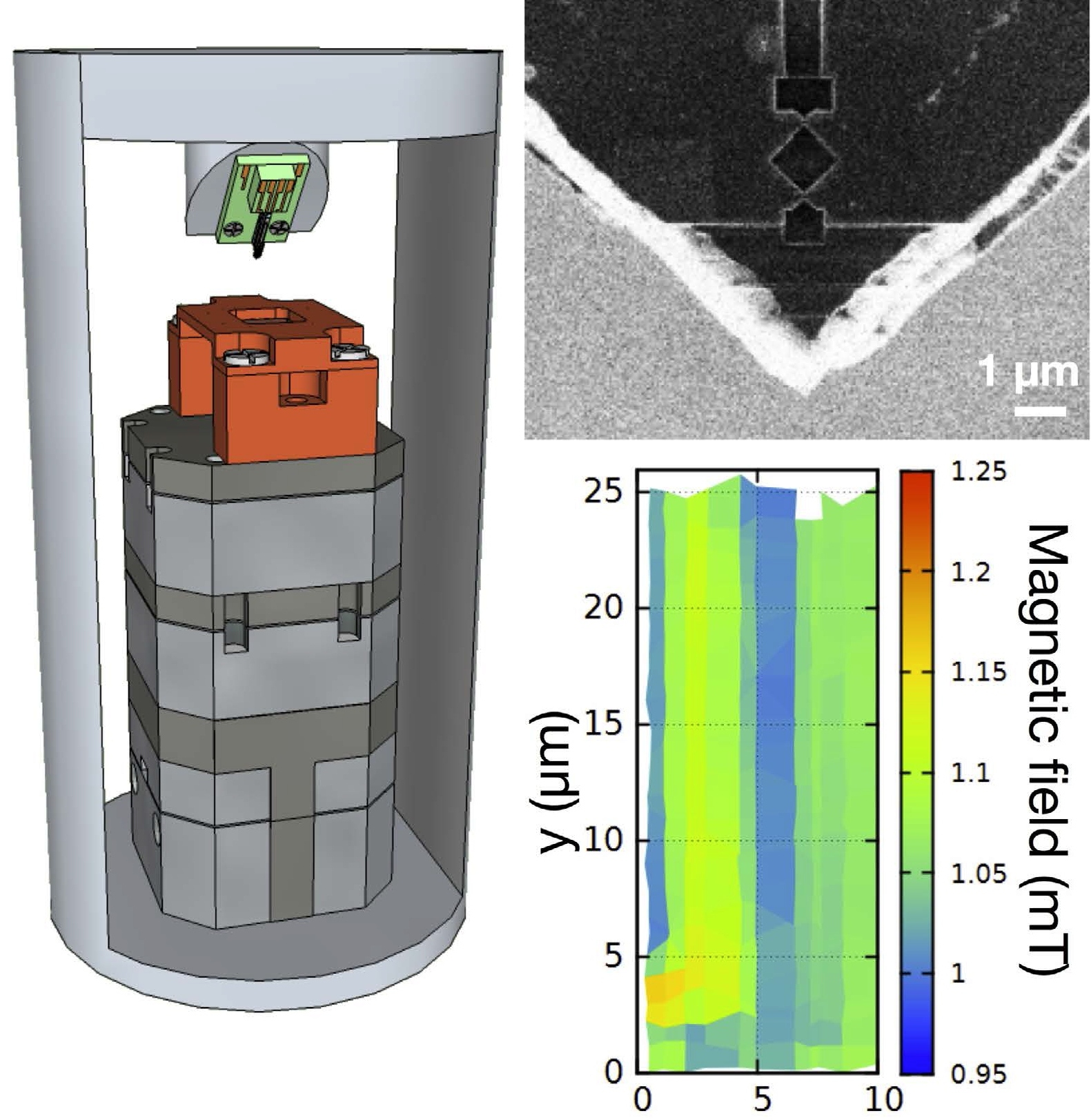
Scanning SQUID microscopy
In condensed matter physics, scanning SQUID microscopy is a technique where a superconducting quantum interference device (SQUID) is used to image surface magnetic field strength with micrometre-scale resolution. A tiny SQUID is mounted onto a tip which is then rastered near the surface of the sample to be measured. As the SQUID is the most sensitive detector of magnetic fields available and can be constructed at submicrometre widths via lithography, the scanning SQUID microscope allows magnetic fields to be measured with unparalleled resolution and sensitivity. The first scanning SQUID microscope was built in 1992 by Black et al. Since then the technique has been used to confirm unconventional superconductivity in several high-temperature superconductors including YBCO and BSCCO compounds.
The scanning SQUID microscope is based upon the thin-film DC SQUID. A DC SQUID consists of superconducting electrodes in a ring pattern connected by two weak-link Josephson junctions (see figure). Above the critical current of the Josephson junctions, the idealized difference in voltage between the electrodes is given by
where R is the resistance between the electrodes, I is the current, I0 is the maximum supercurrent, Ic is the critical current of the Josephson junctions, Φ is the total magnetic flux through the ring, and Φ0 is the magnetic flux quantum.
Hence, a DC SQUID can be used as a flux-to-voltage transducer. However, as noted by the figure, the voltage across the electrodes oscillates sinusoidally with respect to the amount of magnetic flux passing through the device. As a result, alone a SQUID can only be used to measure the change in magnetic field from some known value, unless the magnetic field or device size is very small such that Φ < Φ0. To use the DC SQUID to measure standard magnetic fields, one must either count the number of oscillations in the voltage as the field is changed, which is very difficult in practice, or use a separate DC bias magnetic field parallel to the device to maintain a constant voltage and consequently constant magnetic flux through the loop. The strength of the field being measured will then be equal to the strength of the bias magnetic field passing through the SQUID.
Although it is possible to read the DC voltage between the two terminals of the SQUID directly, because noise tends to be a problem in DC measurements, an alternating current technique is used. In addition to the DC bias magnetic field, an AC magnetic field of constant amplitude, with field strength generating Φ << Φ0, is also emitted in the bias coil. This AC field produces an AC voltage with amplitude proportional to the DC component in the SQUID. The advantage of this technique is that the frequency of the voltage signal can be chosen to be far away from that of any potential noise sources. By using a lock-in amplifier the device can read only the frequency corresponding to the magnetic field, ignoring many other sources of noise.
A Scanning SQUID Microscope is a sensitive near-field imaging system for the measurement of weak magnetic fields by moving a Superconducting Quantum Interference Device (SQUID) across an area. The microscope can map out buried current-carrying wires by measuring the magnetic fields produced by the currents, or can be used to image fields produced by magnetic materials. By mapping out the current in an integrated circuit or a package, short circuits can be localized and chip designs can be verified to see that current is flowing where expected.
As the SQUID material must be superconducting, measurements must be performed at low temperatures. Typically, experiments are carried out below liquid helium temperature (4.2 K) in a helium-3 refrigerator or dilution refrigerator. However, advances in high-temperature superconductor thin-film growth have allowed relatively inexpensive liquid nitrogen cooling to instead be used. It is even possible to measure room-temperature samples by only cooling a high Tc squid and maintaining thermal separation with the sample. In either case, due to the extreme sensitivity of the SQUID probe to stray magnetic fields, in general some form of magnetic shielding is used. Most common is a shield made of mu-metal, possibly in combination with a superconducting "can" (all superconductors repel magnetic fields via the Meissner effect).
The actual SQUID probe is generally made via thin-film deposition with the SQUID area outlined via lithography. A wide variety of superconducting materials can be used, but the two most common are Niobium, due to its relatively good resistance to damage from thermal cycling, and YBCO, for its high Tc > 77 K and relative ease of deposition compared to other high Tc superconductors. In either case, a superconductor with critical temperature higher than that of the operating temperature should be chosen. The SQUID itself can be used as the pickup coil for measuring the magnetic field, in which case the resolution of the device is proportional to the size of the SQUID. However, currents in or near the SQUID generate magnetic fields which are then registered in the coil and can be a source of noise. To reduce this effect it is also possible to make the size of the SQUID itself very small, but attach the device to a larger external superconducting loop located far from the SQUID. The flux through the loop will then be detected and measured, inducing a voltage in the SQUID.
Hub AI
Scanning SQUID microscopy AI simulator
(@Scanning SQUID microscopy_simulator)
Scanning SQUID microscopy
In condensed matter physics, scanning SQUID microscopy is a technique where a superconducting quantum interference device (SQUID) is used to image surface magnetic field strength with micrometre-scale resolution. A tiny SQUID is mounted onto a tip which is then rastered near the surface of the sample to be measured. As the SQUID is the most sensitive detector of magnetic fields available and can be constructed at submicrometre widths via lithography, the scanning SQUID microscope allows magnetic fields to be measured with unparalleled resolution and sensitivity. The first scanning SQUID microscope was built in 1992 by Black et al. Since then the technique has been used to confirm unconventional superconductivity in several high-temperature superconductors including YBCO and BSCCO compounds.
The scanning SQUID microscope is based upon the thin-film DC SQUID. A DC SQUID consists of superconducting electrodes in a ring pattern connected by two weak-link Josephson junctions (see figure). Above the critical current of the Josephson junctions, the idealized difference in voltage between the electrodes is given by
where R is the resistance between the electrodes, I is the current, I0 is the maximum supercurrent, Ic is the critical current of the Josephson junctions, Φ is the total magnetic flux through the ring, and Φ0 is the magnetic flux quantum.
Hence, a DC SQUID can be used as a flux-to-voltage transducer. However, as noted by the figure, the voltage across the electrodes oscillates sinusoidally with respect to the amount of magnetic flux passing through the device. As a result, alone a SQUID can only be used to measure the change in magnetic field from some known value, unless the magnetic field or device size is very small such that Φ < Φ0. To use the DC SQUID to measure standard magnetic fields, one must either count the number of oscillations in the voltage as the field is changed, which is very difficult in practice, or use a separate DC bias magnetic field parallel to the device to maintain a constant voltage and consequently constant magnetic flux through the loop. The strength of the field being measured will then be equal to the strength of the bias magnetic field passing through the SQUID.
Although it is possible to read the DC voltage between the two terminals of the SQUID directly, because noise tends to be a problem in DC measurements, an alternating current technique is used. In addition to the DC bias magnetic field, an AC magnetic field of constant amplitude, with field strength generating Φ << Φ0, is also emitted in the bias coil. This AC field produces an AC voltage with amplitude proportional to the DC component in the SQUID. The advantage of this technique is that the frequency of the voltage signal can be chosen to be far away from that of any potential noise sources. By using a lock-in amplifier the device can read only the frequency corresponding to the magnetic field, ignoring many other sources of noise.
A Scanning SQUID Microscope is a sensitive near-field imaging system for the measurement of weak magnetic fields by moving a Superconducting Quantum Interference Device (SQUID) across an area. The microscope can map out buried current-carrying wires by measuring the magnetic fields produced by the currents, or can be used to image fields produced by magnetic materials. By mapping out the current in an integrated circuit or a package, short circuits can be localized and chip designs can be verified to see that current is flowing where expected.
As the SQUID material must be superconducting, measurements must be performed at low temperatures. Typically, experiments are carried out below liquid helium temperature (4.2 K) in a helium-3 refrigerator or dilution refrigerator. However, advances in high-temperature superconductor thin-film growth have allowed relatively inexpensive liquid nitrogen cooling to instead be used. It is even possible to measure room-temperature samples by only cooling a high Tc squid and maintaining thermal separation with the sample. In either case, due to the extreme sensitivity of the SQUID probe to stray magnetic fields, in general some form of magnetic shielding is used. Most common is a shield made of mu-metal, possibly in combination with a superconducting "can" (all superconductors repel magnetic fields via the Meissner effect).
The actual SQUID probe is generally made via thin-film deposition with the SQUID area outlined via lithography. A wide variety of superconducting materials can be used, but the two most common are Niobium, due to its relatively good resistance to damage from thermal cycling, and YBCO, for its high Tc > 77 K and relative ease of deposition compared to other high Tc superconductors. In either case, a superconductor with critical temperature higher than that of the operating temperature should be chosen. The SQUID itself can be used as the pickup coil for measuring the magnetic field, in which case the resolution of the device is proportional to the size of the SQUID. However, currents in or near the SQUID generate magnetic fields which are then registered in the coil and can be a source of noise. To reduce this effect it is also possible to make the size of the SQUID itself very small, but attach the device to a larger external superconducting loop located far from the SQUID. The flux through the loop will then be detected and measured, inducing a voltage in the SQUID.