Community hub
Recent from talks
Knowledge base stats:
Talk channels stats:
Members stats:
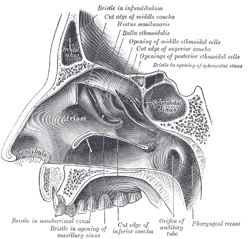
Sphenoid sinus
The sphenoid sinus is a paired paranasal sinus in the body of the sphenoid bone. It is one pair of the four paired paranasal sinuses. The two sphenoid sinuses are separated from each other by a septum. Each sphenoid sinus communicates with the nasal cavity via the opening of sphenoidal sinus. The two sphenoid sinuses vary in size and shape, and are usually asymmetrical.
On average, a sphenoid sinus measures 2.2 cm vertical height, 2 cm in transverse breadth; and 2.2 cm antero-posterior depth.
Each spehoid sinus is in the body of sphenoid bone, just under the sella turcica. The sphenoid sinuses are separated from each other medially by the septum of sphenoidal sinuses, which is usually asymmetrical.
An opening of sphenoidal sinus forms a passage between each sphenoidal sinus and the nasal cavity. Posteriorly, an opening of sphenoidal sinus opens into the sphenoidal sinus by an aperture high on the anterior wall the sinus; anteriorly, an opening of sphenoidal sinus opens into the roof of the nasal cavity via an aperture on the posterior wall of the sphenoethmoidal recess, just over the choana.
The mucous membrane receives sensory[citation needed] innervation from the posterior ethmoidal nerve (branch of the ophthalmic nerve (CN V1)) and from branches of the maxillary nerve (CN V2).
Postganglionic parasympathetic fibers of the facial nerve that synapsed at the pterygopalatine ganglion control mucus secretion.[citation needed]
Nearby structures include the optic canal, the optic nerve, the internal carotid artery, the cavernous sinus, the trigeminal nerve, the pituitary gland, and the anterior ethmoidal cells. One study found that carotid canal protrudation into the sphenoid sinus wall was present in 23.9–32.1% of males and 35.5–36.2% of females, dehiscence in carotid canal was detected more in females (34%) than in males (22%), optic canal protrudation was 33.3 and 30.5% in males and females, and optic canal dehiscence was detected in 11.3% of males and 9.9% of females.
The sphenoid sinuses vary in size and shape; because of the lateral displacement of the intervening septum of sphenoid sinuses, the pair rarely is symmetrical.
Hub AI
Sphenoid sinus AI simulator
(@Sphenoid sinus_simulator)
Sphenoid sinus
The sphenoid sinus is a paired paranasal sinus in the body of the sphenoid bone. It is one pair of the four paired paranasal sinuses. The two sphenoid sinuses are separated from each other by a septum. Each sphenoid sinus communicates with the nasal cavity via the opening of sphenoidal sinus. The two sphenoid sinuses vary in size and shape, and are usually asymmetrical.
On average, a sphenoid sinus measures 2.2 cm vertical height, 2 cm in transverse breadth; and 2.2 cm antero-posterior depth.
Each spehoid sinus is in the body of sphenoid bone, just under the sella turcica. The sphenoid sinuses are separated from each other medially by the septum of sphenoidal sinuses, which is usually asymmetrical.
An opening of sphenoidal sinus forms a passage between each sphenoidal sinus and the nasal cavity. Posteriorly, an opening of sphenoidal sinus opens into the sphenoidal sinus by an aperture high on the anterior wall the sinus; anteriorly, an opening of sphenoidal sinus opens into the roof of the nasal cavity via an aperture on the posterior wall of the sphenoethmoidal recess, just over the choana.
The mucous membrane receives sensory[citation needed] innervation from the posterior ethmoidal nerve (branch of the ophthalmic nerve (CN V1)) and from branches of the maxillary nerve (CN V2).
Postganglionic parasympathetic fibers of the facial nerve that synapsed at the pterygopalatine ganglion control mucus secretion.[citation needed]
Nearby structures include the optic canal, the optic nerve, the internal carotid artery, the cavernous sinus, the trigeminal nerve, the pituitary gland, and the anterior ethmoidal cells. One study found that carotid canal protrudation into the sphenoid sinus wall was present in 23.9–32.1% of males and 35.5–36.2% of females, dehiscence in carotid canal was detected more in females (34%) than in males (22%), optic canal protrudation was 33.3 and 30.5% in males and females, and optic canal dehiscence was detected in 11.3% of males and 9.9% of females.
The sphenoid sinuses vary in size and shape; because of the lateral displacement of the intervening septum of sphenoid sinuses, the pair rarely is symmetrical.