Hubbry Logo
SecretionSecretionMain
Open search
Secretion
Community hub
Secretion
logo
8 pages, 0 posts
0 subscribers
Be the first to start a discussion here.
Contribute something
Secretion
Secretion
from Wikipedia

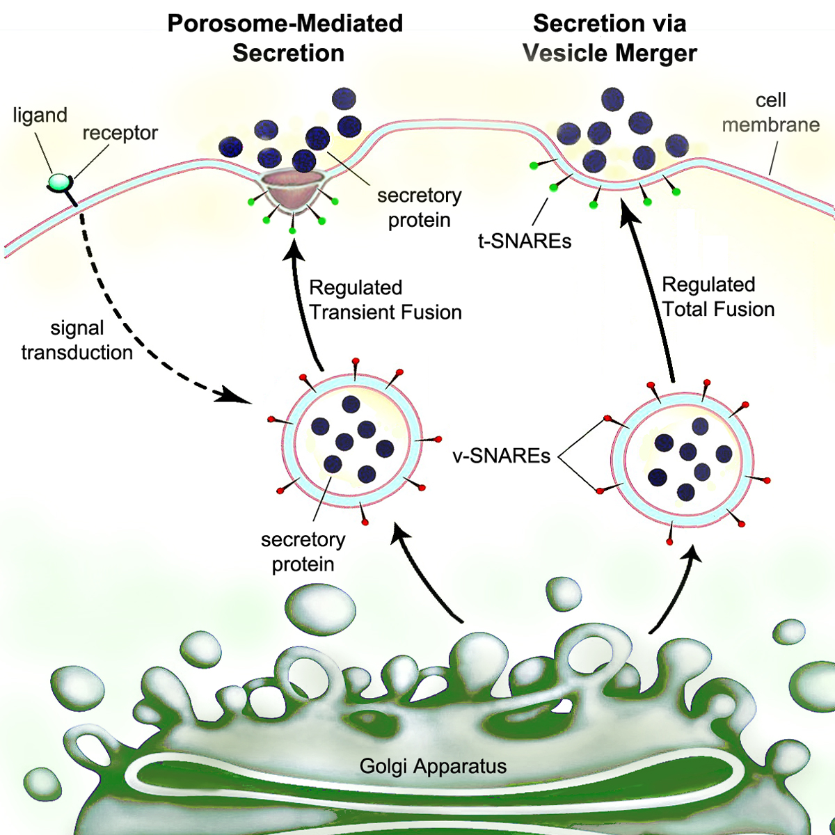
Secretion is the movement of material from one point to another, such as a secreted chemical substance from a cell or gland. In contrast, excretion is the removal of certain substances or waste products from a cell or organism. The classical mechanism of cell secretion is via secretory portals at the plasma membrane called porosomes.[1] Porosomes are permanent cup-shaped lipoprotein structures embedded in the cell membrane, where secretory vesicles transiently dock and fuse to release intra-vesicular contents from the cell.

Secretion in bacterial species means the transport or translocation of effector molecules. For example: proteins, enzymes or toxins (such as cholera toxin in pathogenic bacteria e.g. Vibrio cholerae) from across the interior (cytoplasm or cytosol) of a bacterial cell to its exterior. Secretion is a very important mechanism in bacterial functioning and operation in their natural surrounding environment for adaptation and survival.

In eukaryotic cells

[edit]
Porosome

Mechanism

[edit]

Eukaryotic cells, including human cells, have a highly evolved process of secretion. Proteins targeted for the outside are synthesized by ribosomes docked to the rough endoplasmic reticulum (ER). As they are synthesized, these proteins translocate into the ER lumen, where they are glycosylated and where molecular chaperones aid protein folding. Misfolded proteins are usually identified here and retrotranslocated by ER-associated degradation to the cytosol, where they are degraded by a proteasome. The vesicles containing the properly folded proteins then enter the Golgi apparatus.

In the Golgi apparatus, the glycosylation of the proteins is modified and further post-translational modifications, including cleavage and functionalization, may occur. The proteins are then moved into secretory vesicles which travel along the cytoskeleton to the edge of the cell. More modification can occur in the secretory vesicles (for example insulin is cleaved from proinsulin in the secretory vesicles).

Eventually, there is vesicle fusion with the cell membrane at porosomes, by a process called exocytosis, dumping its contents out of the cell's environment.[2]

Strict biochemical control is maintained over this sequence by usage of a pH gradient: the pH of the cytosol is 7.4, the ER's pH is 7.0, and the cis-golgi has a pH of 6.5. Secretory vesicles have pHs ranging between 5.0 and 6.0; some secretory vesicles evolve into lysosomes, which have a pH of 4.8.

Nonclassical secretion

[edit]

There are many proteins like FGF1 (aFGF), FGF2 (bFGF), interleukin-1 (IL1) etc. which do not have a signal sequence. They do not use the classical ER-Golgi pathway. These are secreted through various nonclassical pathways.

At least four nonclassical (unconventional) protein secretion pathways have been described.[3] They include:

  • direct protein translocation across the plasma membrane likely through membrane transport proteins
  • blebbing
  • lysosomal secretion
  • release via exosomes derived from multivesicular bodies

In addition, proteins can be released from cells by mechanical or physiological wounding[4] and through non-lethal, transient oncotic pores in the plasma membrane induced by washing cells with serum-free media or buffers.[5]

In human tissues

[edit]

Many human cell types have the ability to be secretory cells. They have a well-developed endoplasmic reticulum, and Golgi apparatus to fulfill this function. Tissues that produce secretions include the gastrointestinal tract, which secretes digestive enzymes and gastric acid, the lungs, which secrete surfactants, and sebaceous glands, which secrete sebum to lubricate the skin and hair. Meibomian glands in the eyelid secrete meibum to lubricate and protect the eye.

In gram-negative bacteria

[edit]

Secretion is not unique to eukaryotes – it is also present in bacteria and archaea as well. ATP binding cassette (ABC) type transporters are common to the three domains of life. Some secreted proteins are translocated across the cytoplasmic membrane by the SecYEG translocon, one of two translocation systems, which requires the presence of an N-terminal signal peptide on the secreted protein. Others are translocated across the cytoplasmic membrane by the twin-arginine translocation pathway (Tat). Gram-negative bacteria have two membranes, thus making secretion topologically more complex. There are at least six specialized secretion systems in Gram-negative bacteria.[6]

Type I secretion system (T1SS or TOSS)

[edit]

Type I secretion is a chaperone dependent secretion system employing the Hly and Tol gene clusters. The process begins as a leader sequence on the protein to be secreted is recognized by HlyA and binds HlyB on the membrane. This signal sequence is extremely specific for the ABC transporter. The HlyAB complex stimulates HlyD which begins to uncoil and reaches the outer membrane where TolC recognizes a terminal molecule or signal on HlyD. HlyD recruits TolC to the inner membrane and HlyA is excreted outside of the outer membrane via a long-tunnel protein channel.

Type I secretion system transports various molecules, from ions, drugs, to proteins of various sizes (20 – 900 kDa). The molecules secreted vary in size from the small Escherichia coli peptide colicin V, (10 kDa) to the Pseudomonas fluorescens cell adhesion protein LapA of 520 kDa.[7] The best characterized are the RTX toxins and the lipases. Type I secretion is also involved in export of non-proteinaceous substrates like cyclic β-glucans and polysaccharides.

Type II secretion system (T2SS)

[edit]

Proteins secreted through the type II system, or main terminal branch of the general secretory pathway, depend on the Sec or Tat system for initial transport into the periplasm. Once there, they pass through the outer membrane via a multimeric (12–14 subunits) complex of pore forming secretin proteins. In addition to the secretin protein, 10–15 other inner and outer membrane proteins compose the full secretion apparatus, many with as yet unknown function. Gram-negative type IV pili use a modified version of the type II system for their biogenesis, and in some cases certain proteins are shared between a pilus complex and type II system within a single bacterial species.

Type III secretion system (T3SS or TTSS)

[edit]

It is homologous to the basal body in bacterial flagella. It is like a molecular syringe through which a bacterium (e.g. certain types of Salmonella, Shigella, Yersinia, Vibrio) can inject proteins into eukaryotic cells. The low Ca2+ concentration in the cytosol opens the gate that regulates T3SS. One such mechanism to detect low calcium concentration has been illustrated by the lcrV (Low Calcium Response) antigen utilized by Yersinia pestis, which is used to detect low calcium concentrations and elicits T3SS attachment. The Hrp system in plant pathogens inject harpins and pathogen effector proteins through similar mechanisms into plants. This secretion system was first discovered in Yersinia pestis and showed that toxins could be injected directly from the bacterial cytoplasm into the cytoplasm of its host's cells rather than simply be secreted into the extracellular medium.[8]

Type IV secretion system (T4SS or TFSS)

[edit]
T4SS
Type IV secretion system
Identifiers
SymbolT4SS
PfamPF07996
InterProIPR012991
SCOP21gl7 / SCOPe / SUPFAM
TCDB3.A.7
OPM superfamily215
OPM protein3jqo
Available protein structures:
Pfam  structures / ECOD  
PDBRCSB PDB; PDBe; PDBj
PDBsumstructure summary

It is homologous to conjugation machinery of bacteria, the conjugative pili. It is capable of transporting both DNA and proteins. It was discovered in Agrobacterium tumefaciens, which uses this system to introduce the T-DNA portion of the Ti plasmid into the plant host, which in turn causes the affected area to develop into a crown gall (tumor). Helicobacter pylori uses a type IV secretion system to deliver CagA into gastric epithelial cells, which is associated with gastric carcinogenesis.[9] Bordetella pertussis, the causative agent of whooping cough, secretes the pertussis toxin partly through the type IV system. Legionella pneumophila, the causing agent of legionellosis (Legionnaires' disease) utilizes a type IVB secretion system, known as the icm/dot (intracellular multiplication / defect in organelle trafficking genes) system, to translocate numerous effector proteins into its eukaryotic host.[10] The prototypic Type IVA secretion system is the VirB complex of Agrobacterium tumefaciens.[11]

Protein members of this family are components of the type IV secretion system. They mediate intracellular transfer of macromolecules via a mechanism ancestrally related to that of bacterial conjugation machineries.[12][13]

Function

[edit]

The Type IV secretion system (T4SS) is the general mechanism by which bacterial cells secrete or take up macromolecules. Their precise mechanism remains unknown. T4SS is encoded on Gram-negative conjugative elements in bacteria. T4SS are cell envelope-spanning complexes, or, in other words, 11–13 core proteins that form a channel through which DNA and proteins can travel from the cytoplasm of the donor cell to the cytoplasm of the recipient cell. T4SS also secrete virulence factor proteins directly into host cells as well as taking up DNA from the medium during natural transformation.[14]

Structure

[edit]

As shown in the above figure, TraC, in particular consists of a three helix bundle and a loose globular appendage.[13]

Interactions

[edit]

T4SS has two effector proteins: firstly, ATS-1, which stands for Anaplasma translocated substrate 1, and secondly AnkA, which stands for ankyrin repeat domain-containing protein A. Additionally, T4SS coupling proteins are VirD4, which bind to VirE2.[15]

Type V secretion system (T5SS)

[edit]

Also called the autotransporter system,[16] type V secretion involves use of the Sec system for crossing the inner membrane. Proteins which use this pathway have the capability to form a beta-barrel with their C-terminus which inserts into the outer membrane, allowing the rest of the peptide (the passenger domain) to reach the outside of the cell. Often, autotransporters are cleaved, leaving the beta-barrel domain in the outer membrane and freeing the passenger domain. Some researchers believe remnants of the autotransporters gave rise to the porins which form similar beta-barrel structures.[citation needed] A common example of an autotransporter that uses this secretion system is the Trimeric Autotransporter Adhesins.[17]

Type VI secretion system (T6SS)

[edit]

Type VI secretion systems were originally identified in 2006 by the group of John Mekalanos at the Harvard Medical School (Boston, USA) in two bacterial pathogens, Vibrio cholerae and Pseudomonas aeruginosa.[18][19] These were identified when mutations in the Hcp and VrgG genes in Vibrio cholerae led to decreased virulence and pathogenicity. Since then, Type VI secretion systems have been found in a quarter of all proteobacterial genomes, including animal, plant, human pathogens, as well as soil, environmental or marine bacteria.[20][21] While most of the early studies of Type VI secretion focused on its role in the pathogenesis of higher organisms, more recent studies suggested a broader physiological role in defense against simple eukaryotic predators and its role in inter-bacteria interactions.[22][23] The Type VI secretion system gene clusters contain from 15 to more than 20 genes, two of which, Hcp and VgrG, have been shown to be nearly universally secreted substrates of the system. Structural analysis of these and other proteins in this system bear a striking resemblance to the tail spike of the T4 phage, and the activity of the system is thought to functionally resemble phage infection.[24]

Type VII secretion system (T7SS)

[edit]

Type VIII secretion system (T8SS)

[edit]

Type IX secretion system (T9SS)

[edit]

Release of outer membrane vesicles

[edit]

In addition to the use of the multiprotein complexes listed above, Gram-negative bacteria possess another method for release of material: the formation of bacterial outer membrane vesicles.[25] Portions of the outer membrane pinch off, forming nano-scale spherical structures made of a lipopolysaccharide-rich lipid bilayer enclosing periplasmic materials, and are deployed for membrane vesicle trafficking to manipulate environment or invade at host–pathogen interface. Vesicles from a number of bacterial species have been found to contain virulence factors, some have immunomodulatory effects, and some can directly adhere to and intoxicate host cells. release of vesicles has been demonstrated as a general response to stress conditions, the process of loading cargo proteins seems to be selective.[26]

In gram-positive bacteria

[edit]

In some Staphylococcus and Streptococcus species, the accessory secretory system handles the export of highly repetitive adhesion glycoproteins.

See also

[edit]

References

[edit]

Further reading

[edit]
[edit]
Revisions and contributorsEdit on WikipediaRead on Wikipedia
from Grokipedia
Secretion is the by which living cells produce and release specific molecules, such as proteins, hormones, enzymes, and fluids, from their interior to the or external environment, enabling essential functions like intercellular communication, , and removal. This process is fundamental to cellular across eukaryotes and prokaryotes, occurring through specialized pathways that ensure precise and targeted delivery of secreted products. In multicellular organisms, secretion supports critical systems including the endocrine and exocrine glands, where substances like insulin or are expelled to maintain . At the cellular level, secretion primarily involves the secretory pathway, beginning with synthesis in the rough endoplasmic reticulum (RER), followed by processing and packaging in the Golgi apparatus, and culminating in vesicle transport to the plasma membrane for . can be constitutive, occurring continuously to release proteins for structural or signaling roles, or regulated, triggered by stimuli like calcium ions to discharge contents such as neurotransmitters in neurons. Key proteins that mediate vesicle-plasma membrane fusion include SNARE proteins. In prokaryotes, secretion systems like type III or IV machineries allow to export virulence factors or nutrients, highlighting secretion's role in microbial and . The importance of secretion extends to health and disease; disruptions can lead to conditions such as (impaired insulin secretion) or (defective chloride ion secretion), while therapeutic strategies often target secretory pathways to modulate immune responses or . Examples of secreted products include hormones like adrenaline for stress responses, antibodies from plasma cells for immunity, and exosomes—small vesicles carrying nucleic acids for cell-to-cell signaling. Overall, secretion underscores the dynamic interplay between cellular machinery and organismal function, with ongoing research revealing molecular details through techniques like .

General Concepts

Definition and Classification

Secretion refers to the directed and of substances, such as proteins, , and metabolites, from the interior of a cell to the or external environment, distinguishing it from passive or uncontrolled cell that releases contents indiscriminately. This process is essential for cellular communication, nutrient release, and environmental interaction across all domains of , involving specialized molecular machinery to ensure specificity and energy dependence. In eukaryotes, secretion is broadly classified into constitutive and regulated pathways based on the timing and control of release. Constitutive secretion occurs continuously, delivering proteins and via vesicles that fuse with the plasma without external stimuli, supporting ongoing cellular maintenance such as membrane renewal. In contrast, regulated secretion involves storage of substances in secretory granules, with release triggered by specific signals like calcium influx, enabling rapid responses such as discharge in neurons. Eukaryotic pathways are further categorized as classical or non-classical: classical secretion relies on an N-terminal directing proteins through the (ER) and Golgi apparatus via vesicle-mediated , while non-classical pathways bypass the Golgi and are independent of signal peptides, often involving direct translocation across the plasma . Prokaryotes employ distinct secretion systems, classified from Type I (T1SS) to Type IX (T9SS) or beyond, based on their structural components, energy sources, and substrates translocated across one or both membranes. These systems vary in complexity; for instance, T1SS uses ATP-binding cassette (ABC) transporters to couple energy from directly to protein export across the cell envelope in a single step, common in for exporting toxins or adhesins. Exocytosis, characterized by vesicle fusion with the plasma membrane, serves as a hallmark mechanism in eukaryotes for both constitutive and regulated secretion, exemplified briefly by insulin release from pancreatic beta cells in response to glucose stimulation. The term "secretion" originated in 17th-century from the Latin secretio, meaning separation or release, initially describing glandular functions in animals. Its conceptualization in modern was formalized in the mid-20th century through electron microscopy studies by George Palade in the 1950s, which visualized the vesicular transport pathway and established the foundational model for protein secretion.

Biological Roles and Importance

Secretion plays a pivotal role in acquisition across organisms, as cells release such as amylases, proteases, and lipases into the extracellular environment to break down complex macromolecules into absorbable forms. In , this process is crucial for scavenging s from limited environments, where secreted enzymes enable the of polymers like or proteins, supporting growth under nutrient stress. Similarly, in multicellular , pancreatic and salivary secretions facilitate the of food, ensuring efficient uptake for energy and . Beyond nutrition, secretion underpins intercellular communication through the release of signaling molecules like hormones and neurotransmitters, which coordinate physiological responses over short and long distances. Hormones, secreted by endocrine glands into the bloodstream, regulate diverse processes including , growth, , and stress responses in multicellular organisms. Neurotransmitters, released at synapses, enable rapid neural signaling for functions such as and . In defense mechanisms, organisms secrete antimicrobial compounds and toxins; for instance, bacteria produce antibiotics like to inhibit competitors, while immune cells release cytokines and to combat pathogens. Additionally, secretion contributes to structural integrity by exporting (ECM) components such as collagens, , and proteoglycans, which provide mechanical support, guide , and maintain tissue architecture during development and repair. In multicellular organisms, secretion is essential for maintaining tissue homeostasis and facilitating development, as secreted factors like growth factors and ECM proteins regulate cell proliferation, differentiation, and extracellular signaling to ensure organ function and repair. For microbes, secretion drives pathogenesis by delivering virulence factors that manipulate host cells, promotes symbiosis through nutrient exchange in mutualistic interactions, and enables biofilm formation by secreting adhesins and matrix polysaccharides that protect communities from environmental stresses and antibiotics. Defects in secretion, such as misfolding and impaired trafficking of the cystic fibrosis transmembrane conductance regulator (CFTR) protein, underlie diseases like cystic fibrosis, leading to defective ion and fluid secretion in epithelial tissues and chronic infections. Bacterial secretion systems exacerbate antibiotic resistance by facilitating the horizontal transfer of resistance genes via conjugation and through the action of efflux pumps that expel drugs, contributing to an estimated global economic burden of approximately US$900 billion annually (as of 2019 estimates), including hospital costs and productivity losses from resistant infections. Evolutionarily, secretion systems represent ancient innovations, with bacterial type III systems tracing back over a billion years and diversifying through horizontal gene transfer, which has accelerated adaptation and ecological niches across prokaryotes.

Eukaryotic Secretion

Classical Secretory Pathway

The classical secretory pathway in eukaryotic cells is the primary mechanism for exporting proteins destined for secretion or membrane insertion, involving a series of vesicular transport steps from the (ER) to the plasma membrane. This pathway was elucidated through pioneering pulse-chase experiments in the 1960s using pancreatic exocrine cells, where radiolabeled tracked the movement of secretory proteins from the rough ER through the Golgi apparatus to granules and eventual . Approximately one-third of the eukaryotic enters this pathway, encompassing soluble secreted proteins and transmembrane proteins. Proteins enter the pathway during synthesis on cytosolic ribosomes, where an N-terminal —typically 15-30 hydrophobic amino acids—directs the nascent polypeptide to the ER via the (SRP) and its receptor. In the ER lumen, proteins undergo folding assisted by chaperones like BiP and , along with post-translational modifications such as N-linked glycosylation (addition of core mannose-rich oligosaccharides) and formation of bonds by oxidoreductases like PDI, which stabilize structure and prevent aggregation. Properly folded proteins are then packaged into COPII-ed vesicles at ER exit sites; the assembles via the Sar1 recruiting Sec23/24 (inner for selection) and Sec13/31 (outer for curvature), forming ~60-80 nm vesicles that bud from the ER and fuse with the cis-Golgi via SNARE-mediated docking.81577-9) Within the Golgi, proteins traverse the stacks via cisternal maturation or vesicular , with COPI coats (assembled by ARF1 GTPase and coatomer) mediating intra-Golgi retrograde trafficking to retrieve ER residents and recycle components. At the trans-Golgi network, cargo is sorted into secretory vesicles or post-Golgi carriers, often guided by Rab GTPases (e.g., Rab6 for Golgi exit, Rab8 for plasma membrane targeting) that recruit effectors for tethering and ensure specificity. These vesicles undergo microtubule-based and fuse with the plasma membrane through , driven by trans-SNARE complexes (e.g., v-SNARE VAMP2 on vesicles pairing with t-SNAREs syntaxin-4 and SNAP-23 on the plasma membrane) and triggered by Ca²⁺ influx, which activates synaptotagmin as a clamp to release fusion energy. This regulated fusion releases contents extracellularly, completing the pathway.

Non-Classical Secretion Pathways

Non-classical secretion pathways in eukaryotes enable the export of proteins that bypass the (ER) and Golgi apparatus, distinguishing them from the classical secretory route. These pathways primarily handle leaderless proteins lacking N-terminal signal peptides, allowing direct translocation or vesicular transport to the plasma membrane. Often triggered by cellular stress or specific signals, they rely on energy sources such as or membrane potentials rather than vesicular trafficking through the Golgi. These mechanisms were first identified in the 1990s through studies on proteins like fibroblast growth factor 2 (FGF2) and interleukin-1β (IL-1β), which were found in extracellular spaces despite lacking signal peptides. Non-classical pathways account for a significant portion of secreted proteins lacking classical signals, with estimates of approximately 50% in and varying proportions in other eukaryotes (e.g., mammals). facilitating rapid responses such as or stress adaptation. Four main types of non-classical secretion have been delineated based on their mechanisms. Type I involves ABC transporter-mediated translocation across the plasma membrane, powered by ATP hydrolysis. For instance, the (MIF) is exported via ABC transporters in mammalian cells, while the a-factor uses the Ste6 ABC transporter; the (CFTR), itself an , exemplifies non-classical trafficking to the membrane under stress conditions, though it functions primarily as a .00819-1) Type II secretion occurs through pore-forming mechanisms at the plasma membrane, independent of external energy input and driven by the protein's intrinsic properties. FGF2, for example, binds (PI(4,5)P2) in the inner leaflet, oligomerizes to form transient lipidic pores, and translocates to the with assistance from proteoglycans.31599-3) Similarly, the HIV Tat protein is exported via a comparable direct translocation route, enabling rapid dissemination during infection. In Type III, autophagy-related processes facilitate secretion by engulfing cytosolic proteins into double-membrane vesicles that fuse with the plasma membrane. IL-1β secretion in macrophages during exemplifies this, where the protein is loaded into autophagosomes under inflammatory cues like stimulation, allowing swift release without ER involvement. This pathway highlights the role of non-classical routes in immune responses, with energy derived from autophagic machinery. Type IV utilizes multivesicular bodies (MVBs) derived from endosomes, where proteins are sorted into intraluminal vesicles that are released extracellularly upon MVB fusion with the plasma membrane, often as exosomes. This mechanism secretes leaderless proteins like galectins and , bypassing Golgi processing and enabling targeted delivery in processes such as . A core feature of these pathways is their operation without ER quality control, which poses challenges including the risk of secreting misfolded proteins and unclear selectivity for cargo recognition, as proteins must interact specifically with transporters, pores, or vesicular components.31599-3) Despite these hurdles, non-classical secretion supports essential functions, such as the rapid export of inflammatory mediators, underscoring its physiological importance.

Physiological Examples in Human Tissues

In human physiology, secretion plays a pivotal role in maintaining across various tissues, with the endocrine system exemplifying regulated through insulin release from pancreatic beta cells. Insulin, a synthesized in the and packaged into secretory granules, is secreted in response to elevated blood glucose levels, facilitating in target cells via binding to insulin receptors. This process involves calcium-triggered fusion of granules with the plasma membrane, ensuring precise control over metabolic regulation. Exocrine secretion is illustrated in the salivary glands, where acinar cells release , a that hydrolyzes starches in the oral cavity. This constitutive and stimulated secretion occurs via the classical pathway, with packaged in granules and discharged into ducts to mix with , aiding initial . The further exemplifies exocrine function, secreting approximately 1-2 liters of enzyme-rich fluid daily to support intestinal nutrient breakdown. In the , plasma cells derived from B lymphocytes secrete antibodies, primarily in mucosal tissues like salivary glands, to neutralize pathogens and modulate local immunity. These antibodies are produced through the classical secretory pathway, with high-volume release enabling rapid humoral responses during infection. Neuronal tissues demonstrate ultrafast regulated secretion, as synaptic vesicles in presynaptic terminals release neurotransmitters such as glutamate or upon arrival. This occurs on a timescale, mediated by synaptotagmin as the calcium sensor that synchronizes SNARE complex assembly for rapid vesicle fusion, ensuring precise across synapses. Disruptions in secretion contribute to major disorders; in , autoimmune destruction of beta cells impairs insulin , leading to and metabolic dysregulation. In , amyloid-beta peptides are secreted via non-classical pathways independent of the endoplasmic reticulum-Golgi route, promoting plaque formation and neuroinflammation. arises from CFTR mutations that dysregulate transport, resulting in mucin hypersecretion and viscous airway that obstructs clearance and fosters chronic infections. Classical and non-classical pathways interplay in tissues, particularly during , where leaderless cytokines like interleukin-1β are released via non-classical mechanisms from immune cells, amplifying responses without signal peptides.

Bacterial Secretion

Common Translocation Systems Across Bacteria

Bacterial secretion relies on conserved translocation systems that span the cytoplasmic , enabling the export of proteins essential for cell envelope biogenesis, nutrient acquisition, and environmental adaptation. Among these, the Sec and Tat pathways represent the primary mechanisms for protein translocation across this in both Gram-negative and , as well as . These systems handle the majority of exported proteins, with the Sec pathway accounting for the translocation of unfolded or nascent polypeptides and the Tat pathway specializing in fully folded substrates. Together, they are crucial for the proper localization of approximately 30% of a typical bacterial , underscoring their universal importance in prokaryotic . The Sec pathway facilitates post-translational translocation of proteins across the cytoplasmic membrane through the SecYEG translocon, a heterotrimeric complex embedded in the . Proteins destined for export via this route are synthesized as precursors bearing an N-terminal , typically 20-30 long with a hydrophobic core, which directs the preprotein to the Sec machinery. The chaperone SecB maintains the precursor in an export-competent, unfolded state before handover to SecA, a peripheral that docks onto SecYEG and drives translocation by cycling through ATP-binding and hydrolysis cycles, pushing the polypeptide through the channel in a stepwise manner. then cleaves the upon emergence into the (in Gram-negatives) or (in Gram-positives). The Sec system was first identified in the 1980s through genetic screens in that isolated conditional lethal mutants defective in protein export, such as secA and secY alleles, which accumulate precursors and exhibit pleiotropic secretion defects unless grown under permissive conditions. These mutants are often lethal without supplementation to bypass periplasmic protein requirements, highlighting the pathway's essentiality for viability. In contrast, the Tat pathway translocates fully folded proteins across the energized cytoplasmic membrane, powered primarily by the proton motive force (ΔpH) rather than . Substrates are recognized by a twin-arginine motif (S/T-R-R-x-F-L-K) at their , which engages the TatABC translocon complex, where TatA forms the pore and TatB/C handle substrate binding and quality control. Unlike the Sec system, Tat accommodates proteins that have already assembled cofactors or oligomeric structures in the cytoplasm, such as enzymes with iron-sulfur clusters or molybdopterin, due to its tolerance for folded states and built-in proofreading mechanisms that prevent export of misfolded cargo. This pathway is particularly vital for oxidative stress responses and pathogenesis in certain bacteria, where cofactor maturation must precede translocation. Both the Sec and Tat pathways are highly conserved across and , with homologs present in virtually all sequenced prokaryotic genomes, reflecting their ancient origins and indispensable roles in . The Sec translocon, in particular, is ubiquitous and handles the bulk of and extracellular protein traffic, while Tat provides a complementary route for specialized, folding-dependent exports. Disruptions in either system lead to severe fitness defects, as seen in essentiality studies where SecYEG depletion halts growth, emphasizing their foundational position upstream of more specialized secretion machineries.

Secretion Systems in

possess a double-membrane envelope, consisting of an inner cytoplasmic membrane, a peptidoglycan-containing ic space, and an outer membrane, which necessitates specialized secretion systems to transport proteins across both barriers into the extracellular environment or directly into host cells. These systems, primarily types I through VI, along with types VIII and IX, enable the export of diverse substrates such as toxins, enzymes, and effectors, playing crucial roles in , nutrient acquisition, and interbacterial competition. Unlike the general Sec and Tat pathways that deliver proteins to the , these dedicated systems couple to inner membrane translocons and span the entire envelope, often utilizing or proton motive force (PMF) as energy sources. Secretion systems in are classified as one-step or two-step based on whether they transport substrates directly from the to the exterior (one-step) or via an intermediate in the (two-step). One-step systems, including types I, III, IV, and VI, form continuous conduits across both s, preventing periplasmic exposure and allowing secretion of folded proteins or macromolecules without unfolding. Two-step systems, such as types II, V, VIII, and IX, first utilize the Sec or Tat pathways to translocate unfolded substrates to the , followed by outer translocation. All systems must navigate the layer, often through enzymatic degradation or structural adaptations like secretins that form β-barrel pores in the outer . Pathogenic typically encode multiple such systems, with species like possessing up to five of the six main types (I–VI), sometimes in multiple copies, to coordinate . Recent structural studies using cryo-electron (as of 2024) have revealed detailed mechanisms of type IV and VI systems, including pilus assembly and effector delivery. Key distinctions among these systems lie in their energy sources, substrates, and architectures. The type I secretion system (T1SS) is a one-step ATP-driven ABC exporter that secretes large unfolded proteins like hemolysins and proteases directly across both membranes using a tripartite complex of inner membrane ABC transporter, membrane fusion protein, and outer membrane TolC channel. The type II secretion system (T2SS), a two-step process powered by ATP via a pseudopilus assembly, exports folded periplasmic proteins such as lipases and chitinases through 12–15 core proteins including the outer membrane secretin. The type III secretion system (T3SS) employs a one-step needle-like injectisome energized by ATP and PMF to deliver effector proteins into eukaryotic host cells; it evolved from the flagellar export apparatus through gene duplication and modification of conserved components like the ATPase. The type IV secretion system (T4SS) is versatile, functioning in one- or two-step modes with ATP-driven assembly to transfer DNA (conjugation) or inject proteins like CagA from across membranes via a core complex of 11–12 proteins. Type V (T5SS), a two-step autotransporter, relies on Sec-mediated periplasmic delivery followed by self-insertion of the passenger domain through a β-barrel translocator domain in the outer membrane, secreting adhesins and proteases without additional energy input beyond folding. The type VI secretion system (T6SS), a one-step contractile phage tail-like structure powered by ATP, propels effectors and toxins to puncture neighboring cells or hosts, aiding in competition and . Type VIII (T8SS) facilitates two-step secretion of fibers like curli in , using a chaperone-usher pathway for assembly on the cell surface to promote formation. Type IX (T9SS), prevalent in Bacteroidetes, is a two-step system that translocates diverse proteins to the outer via a PorSS complex, powering and degrading complex substrates like . These systems collectively ensure efficient envelope traversal, with outer membrane factors like lipopolysaccharides influencing assembly and function.

Secretion Systems in Gram-Positive Bacteria

Gram-positive bacteria feature a single cytoplasmic membrane enveloped by a thick peptidoglycan layer, which simplifies protein export compared to the dual-membrane architecture of Gram-negative counterparts, as there is no outer membrane to traverse. Protein secretion primarily relies on the conserved Sec and Tat translocase systems to cross this membrane, with the Sec pathway exporting unfolded preproteins via a signal peptidase-cleaved N-terminal signal peptide, and the Tat pathway handling fully folded proteins, often those with cofactors like molybdenum or iron-sulfur clusters. These systems are essential for nutrient acquisition, cell wall maintenance, and virulence factor deployment. In Firmicutes, a major phylum of , the secretome—comprising proteins destined for export—typically accounts for 12-42% of the , with the majority routed through the Sec pathway and a smaller fraction via Tat. For instance, in , approximately 300 proteins are predicted to use Sec-dependent export for functions like extracellular enzyme production. Post-translocation, proteins must navigate the dense , where sortase enzymes covalently anchor many to using the LPXTG sorting motif, enabling surface display of adhesins, enzymes, and toxins critical for formation and host colonization. Class A sortases, such as SrtA in , recognize this motif to link proteins to lipid II intermediates, which are then incorporated into the wall. Among specialized systems, the Type VII secretion system (T7SS) stands out, particularly in Actinobacteria like mycobacteria and in Firmicutes such as staphylococci and . Discovered in the early 2000s through analysis of the RD1 genomic region in , T7SS exports small WXG100 superfamily proteins, including the factors ESAT-6 and CFP-10, via an -driven mechanism involving core components like EccA (an AAA+ ) and channels formed by EccB-E. In tuberculosis pathogenesis, the ESX-1 variant of T7SS, encoded by the RD1 locus, facilitates phagosomal rupture in macrophages, promoting intracellular survival and formation. Recent 2020s research has identified T7SS variants in S. aureus, where they secrete EsxA-like effectors to inhibit competing and maintain against host antimicrobials like fatty acids, underscoring their role in polymicrobial environments and infection. Accessory secretion mechanisms complement these pathways, including holin proteins that form pores to enable non-lytic export of endolysins or other substrates in tandem with cell wall hydrolases, as observed in species for protein release without compromising cell integrity. Unlike the diverse Type I-VI systems in that navigate two membranes, Gram-positive secretion features fewer dedicated types, prioritizing efficient single- crossing and robust anchoring to support their ecological niches.

Advanced Topics

Outer Membrane Vesicles in

Outer membrane vesicles (OMVs) are nanoscale, spherical structures naturally released by through the budding and fission of the outer membrane, serving as a mechanism for vesicle-based secretion distinct from traditional protein translocon systems. These vesicles encapsulate a diverse array of , including , proteins, nucleic acids, and metabolites, enabling bacteria to interact with their environment without direct cell-cell contact. OMVs typically range in diameter from 20 to 250 nm, with variations influenced by bacterial species and growth conditions. The biogenesis of OMVs involves blebbing of the outer membrane, driven by factors such as (PG) asymmetry in the and (LPS) modifications. Accumulation of misfolded proteins, PG fragments, or other periplasmic components creates that promotes outward bulging of the outer membrane, while reduced cross-linking between the outer membrane and PG layer—mediated by proteins like Lpp or OmpA—facilitates vesicle pinching off. LPS plays a key role by undergoing remodeling, such as deacylation or enrichment with phospholipids, which increases membrane and charge repulsion to drive vesiculation; for instance, in , the quinolone signal (PQS) integrates into the membrane to further induce . This process is non-lytic and occurs constitutively during growth, though production escalates under stress like nutrient limitation or exposure. OMVs fulfill multiple functions critical to bacterial physiology and pathogenesis, including targeted cargo delivery, immune evasion, and horizontal gene transfer. They transport virulence factors, enzymes, and DNA to distant sites, such as host cells or other bacteria, enhancing infectivity; for example, OMVs from pathogenic strains deliver toxins like those in Vibrio cholerae to modulate host responses. In terms of protection from host immunity, OMVs act as decoys by binding antimicrobial peptides or antibodies, shielding the parent bacterium and disseminating immunomodulatory molecules via Toll-like receptor signaling. Additionally, OMVs facilitate horizontal transfer of genetic material, such as plasmids or chromosomal DNA, promoting antibiotic resistance dissemination across bacterial populations. Analogous to eukaryotic exosomes, OMVs enable long-range intercellular communication in prokaryotes. First observed in the through electron microscopy of cultures, OMVs were initially dismissed as cellular debris but later recognized as purposeful secretions. They are particularly prominent in pathogens like , where production rates are high under optimal conditions, aiding formation and chronic infections. OMVs can incorporate proteins from the type V secretion system (T5SS), such as autotransporters, allowing these to be packaged and delivered via vesicles rather than direct translocation, though OMV release remains mechanistically distinct from T5SS-mediated export. Therapeutically, post-2020 studies have highlighted OMVs' potential as vaccine adjuvants; for instance, engineered OMVs from detoxified of LPS have shown enhanced immunogenicity in eliciting protective antibodies against bacterial pathogens, with ongoing trials exploring their use in combination s. As of 2025, meta-analyses indicate approximately 38% effectiveness of OMV-based meningococcal vaccines in preventing infections, supporting their role in broader strategies.

Evolutionary and Comparative Perspectives

The evolutionary origins of secretion systems trace back to the (LUCA), which is inferred to have possessed an ancestral Sec-like system for protein export across . Phylogenetic analyses indicate that components such as SecYEG were present in LUCA, enabling post-translational translocation of unfolded proteins, a mechanism conserved across and eukaryotes. This primordial system likely facilitated basic cellular functions like membrane biogenesis in an anaerobic, prokaryote-grade environment. In prokaryotes, specialized secretion systems evolved from these ancestral elements, with type III secretion systems (T3SS) and type IV secretion systems (T4SS) deriving from flagellar and type IV pilus assemblies, respectively. The non-flagellar T3SS, used for effector injection in pathogens, arose through of flagellar export machinery, adapting components for without . Similarly, T4SSs originated from ancient conjugation machines involving type IV pili, enabling DNA and protein transfer, and diversified through modular assembly in diderm . Eukaryotic classical secretion, involving the (ER) and Golgi, represents a derived , with its membrane-trafficking components emerging autogenously post-endosymbiosis, though the core Sec61 translocon homologs stem from bacterial ancestry acquired during mitochondrial integration. Comparative analyses reveal stark differences between prokaryotic and eukaryotic secretion: prokaryotes typically employ one- or two-step processes, such as direct periplasmic export in Gram-negatives or single-membrane translocation in Gram-positives, allowing rapid, energy-efficient deployment of effectors or adhesins. In contrast, eukaryotic secretion is a multi-organelle cascade involving ER folding, vesicular transport to the Golgi, and exocytosis, which supports complex glycosylation and quality control but increases energetic costs. Convergent evolution is evident in structures like T3SS injectisomes, which mimic molecular syringes for precise host-cell targeting, paralleling eukaryotic vesicle fusion but achieved through simpler, needle-like apparatuses. Horizontal gene transfer has profoundly shaped prokaryotic diversity, with systems like T6SS disseminated widely—present in approximately 25% of Gram-negative bacteria—facilitating niche adaptation and competition. Post-2015 studies further link T9SS evolution to gliding motility in Bacteroidota, where rotary motor components integrate secretion with propulsion via adhesin recycling. Recent cryo-EM structures from the 2020s have unified mechanistic insights across secretion types, revealing conserved energy-coupling motifs like ATP-driven contractions in T4SS and T6SS, which bridge prokaryotic diversity and inform evolutionary relationships. These advances highlight potential for , where engineered T3SS or T4SS deliver therapeutics directly into host cells, bypassing immune barriers for applications in cancer targeting and modulation.

References

Add your contribution
Related Hubs
Contribute something
User Avatar
No comments yet.