Hubbry Logo
SpinalisSpinalisMain
Open search
Spinalis
Community hub
Spinalis
logo
8 pages, 0 posts
0 subscribers
Be the first to start a discussion here.
Spinalis
Spinalis
from Wikipedia
Spinalis
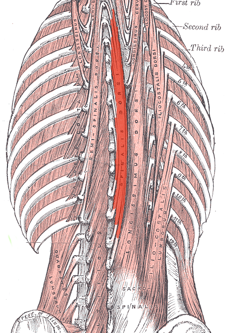
Deep muscles of the back (spinalis dorsi visible at center. Other spinalis muscles not visible).
Details
OriginThoracis: Spinous process of upper lumbar and lower thoracic vertebrae.
Cervicis: nuchal ligament and spinous process of C7.
InsertionThoracis: Spinous process of upper thoracic vertebrae.
Cervicis: Spinous process of cervical vertebrae except atlas.
ArteryLateral sacral artery
NervePosterior branch of spinal nerve
ActionsLaterally: Flex the head and neck to the same side. Bilaterally: Extend the vertebral column.
AntagonistRectus abdominis muscle
Identifiers
Latinmusculus spinalis
TA98A04.3.02.015
TA22267
FMA77179
Anatomical terms of muscle

The spinalis is a portion of the erector spinae, a bundle of muscles and tendons, located nearest to the spine. It is divided into three parts: Spinalis dorsi, spinalis cervicis, and spinalis capitis.

Spinalis dorsi

[edit]

Spinalis dorsi, the medial continuation of the sacrospinalis, is scarcely separable as a distinct muscle. It is situated at the medial side of the longissimus dorsi, and is intimately blended with it; it arises by three or four tendons from the spinous processes of the first two lumbar and the last two thoracic vertebrae: these, uniting, form a small muscle which is inserted by separate tendons into the spinous processes of the upper thoracic vertebrae, the number varying from four to eight.

It is intimately united with the semispinalis dorsi, situated beneath it.

Spinalis cervicis

[edit]

Spinalis cervicis, or spinalis colli, is an inconstant muscle, which arises from the lower part of the nuchal ligament, the spinous process of the seventh cervical, and sometimes from the spinous processes of the first and second thoracic vertebrae, and is inserted into the spinous process of the axis, and occasionally into the spinous processes of the two cervical vertebrae below it.

Spinalis capitis

[edit]

Spinalis capitis (biventer cervicis) is usually inseparably connected with the semispinalis capitis.

Spinalis capitis is not well characterized in modern anatomy textbooks and atlases, and is often omitted from anatomical illustration. However, it can be identified as fibers that extend from the spinous processes of TV1 and CV7 to the cranium, often blending with semispinalis capitis[citation needed]

See also

[edit]

References

[edit]
[edit]
Revisions and contributorsEdit on WikipediaRead on Wikipedia
from Grokipedia
The spinalis is the most medial column of the erector spinae muscle group in the , consisting of a series of interconnected muscle bundles that primarily extend and stabilize the vertebral column. It is typically divided into three parts: the spinalis thoracis in the thoracic region, the spinalis cervicis in the cervical region, and the spinalis capitis extending to the head, though the latter two are often underdeveloped, blended with adjacent muscles, or absent in many individuals. The spinalis thoracis originates from the spinous processes of the upper and lower (T11 to L2) and inserts into the spinous processes of the upper (T1 to T4), often blending laterally with the thoracis muscle. The spinalis cervicis arises from the spinous process of C7 and the ligamentum nuchae, inserting into the spinous processes of the axis (C2) and C3 to C4 vertebrae, while the spinalis capitis, when present as a distinct structure, originates from C7 and T1 spinous processes and inserts near the occiput, though it is frequently represented only by a few fibers of the semispinalis capitis. Collectively, these components function to extend the spine, laterally flex the trunk and , and provide postural support, with innervation supplied by the dorsal primary rami of spinal nerves from C2 to L3 levels. Blood supply to the spinalis derives from the deep cervical, posterior intercostal, subcostal, and arteries. Anatomical variability is a notable feature of the spinalis, particularly in the cervicis and capitis portions, which may exist as independent units, blend with the semispinalis muscles, or be entirely absent, with no correlation to sex or body side observed in studies. This variability can influence the precise contributions to head and neck movements, though the muscle's overall role remains centered on spinal extension and stability rather than generating significant force in the cervical region due to its small size.

Overview

Definition and General Anatomy

The spinalis muscle is defined as the most medial column of the erector spinae muscle group, a key component of the intrinsic back muscles responsible for spinal extension and stabilization. It consists of three distinct parts—spinalis capitis, spinalis cervicis, and spinalis thoracis (also known as spinalis dorsi)—that collectively extend along the length of the vertebral column. The erector spinae, including the spinalis, originates from a broad tendinous mass in the lower back and ascends bilaterally along the posterior trunk. In terms of general location, the spinalis runs parallel and in close proximity to the spinous processes of the vertebrae, spanning from the upper and lower thoracic regions upward toward the , forming a narrow, tendinous muscle mass that lies deep to the . This positioning situates it posteromedially within the erector spinae complex, adjacent to the midline of the back. As part of the broader intrinsic back musculature, which extends from the to the , the spinalis contributes to the overall longitudinal support of the spine. Regarding gross morphology, the spinalis is characterized by slender, flat fascicles of varying lengths that often appear underdeveloped or tendinous rather than robustly muscular, particularly in its superior portions. It frequently blends with adjacent erector spinae components, such as the longissimus and semispinalis muscles, making its boundaries indistinct in many cases. Anatomical variations are common, with the spinalis cervicis and capitis portions often poorly developed, absent, or exhibiting three primary morphological forms (continuous, discontinuous, or rudimentary) in a significant proportion of individuals. The nomenclature "spinalis" derives from the Latin "spina," meaning spine, reflecting its close association with the vertebral spinous processes.

Embryological Development

The spinalis muscle originates from the paraxial mesoderm during the fourth week of embryogenesis, when this mesodermal layer segments into paired somites along the neural tube. These somites further differentiate into dermatomes, sclerotomes, and myotomes, with the myotomes giving rise to the skeletal musculature of the trunk and limbs. The dorsal portion of each myotome, known as the epimere or epaxial division, specifically contributes to the deep back muscles, including the spinalis as part of the erector spinae group. During weeks 6 to 8 of development (corresponding to 16–20), the epaxial myotomes elongate and fuse longitudinally to form the of the erector spinae musculature. The emerges from the medialmost fibers of this , segregating into the innermost column alongside the , while the intermediate and lateral columns develop into the and , respectively. This medial positioning reflects the segmental migration and condensation of myotomal cells adjacent to the vertebral column. Hox genes play a critical role in regulating the segmental patterning of the paraxial mesoderm and vertebral column, influencing the precise rostrocaudal identity of somites and myotomes that form the spinalis. Disruptions in expression can lead to homeotic transformations, resulting in asymmetries or segmental absences in spinalis development along the cervical, thoracic, or regions. Congenital variations in the spinalis are common due to incomplete fusion or migration of myotomes during epaxial differentiation. For instance, the spinalis cervicis is absent in approximately 36% of cases and blended with adjacent muscles in 18%, while the spinalis capitis shows even higher variability, with absence in about 70% and distinct formation in only 1%. These hypoplastic or aplastic forms often arise from aberrant myotomal segmentation but typically do not impair overall back musculature function.

Anatomical Structure

Origin and Insertion Points

The spinalis muscles originate from the spinous processes of the upper and lower (primarily T11 to L2 for the spinalis thoracis), with the spinalis cervicis arising from the spinous process of C7 and the ligamentum nuchae, and the spinalis capitis from the spinous processes of C7 and T1 when present as a distinct . This attachment often occurs via a shared with the and muscles of the erector spinae group. They insert into the spinous processes of the upper (T2 to T8), (C2 to C4), and the occiput along the midline. The spinalis exists as a pair of muscles positioned bilaterally along the spinal midline, with longitudinally oriented fibers that contribute to the medial column of the erector spinae. Anatomical variations may include blending of the superior insertions or origins with the in certain individuals, particularly in the upper divisions.

Composition and Relations to Adjacent Structures

The spinalis is composed primarily of type I slow-twitch fibers, which predominate in the erector spinae group to facilitate sustained postural activity. In cross-sectional view, the spinalis presents as the smallest and most medial column of the erector spinae, forming a narrow band adjacent to the midline and lateral to the deeper transversospinalis muscles. It often blends laterally with the longissimus thoracis, contributing to the integrated structure of the erector spinae, and the upper portions (cervicis and capitis) may be underdeveloped, blended with adjacent muscles, or absent. The spinalis lies deep to the , rhomboids, and serratus posterior inferior in the thoracic region, while overlying the . It is positioned superficial to the and may blend with them in select areas, such as the cervical region. The muscle is enveloped by the , which covers the erector spinae group, and is separated from the lateral by intermuscular septa within this fascial layer.

Divisions

Spinalis Capitis

The spinalis capitis constitutes the uppermost portion of the spinalis muscle, extending cranially as part of the erector spinae group. It arises from the spinous processes of the C7 and T1 vertebrae, with variations including origins from the lower (such as C6) to upper thoracic levels (up to T2) or the ligamentum nuchae. These tendinous fibers frequently blend with the semispinalis capitis, forming an inseparable connection in many cases. The muscle inserts into the midline of the between the superior and inferior . Distinguished as the smallest and most variable segment of the spinalis, the spinalis capitis is present in approximately 30% of individuals—either as a distinct entity (1%) or blended with the semispinalis capitis (29%)—and is often rudimentary or absent altogether. Its structure is highly tendinous, featuring an incomplete tendinous intersection that earns it the synonym biventer cervicis, with fibers inserting through a broad continuous with the erector spinae. When arising from the ligamentum nuchae, it supports tension in this midline structure.

Spinalis Cervicis

The spinalis cervicis constitutes the intermediate division of the spinalis muscle within the erector spinae group, occupying a position between the thoracic and cranial portions. This slender, elongated muscle spans the cervicothoracic transition, featuring fibers that run obliquely from caudal to cranial directions. Its origin is typically from the spinous processes of the C7 to T1 or T2 vertebrae, often arising as a narrow tendinous extension shared with the underlying spinalis dorsi (also known as spinalis thoracis), which provides continuity across the thoracic-cervical boundary. The muscle's proximal attachment reflects its role as a segmental continuation of the medial erector spinae column, blending with adjacent tendinous structures in many individuals. The spinalis cervicis inserts primarily into the spinous process of the axis (C2), with variable fibers extending to the spinous processes of C3 and C4. This muscle exhibits significant variability, appearing thin and poorly developed, and is frequently incomplete or substituted by fibrous bands in about 40% of cases, sometimes blending indistinguishably with nearby structures like the semispinalis cervicis.

Spinalis Dorsi

The spinalis dorsi, also known as the spinalis thoracis, represents the thoracic division of the spinalis muscle and constitutes the most medial and prominent component of the erector spinae group in this region. It arises primarily from the spinous processes of the lower thoracic and upper , specifically T11 to L2, and shares a common origin with the broader erector spinae via its tendon, which attaches to the posterior aspect of the , , sacroiliac ligament, and . This arrangement allows the muscle to anchor firmly to the lower spine and pelvic structures, providing a stable base for its ascending fibers. The muscle inserts into the spinous processes of the upper , ranging from T1 to T8, creating a segmented, column-like structure that runs parallel to the vertebral column. These insertions form overlapping bands of muscle fibers, enabling a continuous reinforcement along the posterior thoracic spine. Laterally, the spinalis dorsi frequently blends or fuses with the adjacent thoracis, enhancing the overall cohesion of the erector spinae complex. As the largest and most consistently developed portion of the spinalis, the spinalis dorsi exhibits a robust, well-organized form with slender, flat fascicles that vary in length, the medial ones being shorter. Its bilateral masses typically converge at the midline, forming a paired yet interconnected structure that underscores its role in midline spinal support. While the upper divisions of the spinalis may show greater variability, the dorsi remains reliably present and substantial across individuals. It serves briefly as the foundational base from which the spinalis cervicis and capitis may extend superiorly.

Function and Biomechanics

Primary Movements and Roles

The spinalis muscle contributes to the extension of the vertebral column, particularly in the thoracic and cervical regions, through bilateral contraction of its components. When both sides activate simultaneously, the muscle elevates the spinous processes and extends the head, , and upper back, facilitating movements such as standing from a flexed position or arching the spine backward. This action is essential for overall spinal alignment. Unilateral contraction of the spinalis produces lateral flexion toward the ipsilateral side in the cervical and thoracic spine, aiding in side-bending motions. Due to its medial location close to the midline, the muscle generates minimal during these actions, distinguishing it from more lateral erector spinae components. This unilateral role supports balanced lateral movements while preserving spinal stability. In postural functions, the spinalis maintains upright posture by countering gravitational forces and resisting anterior , thereby preserving the natural lordotic curve of the spine during standing and walking. It provides continuous low-level tone to stabilize the vertebral column against forward-leaning tendencies. As part of the erector spinae group, it briefly integrates to enhance overall back extension and support. Cadaveric analyses reveal that the spinalis thoracis has a of approximately 1.6 cm². This force capacity underscores its role in sustaining holds against moderate loads in the upper back.

Interaction with Erector Spinae Group

The spinalis muscle, as the most medial component of the erector spinae group, plays a key synergistic role in spinal extension by providing midline stability, complementing the lateral pull from the and the rib motion control by the . During bilateral contraction, these muscles collectively extend the cervical, thoracic, and regions, with the spinalis anchoring the spinous processes to resist excessive lateral deviation. This medial positioning allows the spinalis to counterbalance the broader forces generated by its lateral counterparts, ensuring coordinated vertebral alignment. In load-bearing tasks such as lifting, the spinalis contributes to the erector spinae group's overall production by resisting shear forces along the spinal midline, helping distribute mechanical stress across the vertebral column. Biomechanical models indicate that the erector spinae, including the spinalis, generate substantial extensor moments to counteract forward bending loads, with the medial spinalis aiding in centralized force transmission to prevent disc shear. This interaction enhances spinal stability under gravitational demands, where the spinalis's attachments to spinous processes facilitate efficient midline load sharing without isolated dominance. Electromyographic (EMG) studies reveal differential activation patterns within the erector spinae during various movements, with the spinalis thoracis demonstrating intense activity in pure trunk extension compared to , where the thoracis often predominates. For instance, in orthostatic extension, the lumborum shows very strong potentials, but the spinalis contributes significantly to midline engagement, whereas homolateral elicits balanced intense responses across all three muscles, highlighting the 's rotational emphasis. These patterns underscore the spinalis's preferential role in stability over transverse rotations. Compensatory mechanisms within the erector spinae arise in response to weakness or , where components such as the may to preserve overall spinal balance and extensor function. This helps maintain postural control and load distribution, as the compensates for deficits in support by increasing its cross-sectional area and activation. Such ensures continued synergy, preventing overload on remaining group members during extension tasks.

Innervation and Vascular Supply

Neural Innervation

The spinalis muscle, as part of the erector spinae group, is primarily innervated by the dorsal rami of the spinal nerves from levels C2 to L3, providing both motor and sensory supply to its fibers. These dorsal rami branch into medial and lateral divisions, with the lateral branches specifically targeting the long extensor muscles of the back, including the spinalis. This segmental innervation ensures coordinated activation along the length of the spine, supporting extension and stabilization. Specific divisions of the spinalis receive innervation from corresponding segmental levels: the spinalis capitis is supplied by the lateral branches of the dorsal rami of the upper cervical spinal (C1 to C3), while the spinalis cervicis receives input from the dorsal rami of the lower cervical and upper thoracic spinal , and the spinalis dorsi from T1 to L2. This pattern aligns with the muscle's craniocaudal distribution, allowing precise control over cervical, thoracic, and upper regions. The neural pathways involve the dorsal rami emerging from the intervertebral foramina and piercing the to access the deep surface of the erector spinae, where they ramify into a that distributes branches along the muscle's length. This deep penetration facilitates direct contact with the muscle bellies, minimizing superficial interference. Additionally, the innervation incorporates sensory components, including proprioceptive fibers from Golgi tendon organs embedded at the musculotendinous junctions, which relay information on muscle tension and spinal positioning to the for reflex adjustments. Anatomical variations in the spinalis innervation are noted, particularly in the lower segments. Such variations are infrequent but can influence clinical assessments of back muscle function.

Blood Supply

The arterial supply to the spinalis muscle is segmental, corresponding to its divisions along the spinal column. The spinalis capitis receives blood primarily from muscular branches of the vertebral artery, deep cervical artery, and descending branches of the occipital artery. The spinalis cervicis is supplied by muscular branches of the vertebral and deep cervical arteries. For the spinalis dorsi (also known as spinalis thoracis), the dorsal branches of the posterior intercostal arteries (for thoracic levels) and lumbar arteries (for lower extensions) provide the main supply, with additional contributions from subcostal arteries. These segmental arteries form interconnections, providing redundancy in perfusion across the muscle's length. Venous drainage of the spinalis mirrors its arterial supply and occurs via corresponding segmental veins that empty into the external vertebral venous , a valveless network surrounding the vertebral column. In the cervical region, drainage proceeds to the external jugular and vertebral veins, while the thoracic portions connect to the azygos and hemiazygos systems. This facilitates extensive anastomoses, supporting efficient venous return despite positional changes. Anatomical variations in the spinalis blood supply occur between individuals, particularly in the precise origins and branching patterns of the feeding arteries. The tendinous regions of the muscle, such as intersegmental connections, generally exhibit lower vascular density compared to the fleshy bellies, reflecting typical patterns in architecture.

Clinical Significance

Associated Pathologies and Injuries

The spinalis muscles, components of the erector spinae group, are prone to strains resulting from hyperextension injuries, such as those occurring in whiplash trauma, where sudden forceful stretching can lead to microtears in the tendinous fibers and muscle bellies. These injuries often manifest as acute pain and tenderness along the paravertebral region, with reduced capacity for spinal extension due to and protective guarding. In the context of broader presentations, paraspinal strains, including those affecting the spinalis, contribute to a notable portion of cases, with spine-related injuries accounting for approximately 5-10% of athletic musculoskeletal complaints. In , particularly adolescent idiopathic forms, unilateral and develop in the paraspinal muscles on the concave side of the curve, leading to asymmetric loading and progression of spinal ; studies reveal significant fatty infiltration and cross-sectional area reduction in these muscles compared to the convex side. In degenerative scoliosis, paraspinal muscle degeneration shows fatty infiltration that is often higher on the concave side at the apex, though muscle volume may be larger on the concave side compared to the convex. Similarly, induces paraspinal muscle spasms and , exacerbating spinal rigidity and contributing to chronic beyond mere disuse , as evidenced by histopathological analyses showing excessive deposition. These changes in the spinalis and adjacent paraspinals underlie broader erector spinae dysfunction, impairing postural stability. Symptoms of spinalis involvement typically include localized, sharp or aching along the midline spine, diminished strength in back extension, and, when the spinalis capitis is affected, radiating discomfort to the posterior and occiput. Epidemiologically, such pathologies show elevated prevalence among athletes, with weightlifters experiencing incidence rates up to 40.8%, often linked to repetitive loading that strains the erector spinae components like the spinalis dorsi. In the elderly, degenerative processes amplify risk, with chronic affecting over 20% of those aged 65 and older, frequently involving paraspinal muscle degeneration including and impaired extensibility.

Surgical and Therapeutic Considerations

The spinalis muscles, as components of the erector spinae group, are frequently encountered and at risk during posterior spinal surgeries such as and procedures. In these approaches, the paraspinal musculature is dissected and retracted to access the posterior elements of the spine, potentially leading to through injury to the posterior rami of spinal nerves that innervate these muscles. Such can result in and fatty degeneration, with studies reporting up to 36.8% cross-sectional area reduction in adjacent paraspinal muscles following open posterior lumbar interbody fusion. Minimally invasive techniques, including muscle-splitting and tubular retractors, mitigate these risks by preserving muscle integrity and reducing retraction time, thereby lowering the incidence of postoperative muscle dysfunction compared to traditional open methods. Therapeutic management of spinalis-related issues, such as strains or spasms within the erector spinae, often begins with conservative interventions like focused on isometric extension exercises to strengthen the muscle without exacerbating . These exercises, such as prone trunk extensions held at submaximal effort, target the spinalis capitis and spinalis thoracis to improve endurance and stability along the spine. For acute spasms, fluoroscopy-guided injections into the erector spinae plane provide targeted analgesia and muscle relaxation, with techniques involving local anesthetics or corticosteroids to block nociceptive pathways while minimizing systemic effects. This approach ensures precise delivery to the paraspinal compartment. Rehabilitation protocols for spinalis involvement emphasize progressive strengthening, starting at 20-50% of maximum voluntary contraction (MVC) to avoid overload, and incorporating electromyographic (EMG) for isolated activation of the . devices monitor muscle activity in real-time, enabling patients to achieve targeted recruitment of the spinalis during isometric holds or controlled extensions, which enhances neuromuscular control and reduces compensatory patterns. Protocols typically advance from isometric phases to over 6-12 weeks, integrating core stabilization to support spinalis function. Recent studies (post-2020) on exercise-based therapies for chronic erector spinae-related report reductions of 10-26 mm on the visual analog scale (VAS) and disability improvements of 4-8 points on the Disability Index (ODI) at 3-12 months, reflecting 50-80% of patients achieving clinically meaningful gains. However, recurrence rates can reach 25% within 12 months without ongoing core training, underscoring the need for long-term adherence to preventive exercises.

References

Add your contribution
Related Hubs
User Avatar
No comments yet.