Hubbry Logo
Cortical reactionCortical reactionMain
Open search
Cortical reaction
Community hub
Cortical reaction
logo
8 pages, 0 posts
0 subscribers
Be the first to start a discussion here.
Cortical reaction
Cortical reaction
from Wikipedia
Cortical reaction
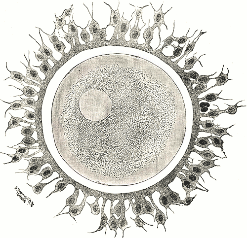
Human ovum. The zona pellucida is seen as a thick clear girdle surrounded by the cells of the corona radiata.
Anatomical terminology

The cortical reaction is a process initiated during fertilization that prevents polyspermy, the fusion of multiple sperm with one egg. In contrast to the fast block of polyspermy which immediately but temporarily blocks additional sperm from fertilizing the egg, the cortical reaction gradually establishes a permanent barrier to sperm entry and functions as the main part of the slow block of polyspermy in many animals.

To create this barrier, cortical granules, specialized secretory vesicles located within the egg's cortex (the region directly below the plasma membrane), are fused with the egg's plasma membrane. This releases the contents of the cortical granules outside the cell, where they modify an existing extracellular matrix to make it impenetrable to sperm entry. The cortical granules contain proteases that clip perivitelline tether proteins, peroxidases that harden the vitelline envelope, and glycosaminoglycans that attract water into the perivitelline space, causing it to expand and form the hyaline layer. The trigger for the cortical granules to exocytose is the release of calcium ions from cortical smooth endoplasmic reticulum in response to sperm binding to the egg.

The migration of cortical granules from their synthesis in the Golgi apparatus to the cortex region has been shown to be mediated by actin filaments in frogs and mice, and by microtubules in other mammals.[1] This migration is commonly used to assess and classify the maturity of developing oocytes.[2]

In most animals, the extracellular matrix present around the egg is the vitelline envelope which becomes the fertilization membrane following the cortical reaction. In mammals, however, the extracellular matrix modified by the cortical reaction is the zona pellucida. This modification of the zona pellucida is known as the zona reaction or zona hardening. Although highly conserved across the animal kingdom, the cortical reaction shows great diversity between species. While much has been learned about the identity and function of the contents of the cortical granules in the highly accessible sea urchin, little is known about the contents of cortical granules in mammals.

The cortical reaction within the egg is analogous to the acrosomal reaction within the sperm, where the acrosome, a specialized secretory vesicle that is homologous to cortical granules, is fused with the plasma membrane of the sperm cell to release its contents which degrade the egg's tough coating and allow the sperm to bind to and fuse with the egg.

Discovery

[edit]

The cortical reaction and cortical granules were first observed by Ethel Brown Harvey in 1910 in sea urchins. The fertilization membrane had been previously defined by Derbès when he observed the fusion of sperm and eggs of sea urchins; however, scientists believed that seminal fluid or other chemicals that the sperm brought into the egg caused the hardening of the fertilization membrane.[3] E. B. Harvey studied the mechanisms after sperm contact that lead to calcium release and formation of the fertilization membrane.[4] Cortical granules were discovered in vertebrates first in hamster oocytes in 1956 by C. R. Austin using a phase contrast microscope.[1]  

Echinoderms

[edit]
Cortical reaction in echinoderms (sea urchins). After sperm contact with egg, cortical granules release contents into perivitelline space, causing vitelline envelope to become the fertilization envelope.

In the well-studied sea urchin model system, the granule contents modify a protein coat on the outside of the plasma membrane (the vitelline layer) so that it is released from the membrane. The released cortical granule proteins exert a colloid osmotic pressure causing water to enter the space between the plasma membrane and the vitelline layer, and the vitelline layer expands away from the egg surface. This is easily visible through a microscope and is known as "elevation of the fertilization envelope". Some of the former granule contents adhere to the fertilization envelope, and it is extensively modified and cross-linked. As the fertilization envelope elevates, non-fertilizing sperm are lifted away from the egg plasma membrane, and as they are not able to pass through the fertilization envelope, they are prevented from entering the egg. Therefore, the cortical reaction prevents polyspermic fertilization, a lethal event. Another cortical granule component, polysaccharide-rich hyalin, remains adherent to the outer surface of the plasma membrane, and becomes part of the hyaline layer.

Mammals

[edit]

Although various mammals have been studied, mice represent the best studied animal models for understanding the cortical reaction in mammals. Most mammalian cortical granules are 0.2 - 0.6 um in diameter.[2] The cortical granules associate with the oocyte membrane due to SNARE proteins, which form a stabilizing complex with other proteins.[2][5] Upon contact of the sperm with the oocyte membrane, a calcium wave is induced through the PIP2 pathway involving IP3 production. IP3 then binds to its receptor on the endoplasmic reticulum which triggers a release of calcium into the cytoplasm.[2] The release of calcium triggers a change in the SNARE protein complex, and the conformation facilitates the fusion of the cortical granule with the oocyte membrane, releasing granules into the perivitelline space. The cortical reaction leads to a modification of the zona pellucida that blocks polyspermy; enzymes released by cortical granules digest sperm receptor glycoproteins ZP2 and ZP3 so that they can no longer bind spermatozoon. This is often referred to as zona hardening.

See also

[edit]

References

[edit]
[edit]
Revisions and contributorsEdit on WikipediaRead on Wikipedia
from Grokipedia
The cortical reaction is a crucial exocytotic process in fertilization across many , particularly mammals, where specialized secretory vesicles known as cortical granules, located just beneath the plasma membrane, undergo calcium-triggered fusion with the surface following penetration, releasing enzymes that modify the to prevent additional entry and ensure monospermic fertilization. This reaction is initiated almost immediately after the first fuses with the membrane, generating a propagating wave of intracellular calcium ions (Ca²⁺) that spreads across the , activating the of thousands of cortical granules within seconds to minutes. The granules, which measure approximately 0.2 to 0.6 micrometers in diameter and are synthesized exclusively in female germ cells during , contain a diverse array of contents including proteases, glycosidases, and peroxidases that are discharged into the perivitelline space. In mammalian fertilization, the primary target of this release is the zona pellucida, the glycoprotein-rich extracellular coat surrounding the ; enzymes such as those that cleave zona pellucida protein 2 (ZP2) and hydrolyze oligosaccharides on ZP3 alter its structure, hardening it and eliminating sperm-binding sites to establish a persistent block against . This serves as the primary mechanism preventing in mammals, ensuring the genetic integrity of the by blocking supernumerary incorporation, which could lead to lethal chromosomal imbalances. The mechanism relies on SNARE proteins, such as SNAP-25 and syntaxin, for vesicle docking and fusion, with the calcium signal orchestrating the synchrony of the reaction over the surface; disruptions in this process, as observed in certain models, underscore its essential role in . While the cortical reaction is conserved evolutionarily, variations exist—such as in sea urchins, where it also contributes to fertilization envelope formation—but in mammals, it is irreversible, with granules not replenished post-release.

Introduction

Definition

The cortical reaction is a fundamental process in activation during fertilization, characterized by the exocytosis of cortical granules from the periphery of the egg cytoplasm (the cortex) in response to - fusion. This calcium-dependent secretory event releases the granules' contents into the surrounding the , initiating modifications that establish a barrier to additional entry. Cortical granules are specialized, membrane-bound organelles, typically ranging from 0.2 to 1 µm in diameter, that are uniformly distributed just beneath the egg's plasma membrane during oocyte maturation. These vesicles contain a diverse array of components, including proteolytic enzymes (such as trypsin-like proteinases), peroxidases (like ovoperoxidase), mucopolysaccharides, and other regulatory factors such as and peptidylarginine deiminase, which collectively enable structural alterations to the egg's protective layers. The process is triggered by the fusion of with the egg's plasma membrane, which introduces the sperm-specific enzyme phospholipase C zeta (PLCζ). PLCζ hydrolyzes (PIP₂) to produce inositol 1,4,5-trisphosphate (IP₃), which binds to receptors on the , releasing stored calcium ions into the and generating a propagating calcium wave that orchestrates the synchronous fusion of cortical granules with the plasma membrane.

Biological Importance

The cortical reaction serves as a primary mechanism for establishing a slow and permanent block to during fertilization, thereby ensuring that only a single nucleus fuses with the to form a diploid . In many , including sea urchins and mammals, this process is triggered immediately upon the first sperm- fusion, releasing contents from cortical granules that modify the egg's plasma membrane and to deter additional penetration. This rapid response is critical because can lead to abnormal numbers, such as triploidy, which often results in embryonic lethality or spontaneous abortion, as observed in approximately 10% of fertilized human eggs. Beyond preventing multiple fertilizations, the cortical reaction contributes to species-specific reproductive barriers by altering the egg's extracellular coat in ways that inhibit heterospecific binding. In mammals, for instance, the released enzymes cleave proteins like ZP2, hardening the zona and reducing its affinity for from other species while maintaining compatibility with conspecific gametes. This modification not only reinforces the block to but also enhances , promoting genetic integrity across populations. The cortical reaction is integral to activation, linking the initial entry to downstream developmental events such as the resumption of and the extrusion of the second . This process, initiated by a calcium wave that propagates across the , ensures that the developmental program advances only after the first successful fusion, preventing further entries even as the transitions to embryonic stages.

Historical Discovery

Early Observations in Invertebrates

The cortical reaction was first observed in eggs by Ethel Brown Harvey in 1910, who described the rapid release of small cortical granules from the egg's peripheral upon fertilization, coinciding with the elevation and hardening of the vitelline membrane to form the fertilization envelope. This observation established the reaction as a key post-fertilization event in echinoderms, preventing by modifying the egg surface. Harvey's work built on prior descriptions of membrane elevation but uniquely linked it to intracellular granule discharge, using unfertilized and fertilized eggs of Arbacia punctulata as the model. Early microscopy techniques relied on light microscopy to visualize the cortical reaction in echinoderm eggs, revealing cortical granules as refractile, membrane-bound vesicles approximately 0.5–1 μm in diameter aligned just beneath the plasma membrane. These methods, including phase-contrast and bright-field illumination, allowed researchers to track granule distribution and exocytosis in living eggs without fixation, highlighting their uniform cortical positioning in mature oocytes of species like Strongylocentrotus purpuratus. Vital dyes such as neutral red were occasionally employed to enhance contrast, staining acidic compartments within the granules for better resolution under compound microscopes. In the 1930s and , foundational experiments confirmed the exocytotic nature of cortical granule release through vital staining and preparatory techniques akin to early electron microscopy. Ethel Brown Harvey's centrifugation studies in stratified egg components, isolating the cortical layer and demonstrating granule involvement in membrane formation by observing their selective retention and discharge in merogones. Complementing this, Y. Endo's 1952 work used neutral red vital staining on Hemicentrotus pulcherrimus eggs to show that cortical granules fuse directly with the plasma membrane during , releasing enzymes and structural proteins that elevate and harden the vitelline membrane into a protective barrier. These staining approaches, combined with time-lapse observations, provided evidence of the reaction's wave-like propagation from the sperm entry point, laying groundwork for later ultrastructural confirmation.

Identification in Vertebrates

The identification of the cortical reaction in vertebrate eggs began with the observation of cortical granules in mammalian oocytes. In 1956, C.R. Austin first described these structures in oocytes using , noting their distribution in the cortical region beneath the plasma membrane and their potential role in fertilization processes. This discovery marked the initial recognition of cortical granules as specialized organelles in vertebrates, analogous to those previously observed in . During the and , researchers extended these findings to other vertebrates, confirming the cortical reaction's involvement in structural changes to the egg's . In amphibians, such as laevis, studies demonstrated that fertilization triggers cortical granule , leading to modifications in the vitelline envelope that prevent . For instance, work by Wyrick, Nishihara, and Hedrick in 1974 showed agglutination of cortical granule components with jelly coat materials, resulting in envelope hardening shortly after sperm entry. Similarly, D.P. Wolf identified the cortical granule reaction and associated protease activities in released contents that contribute to these envelope alterations in eggs. In mammals, investigations in rabbits revealed comparable modifications; exposure of rabbit eggs to spermatozoa led to the loss of cortical granules and subsequent zona hardening, as observed through light microscopy and biochemical assays. These changes were quantified by reduced sperm penetration rates post-fertilization, establishing the cortical reaction's role in the zona reaction across mammalian species. By the 1980s, electron microscopy provided direct ultrastructural evidence of cortical granule fusion in mouse eggs, solidifying the mechanism in vertebrates. High-resolution imaging captured the process, showing cortical granules docking and fusing with the plasma membrane within minutes of , releasing contents into the perivitelline space. This visualization confirmed the granules' membrane-bound nature and their rapid discharge, linking Austin's initial observations to a conserved block in mammalian fertilization.

General Mechanism

Cortical Granule Formation and Distribution

Cortical granules form during , originating from the Golgi apparatus in the , where small vesicles coalesce into larger secretory organelles that subsequently migrate to the cortical region beneath the plasma membrane. This biogenesis begins in early follicular stages, such as the unilaminar phase in or multilayered follicles in humans, and involves cytoskeletal elements like and filaments to facilitate the directed transport and positioning of the granules. At the ultrastructural level, cortical granules are membrane-bound organelles often appearing as multivesicular bodies containing electron-dense cores surrounded by a less dense matrix, as observed via electron microscopy in various . In mammals, these granules exhibit morphological similarity, with diameters typically ranging from 0.2 to 0.6 µm, though some display heterogeneous contents including both electron-dense and electron-lucent forms. Prior to fertilization, cortical granules align uniformly along the egg cortex in most , forming a dense layer several micrometers below the plasma membrane to ensure rapid upon activation. For instance, eggs contain approximately 15,000 to 18,000 cortical granules per cell, distributed in interlaced rows that maintain this peripheral positioning throughout . This organized distribution optimizes the granules' role in the fertilization response while minimizing interference with other cellular processes.

Exocytosis and Calcium Signaling

The cortical reaction is initiated upon sperm-egg fusion, which introduces a sperm-derived factor leading to an increase in intracellular concentration. The nature of this factor varies across species; in non-mammalian models like sea urchins, it involves different soluble factors or direct plasma membrane signaling, while in mammals, the sperm delivers phospholipase C zeta (PLCζ), a sperm-specific , into the . This triggers the of phosphatidylinositol 4,5-bisphosphate (PIP2) in the egg's plasma membrane, generating inositol 1,4,5-trisphosphate (IP3). IP3 then binds to IP3 receptors on the (ER), opening calcium channels and releasing stored Ca²⁺ into the , thereby elevating the intracellular free concentration ([Ca²⁺]ᵢ). This initial Ca²⁺ release creates a localized increase at the site of sperm entry, which propagates as a wave across the cortex, typically at speeds of 10–30 µm/s in eggs. The resting [Ca²⁺]ᵢ of approximately 100 nM rises sharply to 1–10 µM during this wave, providing the necessary signal for downstream events. In mammalian , the Ca²⁺ signal manifests as repeated oscillations rather than a single wave, sustaining over minutes to hours. These oscillations ensure coordinated release of cortical granule contents, such as enzymes that modify the to prevent . The elevated [Ca²⁺]ᵢ directly triggers cortical granule through calcium-dependent activation of the SNARE complex, which mediates the fusion of granule membranes with the plasma membrane. SNARE proteins, including syntaxin, SNAP-25, and VAMP2, form a core complex that docks granules at the cortex and drives bilayer fusion upon Ca²⁺ binding to synaptotagmin sensors. Alpha-SNAP facilitates the assembly and disassembly of this SNARE complex, ensuring efficient and regulated during the propagating Ca²⁺ wave. This process releases the granules' contents extracellularly within seconds of the Ca²⁺ signal onset, completing the cortical reaction.

Implementation in Model Organisms

In Echinoderms

In echinoderms, such as sea urchins, the cortical reaction is a rapid exocytotic event triggered by a propagating calcium wave that releases the contents of cortical granules into the perivitelline space shortly after sperm- fusion. This process modifies the egg's extracellular investments to establish barriers against additional sperm penetration. The , a layer surrounding the unfertilized , elevates and transforms into the fertilization envelope due to the osmotic influx of driven by granule-derived mucopolysaccharides. A critical component of this transformation is ovoperoxidase, an enzyme stored in cortical granules and released during . Ovoperoxidase catalyzes the formation of dityrosine cross-links between proteins in the elevating envelope, hardening it into a rigid, protective fertilization membrane within approximately one minute post-fertilization. This cross-linking, confirmed through biochemical analysis showing one dityrosine bond per 55,000 daltons of protein, renders the envelope impermeable and resistant to sperm access. Concurrent with envelope hardening, cortical granule release contributes to the formation of the , an that assembles beneath the fertilization . Hyalin, a 330-kDa fibrillar secreted from the granules, polymerizes in the presence of calcium to form this gel-like layer, which provides structural integrity and mechanical support to the during subsequent cleavage stages. and biochemical studies have localized hyalin primarily to the translucent compartment of cortical granules, confirming its role in layer assembly without integration into the fertilization envelope itself. Experimental evidence from classic sea urchin insemination assays underscores the efficacy of these changes in blocking polyspermy. In controlled conditions with moderate sperm densities, the cortical reaction establishes a permanent physical barrier that prevents additional sperm fusions in the vast majority of cases, ensuring monospermic fertilization and normal development.

In Mammals

In mammals, the cortical reaction serves as a critical mechanism to block polyspermy by modifying the zona pellucida (ZP), the glycoprotein matrix enveloping the oocyte. Following sperm-oocyte fusion, cortical granules undergo exocytosis, releasing contents including proteases, glycosidases, and other enzymes that target ZP glycoproteins such as ZP2 and ZP3. These modifications inactivate sperm-binding sites on ZP3, a key receptor for sperm adhesion, and cross-link ZP proteins to induce zona hardening, thereby preventing additional sperm penetration. This is triggered by prolonged calcium (Ca²⁺) oscillations in the , typically comprising 5–10 transient waves occurring over several hours, which contrast with the single, rapid Ca²⁺ wave observed in many . The oscillations are initiated by sperm-derived phospholipase C zeta (PLCζ), a soluble factor injected into the that hydrolyzes (PIP₂) to generate inositol 1,4,5-trisphosphate (IP₃), stimulating Ca²⁺ release from stores via IP₃ receptors. Extensive research in and models has demonstrated that these oscillations are essential for sustained cortical granule release and ZP alterations, with disruptions in PLCζ activity leading to fertilization failure. Confirmation of this in humans comes from studies on , where cortical granule was observed to increase in mature oocytes, contributing to ZP hardening and monospermy. A investigation by Rojas et al. utilized advanced to verify granule release during Ca²⁺-dependent in human oocytes, highlighting similarities with rodent models while noting species-specific variations in oscillation patterns. Cortical granules in mammalian oocytes form during through Golgi-derived biogenesis, positioning them subcortically for rapid response.

Variations Across Species

In Other Invertebrates

In ascidians, fertilization induces the of cortical granules from the cortex, releasing contents including chymotrypsin-like enzymes that promote the elevation and hardening of the vitelline coat, thereby establishing a physical and chemical barrier to additional penetration and preventing . This process parallels the cortical reaction in echinoderms but results in a more modest structural modification of the coat. In insects like Drosophila melanogaster, egg activation occurs prior to sperm entry and involves a single rapid calcium wave, but the species lacks classical cortical granules. Polyspermy is prevented primarily by the single micropyle entry point and post-entry mechanisms, with low incidence under natural conditions; the calcium signaling coordinates cellular events like meiosis resumption rather than granule exocytosis. Across other , including certain mollusks, the cortical reaction exhibits variable efficiency in blocking , with higher occurring at elevated densities.

In Non-Mammalian Vertebrates

In amphibians, such as Xenopus laevis, the cortical reaction is initiated upon -egg fusion, triggering calcium waves that induce of cortical granules located beneath the plasma membrane. This discharge releases proteolytic enzymes, such as trypsin-like and chymotrypsin-like serine proteases, and other components into the perivitelline space, causing the vitelline membrane to elevate and transform into a hardened fertilization envelope that prevents by blocking additional penetration. These proteases modify envelope glycoproteins, enhancing the structural barrier. The process involves calcium oscillations similar to those in mammals. In teleost fish, exemplified by medaka (Oryzias latipes), the cortical reaction follows entry through the single , a specialized opening in the that guides the fertilizing . Cortical alveoli—analogous to cortical granules—undergo , releasing alveolin, an oocyte-specific astacin-family metalloproteinase, into the perivitelline space to initiate hardening. Alveolin proteolytically modifies glycoproteins, promoting cross-linking and structural reinforcement that seals the and impedes further access, contrasting with the more uniform modifications in mammals. While hatching enzymes like choriolysin are later secreted by the embryo to soften the for , the initial post-fertilization hardening relies on alveolin-mediated changes. In birds, such as chickens (Gallus gallus domesticus), the cortical reaction is subdued compared to other vertebrates, with being physiologically tolerated as multiple penetrate the , but only one achieves syngamy while others degenerate. The perivitelline layer, composed of homologs ZP1 and ZP3, undergoes modification during fertilization primarily through -derived proteases that act as lysins to facilitate initial penetration at the germinal disc. These lysins hydrolyze components of the inner perivitelline layer, creating holes for sperm entry, after which structural alterations in the layer limit excessive penetration and support early embryonic development, differing from the robust enzymatic hardening in and envelopes. This mechanism emphasizes a post-penetration block rather than a pre-fusion barrier, allowing physiological unique to avian .

Molecular Components

Contents of Cortical Granules

Cortical granules are specialized secretory vesicles in the egg cortex that store a diverse array of biochemical components, primarily enzymes, structural proteins, and , which are released during upon fertilization to modify the egg's extracellular environment. In echinoderms like sea urchins, these granules contain a heterogeneous of proteins, including structural molecules and glycosaminoglycans, which contribute to the formation of protective barriers. Among the enzymatic contents, peroxidases such as ovoperoxidase are prominent, catalyzing dityrosine cross-links in structural proteins to harden the fertilization envelope in sea urchins. Proteases, including serine proteases, are also key; in mammals, ovastacin (a metalloendoprotease encoded by the Astl gene) is released to cleave ZP2 in the zona pellucida, thereby preventing polyspermy by altering sperm binding sites. Glycosidases, such as β-glucuronidase and N-acetylglucosaminidase (β-hexosaminidase B), function in matrix modification by removing carbohydrate residues, with the latter identified in mouse oocytes where it contributes to the zona reaction. Structural components include mucopolysaccharides, which are carbohydrate-rich and detected via periodic acid-Schiff (PAS) staining in both mammalian and eggs; these absorb water to elevate and stiffen the vitelline or fertilization envelope. proteins, such as the major hyaline protein in sea urchins and immunologically related p62/p56 proteins in oocytes, provide structural support for the layer or cortical granule envelope. Other factors encompass protease inhibitors, like those binding soybean trypsin inhibitor in mouse and hamster eggs, which modulate enzymatic activity post-release. Antimicrobial peptides have been suggested in some species but remain unconfirmed in detailed catalogs. Recent research has identified distinct populations of cortical granules in mouse oocytes, including those releasing zinc sparks and others secreting ovastacin, revealing compartmentalized functions. Despite advances, the full molecular inventory of cortical granules, particularly in mammals, remains incomplete, with estimates of only 4-14 major proteins identified and species-specific heterogeneity poorly characterized.

Key Proteins and Pathways

The of cortical granules during the cortical reaction is facilitated by SNARE proteins, which form a core fusion machinery between granule and plasma membranes. Key components include the t-SNAREs syntaxin 4 and SNAP-23, localized on the plasma membrane, and the v-SNAREs synaptobrevin isoforms VAMP1 and VAMP3, present on cortical granule membranes; these proteins zipper together in a calcium-dependent manner to drive membrane fusion. SNAP-23 is particularly critical, as its expression in unfertilized oocytes enables granule , and targeted disruption or inhibition abolishes this process. In mammals, α-SNAP (alpha-soluble NSF attachment protein) plays a regulatory role by binding to the SNARE complex post-fusion, recruiting NSF (N-ethylmaleimide-sensitive factor) to disassemble it via , thereby allowing recycling and sustained during calcium waves. Sperm-derived factors initiate the signaling cascade leading to cortical granule . In mammals, phospholipase C zeta (PLCζ), a sperm-specific introduced upon fusion, serves as the primary soluble oscillator; it hydrolyzes (PIP2) in the to produce inositol 1,4,5-trisphosphate (IP3), triggering repetitive calcium oscillations essential for egg activation and the cortical reaction. This discovery in the early 2000s resolved long-standing questions about the sperm factor , confirming PLCζ's potency at physiological concentrations mimics fertilization-induced calcium transients. Downstream pathways propagate calcium signals to coordinate . IP3 binds to IP3 receptors (IP3R, primarily IP3R1 in oocytes), ligand-gated calcium channels on the (ER), releasing stored Ca²⁺ into the to initiate propagating waves that sensitize the fusion machinery. (sarco/endoplasmic reticulum Ca²⁺-ATPase) pumps, such as SERCA2b, counterbalance this by actively sequestering Ca²⁺ back into the ER, sustaining oscillatory dynamics and preventing depletion of stores during wave propagation. Recent 2021 analyses of human cases have identified PLCZ1 variants, such as frameshift mutations, that disrupt IP3 production and calcium release, resulting in failed cortical granule and total fertilization failure during assisted reproduction.

Significance and Implications

Role in Preventing Polyspermy

The cortical reaction serves as the primary slow block to , a permanent mechanism that complements the transient fast block mediated by membrane depolarization. In sea urchins, upon entry, the cortical granules undergo calcium-triggered , releasing enzymes such as proteases, mucopolysaccharides, and peroxidases that transform the vitelline into a hardened fertilization envelope. This alteration physically impedes additional penetration and removes already bound , ensuring monospermy. In mammals, the reaction similarly modifies the through the release of proteases like ovastacin, which cleaves ZP2 into ZP2f, thereby reducing binding sites and creating a biochemical barrier to further fusion. Experimental evidence demonstrates that inducing the cortical reaction artificially mimics its polyspermy-preventing effects. In sea urchins, treatment with the calcium A23187 triggers granule and envelope elevation without fusion, resulting in eggs resistant to subsequent , as observed through direct microscopic examination of fertilization outcomes. Similarly, in mammalian oocytes, such as those from mice and pigs, ionophore-induced activation leads to hardening and decreased penetration in assays, confirming the reaction's causal role independent of other fertilization events. These studies highlight the reaction's specificity, as incomplete correlates with higher rates. Quantitatively, the cortical reaction dramatically lowers incidence. In sea urchins, under conditions of excess , 85-95% of eggs achieve monospermy due to the robust fertilization formed by approximately 15,000 cortical granules per egg. In mice, the reaction reduces bound from an average of 52 per egg to 13 post-exocytosis, contributing to rates below 5% in optimized and settings, compared to over 50% in scenarios where the reaction is inhibited or absent, such as in certain IVF protocols with high concentrations. This efficacy underscores the reaction's essential role in reproductive success across species.

Evolutionary and Pathophysiological Aspects

The cortical reaction, characterized by calcium-triggered of cortical granules, is a highly conserved mechanism across most metazoans, from such as sea urchins and to vertebrates including mammals, serving as a fundamental barrier to during fertilization. This core process of calcium-dependent relies on evolutionarily preserved molecular components, including SNARE complexes (e.g., and synaptobrevin), Rab proteins, and synaptotagmin-1, which facilitate granule fusion with the plasma membrane and are distributed widely among animal taxa. The evolutionary origin of cortical granules is closely linked to , where they arise from the Golgi apparatus early in development, accumulating progressively in the cortex through a process that parallels the emergence of regulated secretion in early metazoan reproduction. Pathophysiological disruptions in the cortical reaction contribute to reproductive failures, particularly in assisted reproductive technologies like in vitro fertilization (IVF), where defects in granule exocytosis can lead to polyspermy and fertilization arrest. Approximately 7% of fertilized human oocytes in IVF exhibit polyspermy, often due to incomplete cortical reactions in immature or overmature eggs, resulting in triploid embryos with reduced viability and increased risk of genetic abnormalities. Recent studies in the 2020s have identified mutations in phospholipase C zeta (PLCζ), the sperm-derived factor that initiates calcium oscillations necessary for triggering the cortical reaction, as a key cause of male-factor infertility; for instance, PLCZ1 mutations such as c.588C>A (p.Cys196Ter) abolish calcium signaling in over 95% of affected spermatozoa, leading to delayed or absent granule exocytosis, excessive sperm-zona binding, and polyspermy rates exceeding 50% in conventional IVF. Similarly, gain-of-function mutations like L277P in PLCζ hyperactivate calcium oscillations, prolonging egg activation but impairing normal cortical granule release and contributing to fertilization failure. Artificial oocyte activation via intracytoplasmic sperm injection has successfully rescued outcomes in such cases, yielding live births. Despite this conservation, significant gaps persist in understanding the of cortical granule composition and species-specific adaptations, with limited comparative genomic data on how granule contents (e.g., proteases like ovastacin in mammals versus transglutaminases in teleosts) diverged to suit diverse extracellular matrices. These knowledge gaps hinder insights into how environmental or genetic factors might exacerbate pathophysiological risks across taxa.

References

Add your contribution
Related Hubs
User Avatar
No comments yet.