Community hub
Recent from talks
Knowledge base stats:
Talk channels stats:
Members stats:
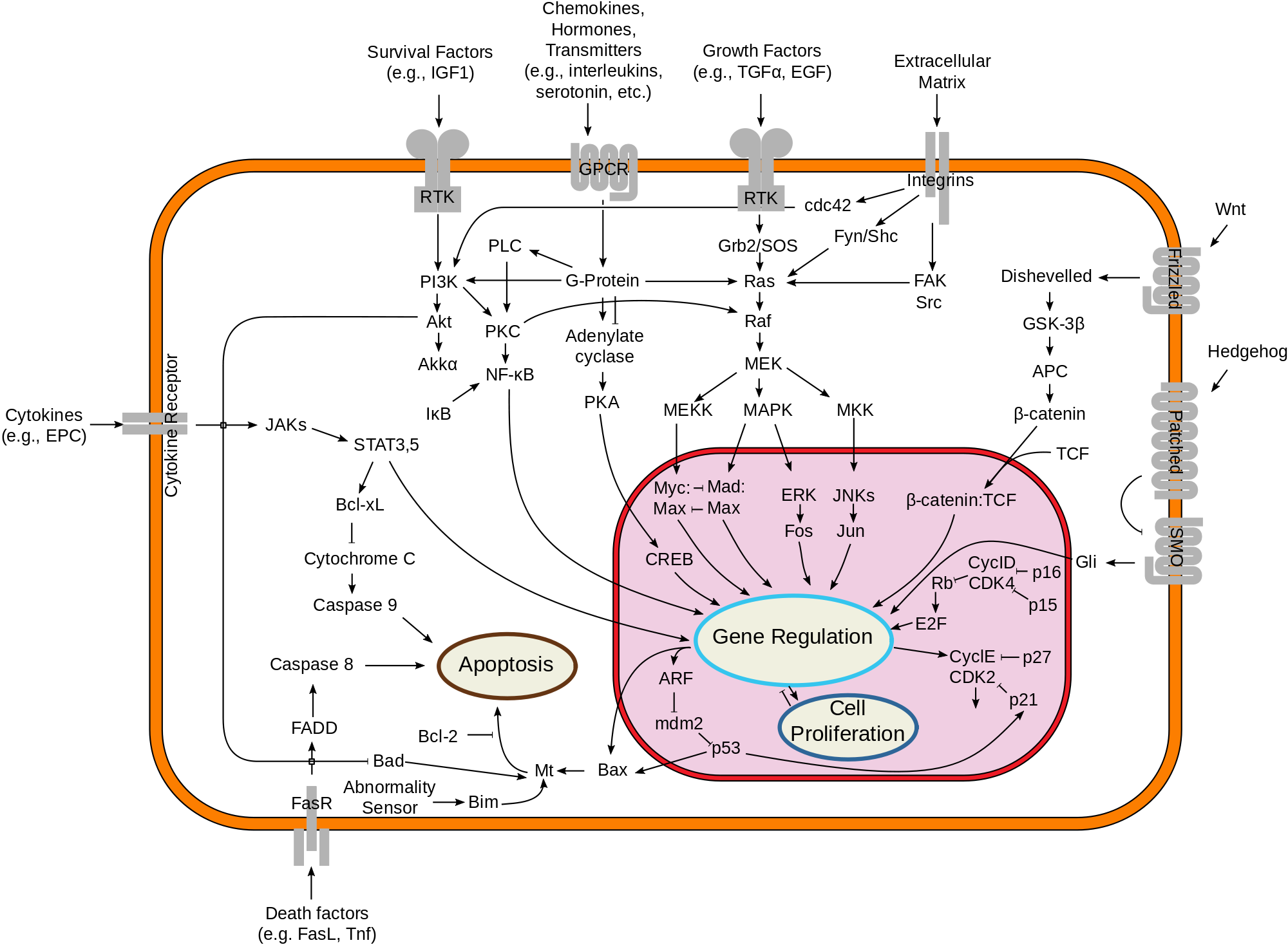
Cell death
Cell death is the event of a biological cell ceasing to carry out its functions. This may be the result of the natural process of old cells dying and being replaced by new ones, as in programmed cell death, or may result from factors such as diseases, localized injury, or the death of the organism of which the cells are part. Apoptosis or Type I cell-death, and autophagy or Type II cell-death are both forms of programmed cell death, while necrosis is a non-physiological process that occurs as a result of infection or injury.
The term "cell necrobiology" has been used to describe the life processes associated with morphological, biochemical, and molecular changes which predispose, precede, and accompany cell death, as well as the consequences and tissue response to cell death. The word is derived from the Greek νεκρό meaning "death", βìο meaning "life", and λόγος meaning "the study of". The term was initially coined to broadly define investigations of the changes that accompany cell death, detected and measured by multiparameter flow- and laser scanning- cytometry. It has been used to describe the real-time changes during cell death, detected by flow cytometry.
Programmed cell death (PCD) sometimes referred to as cell, or cellular suicide is the death of a cell as a result of events inside of a cell, such as apoptosis or autophagy. PCD is carried out in a biological process, which usually confers advantage during an organism's lifecycle. For example, the differentiation of fingers and toes in a developing human embryo occurs because cells between the fingers apoptose; the result is that the digits are separate. PCD serves fundamental functions during both plant and animal tissue development.
Programmed cell death (PCD) is cell death mediated by an intracellular program. PCD is carried out in a regulated process, which usually confers advantage during an organism's life-cycle. For example, the differentiation of fingers and toes in a developing human embryo occurs because cells between the fingers apoptose; the result is that the digits separate. PCD serves fundamental functions during both plant and metazoa (multicellular animals) tissue development.
Apoptosis is the processor of programmed cell death (PCD) that may occur in multicellular organisms. Biochemical events lead to characteristic cell changes (morphology) and death. These changes include blebbing, cell shrinkage, nuclear fragmentation, chromatin condensation, and chromosomal DNA fragmentation. It is now thought that – in a developmental context – cells are induced to positively commit suicide whilst in a homeostatic context; the absence of certain survival factors may provide the impetus for suicide. There appears to be some variation in the morphology and indeed the biochemistry of these suicide pathways; some treading the path of "apoptosis", others following a more generalized pathway to deletion, but both usually being genetically and synthetically motivated. There is some evidence that certain symptoms of "apoptosis" such as endonuclease activation can be spuriously induced without engaging a genetic cascade, however, presumably true apoptosis and programmed cell death must be genetically mediated. It is also becoming clear that mitosis and apoptosis are toggled or linked in some way and that the balance achieved depends on signals received from appropriate growth or survival factors.
Certain key proteins primarily employed in the repair of DNA damage can also induce apoptosis when DNA damage exceeds the cell’s repair capability. These dual role proteins protect against proliferation of unstable damaged cells that might lead to cancer.
Autophagy is cytoplasmic, characterized by the formation of large vacuoles that eat away organelles in a specific sequence prior to the destruction of the nucleus. Macroautophagy, often referred to as autophagy, is a catabolic process that results in the autophagosomic-lysosomal degradation of bulk cytoplasmic contents, abnormal protein aggregates, and excess or damaged organelles. Autophagy is generally activated by conditions of nutrient deprivation but has also been associated with physiological as well as pathological processes such as development, differentiation, neurodegenerative diseases, stress, infection and cancer.
Other pathways of programmed cell death have been discovered. Called "non-apoptotic programmed cell-death" (or "caspase-independent programmed cell-death"), these alternative routes to death are as efficient as apoptosis and can function as either backup mechanisms or the main type of PCD.
Hub AI
Cell death AI simulator
(@Cell death_simulator)
Cell death
Cell death is the event of a biological cell ceasing to carry out its functions. This may be the result of the natural process of old cells dying and being replaced by new ones, as in programmed cell death, or may result from factors such as diseases, localized injury, or the death of the organism of which the cells are part. Apoptosis or Type I cell-death, and autophagy or Type II cell-death are both forms of programmed cell death, while necrosis is a non-physiological process that occurs as a result of infection or injury.
The term "cell necrobiology" has been used to describe the life processes associated with morphological, biochemical, and molecular changes which predispose, precede, and accompany cell death, as well as the consequences and tissue response to cell death. The word is derived from the Greek νεκρό meaning "death", βìο meaning "life", and λόγος meaning "the study of". The term was initially coined to broadly define investigations of the changes that accompany cell death, detected and measured by multiparameter flow- and laser scanning- cytometry. It has been used to describe the real-time changes during cell death, detected by flow cytometry.
Programmed cell death (PCD) sometimes referred to as cell, or cellular suicide is the death of a cell as a result of events inside of a cell, such as apoptosis or autophagy. PCD is carried out in a biological process, which usually confers advantage during an organism's lifecycle. For example, the differentiation of fingers and toes in a developing human embryo occurs because cells between the fingers apoptose; the result is that the digits are separate. PCD serves fundamental functions during both plant and animal tissue development.
Programmed cell death (PCD) is cell death mediated by an intracellular program. PCD is carried out in a regulated process, which usually confers advantage during an organism's life-cycle. For example, the differentiation of fingers and toes in a developing human embryo occurs because cells between the fingers apoptose; the result is that the digits separate. PCD serves fundamental functions during both plant and metazoa (multicellular animals) tissue development.
Apoptosis is the processor of programmed cell death (PCD) that may occur in multicellular organisms. Biochemical events lead to characteristic cell changes (morphology) and death. These changes include blebbing, cell shrinkage, nuclear fragmentation, chromatin condensation, and chromosomal DNA fragmentation. It is now thought that – in a developmental context – cells are induced to positively commit suicide whilst in a homeostatic context; the absence of certain survival factors may provide the impetus for suicide. There appears to be some variation in the morphology and indeed the biochemistry of these suicide pathways; some treading the path of "apoptosis", others following a more generalized pathway to deletion, but both usually being genetically and synthetically motivated. There is some evidence that certain symptoms of "apoptosis" such as endonuclease activation can be spuriously induced without engaging a genetic cascade, however, presumably true apoptosis and programmed cell death must be genetically mediated. It is also becoming clear that mitosis and apoptosis are toggled or linked in some way and that the balance achieved depends on signals received from appropriate growth or survival factors.
Certain key proteins primarily employed in the repair of DNA damage can also induce apoptosis when DNA damage exceeds the cell’s repair capability. These dual role proteins protect against proliferation of unstable damaged cells that might lead to cancer.
Autophagy is cytoplasmic, characterized by the formation of large vacuoles that eat away organelles in a specific sequence prior to the destruction of the nucleus. Macroautophagy, often referred to as autophagy, is a catabolic process that results in the autophagosomic-lysosomal degradation of bulk cytoplasmic contents, abnormal protein aggregates, and excess or damaged organelles. Autophagy is generally activated by conditions of nutrient deprivation but has also been associated with physiological as well as pathological processes such as development, differentiation, neurodegenerative diseases, stress, infection and cancer.
Other pathways of programmed cell death have been discovered. Called "non-apoptotic programmed cell-death" (or "caspase-independent programmed cell-death"), these alternative routes to death are as efficient as apoptosis and can function as either backup mechanisms or the main type of PCD.