Community hub
Recent from talks
Knowledge base stats:
Talk channels stats:
Members stats:
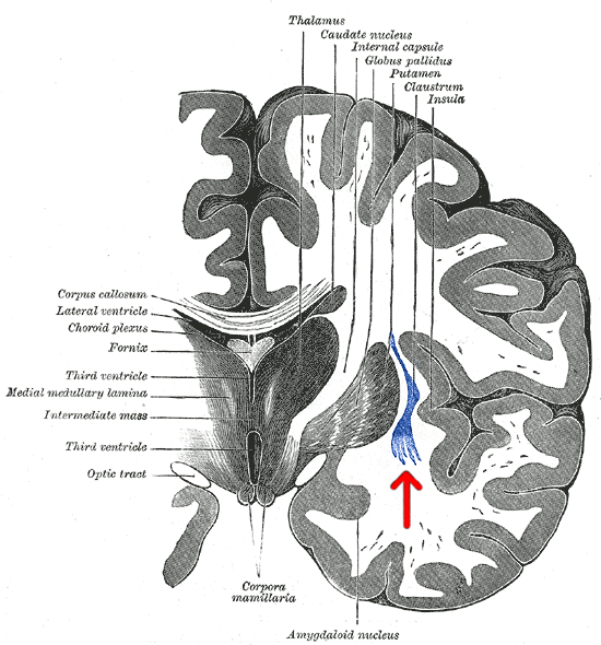
Claustrum
The claustrum (Latin, meaning "to close" or "to shut") is a thin sheet of neurons and supporting glial cells in the brain, that connects to the cerebral cortex and subcortical regions including the amygdala, hippocampus and thalamus. It is located between the insular cortex laterally and the putamen medially, encased by the extreme and external capsules respectively. Blood to the claustrum is supplied by the middle cerebral artery. It is considered to be the most densely connected structure in the brain, and thus hypothesized to allow for the integration of various cortical inputs such as vision, sound and touch, into one experience. Other hypotheses suggest that the claustrum plays a role in salience processing, to direct attention towards the most behaviorally relevant stimuli amongst the background noise. The claustrum is difficult to study given the limited number of individuals with claustral lesions and the poor resolution of neuroimaging.
The claustrum is made up of various cell types differing in size, shape and neurochemical composition. Five cell types exist, and a majority of these cells resemble pyramidal neurons found in the cortex. Within the claustrum, there is no laminar organization of cell types as in the cortical layers, and the cell bodies can be a pyramidal, fusiform or circular. The principal cell type found in the claustrum is the Golgi type I neuron, which is a large cell with dendrites covered in spines.
Through interhemispheric connections, the claustrum is believed to play a role in synchronizing activity in widely separated, but functionally related, parts of the brain such as between frontal eye fields and the visual cortex. As such, the claustrum is thought to play a role in combining different information modalities, potentially to support consciousness itself. Another proposed function of the claustrum is to differentiate between relevant and irrelevant information so that the latter can be ignored.
Cortical components of consciousness include the fronto-parietal cortex, cingulate and precuneus. Due to the claustrum's widespread connectivity to these areas, it is suggested that it may play a role in both attention and consciousness. The neural networks that mediate sustained attention and consciousness send inputs to the claustrum, and one case report in humans suggests that electrical stimulation near the claustrum reversibly disrupted the patient's conscious state.
The claustrum is a small bilateral gray matter structure (comprising roughly 0.25% of the cerebral cortex) located deep to the insular cortex and extreme capsule, and superficial to the external capsule and basal ganglia.
Its name means "hidden away", and was first identified in 1672, with more detailed descriptions coming later on during the 19th century. Although the regional neuroanatomical boundaries of the claustrum have been defined, there remains a lack of consensus in the literature when defining its precise margins, though a meeting in 2019 of experts has posited a framework by which to refer to the structures across species.
An early summary of reports from the 20th century emphasized cortical inputs and outputs. However, later work has suggested the claustrum has extensive connections to cortical and subcortical regions. More specifically, electrophysiological studies show extensive connections to thalamic nuclei and the basal ganglia, while isotopological reports have linked the claustrum with the prefrontal, frontal, parietal, temporal and occipital cortices. Additional studies have also looked at the relationship of the claustrum to well-described subcortical white matter tracts. Structures such as the corona radiata, occipitofrontal fasciculus and uncinate fasciculus project to the claustrum from frontal, pericentral, parietal and occipital regions. Reciprocal connections also exist with motor, somatosensory, auditory and visual cortical regions. Altogether, these findings leave the claustrum as the most highly connected structure per regional volume in the brain and suggest that it may serve as a hub to coordinate activity of cerebral circuits. Even with this extensive connectivity, most projections to and from the claustrum are ipsilateral (although there are still contralateral projections), and little evidence exists to describe its afferent or efferent connections with the brainstem and spinal cord. In summary, the cortical and subcortical connectivity of the claustrum implies that it is most involved with processing sensory information, as well as the physical and emotional state of an animal.
Inputs to the claustrum are organized by modality, which include prefrontal, visual, auditory and somatomotor processing areas. In the same way that the morphology of neurons in the Rexed laminae of the spinal cord is indicative of function, the visual, auditory and somatomotor regions within the claustrum share similar neurons with specific functional characteristics. For example, the portion of the claustrum that processes visual information (primarily synthesizing afferent fibers concerned with our peripheral visual field) is comprised by a majority of binocular cells that have "elongated receptive fields and no orientation selectivity".
Hub AI
Claustrum AI simulator
(@Claustrum_simulator)
Claustrum
The claustrum (Latin, meaning "to close" or "to shut") is a thin sheet of neurons and supporting glial cells in the brain, that connects to the cerebral cortex and subcortical regions including the amygdala, hippocampus and thalamus. It is located between the insular cortex laterally and the putamen medially, encased by the extreme and external capsules respectively. Blood to the claustrum is supplied by the middle cerebral artery. It is considered to be the most densely connected structure in the brain, and thus hypothesized to allow for the integration of various cortical inputs such as vision, sound and touch, into one experience. Other hypotheses suggest that the claustrum plays a role in salience processing, to direct attention towards the most behaviorally relevant stimuli amongst the background noise. The claustrum is difficult to study given the limited number of individuals with claustral lesions and the poor resolution of neuroimaging.
The claustrum is made up of various cell types differing in size, shape and neurochemical composition. Five cell types exist, and a majority of these cells resemble pyramidal neurons found in the cortex. Within the claustrum, there is no laminar organization of cell types as in the cortical layers, and the cell bodies can be a pyramidal, fusiform or circular. The principal cell type found in the claustrum is the Golgi type I neuron, which is a large cell with dendrites covered in spines.
Through interhemispheric connections, the claustrum is believed to play a role in synchronizing activity in widely separated, but functionally related, parts of the brain such as between frontal eye fields and the visual cortex. As such, the claustrum is thought to play a role in combining different information modalities, potentially to support consciousness itself. Another proposed function of the claustrum is to differentiate between relevant and irrelevant information so that the latter can be ignored.
Cortical components of consciousness include the fronto-parietal cortex, cingulate and precuneus. Due to the claustrum's widespread connectivity to these areas, it is suggested that it may play a role in both attention and consciousness. The neural networks that mediate sustained attention and consciousness send inputs to the claustrum, and one case report in humans suggests that electrical stimulation near the claustrum reversibly disrupted the patient's conscious state.
The claustrum is a small bilateral gray matter structure (comprising roughly 0.25% of the cerebral cortex) located deep to the insular cortex and extreme capsule, and superficial to the external capsule and basal ganglia.
Its name means "hidden away", and was first identified in 1672, with more detailed descriptions coming later on during the 19th century. Although the regional neuroanatomical boundaries of the claustrum have been defined, there remains a lack of consensus in the literature when defining its precise margins, though a meeting in 2019 of experts has posited a framework by which to refer to the structures across species.
An early summary of reports from the 20th century emphasized cortical inputs and outputs. However, later work has suggested the claustrum has extensive connections to cortical and subcortical regions. More specifically, electrophysiological studies show extensive connections to thalamic nuclei and the basal ganglia, while isotopological reports have linked the claustrum with the prefrontal, frontal, parietal, temporal and occipital cortices. Additional studies have also looked at the relationship of the claustrum to well-described subcortical white matter tracts. Structures such as the corona radiata, occipitofrontal fasciculus and uncinate fasciculus project to the claustrum from frontal, pericentral, parietal and occipital regions. Reciprocal connections also exist with motor, somatosensory, auditory and visual cortical regions. Altogether, these findings leave the claustrum as the most highly connected structure per regional volume in the brain and suggest that it may serve as a hub to coordinate activity of cerebral circuits. Even with this extensive connectivity, most projections to and from the claustrum are ipsilateral (although there are still contralateral projections), and little evidence exists to describe its afferent or efferent connections with the brainstem and spinal cord. In summary, the cortical and subcortical connectivity of the claustrum implies that it is most involved with processing sensory information, as well as the physical and emotional state of an animal.
Inputs to the claustrum are organized by modality, which include prefrontal, visual, auditory and somatomotor processing areas. In the same way that the morphology of neurons in the Rexed laminae of the spinal cord is indicative of function, the visual, auditory and somatomotor regions within the claustrum share similar neurons with specific functional characteristics. For example, the portion of the claustrum that processes visual information (primarily synthesizing afferent fibers concerned with our peripheral visual field) is comprised by a majority of binocular cells that have "elongated receptive fields and no orientation selectivity".