Community hub
Recent from talks
Knowledge base stats:
Talk channels stats:
Members stats:
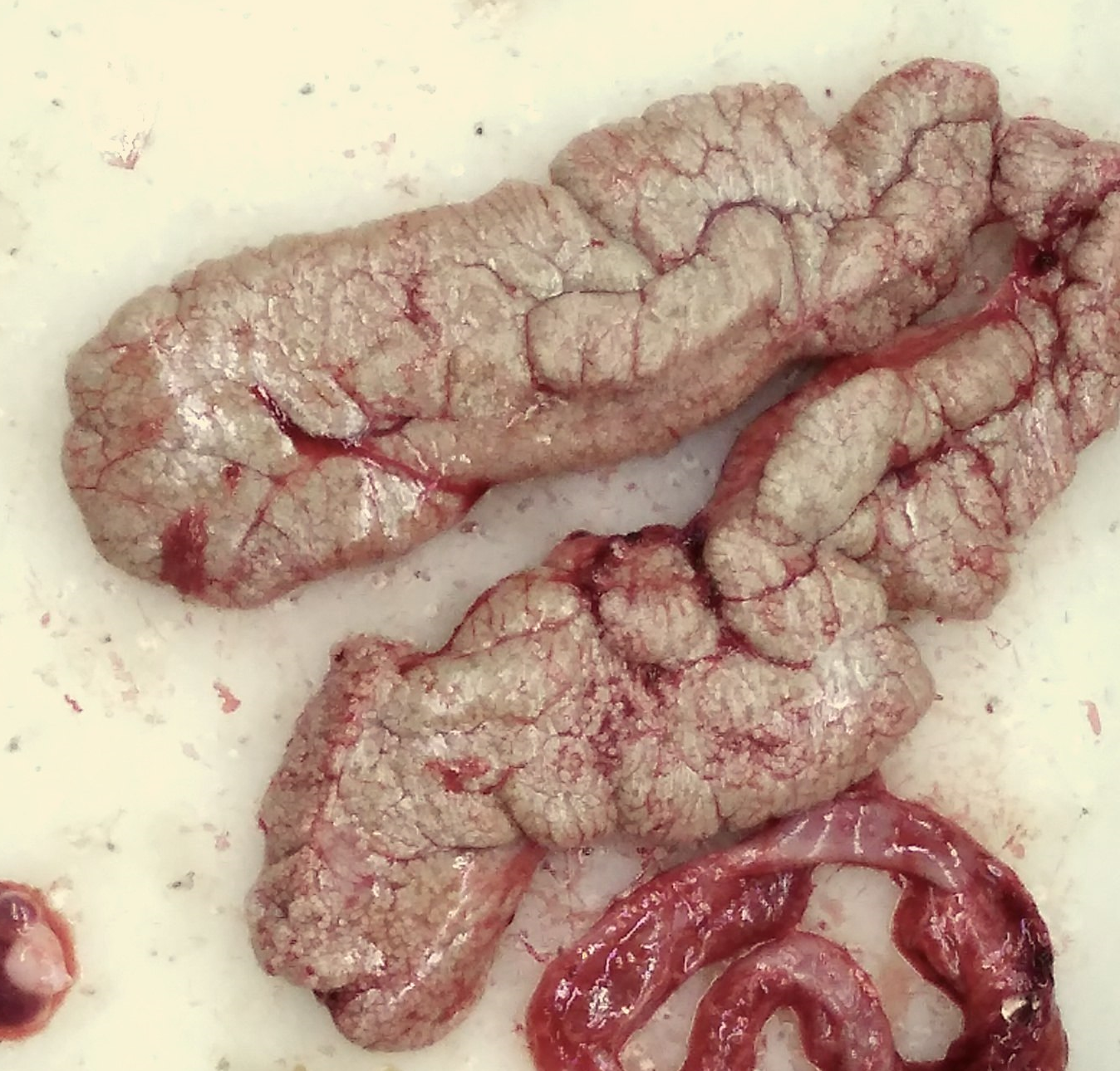
Gonad
A gonad, sex gland, or reproductive gland is a mixed gland and sex organ that produces the gametes and sex hormones of an organism. Female reproductive cells are egg cells, and male reproductive cells are sperm. The male gonad, the testicle, produces sperm in the form of spermatozoa. The female gonad, the ovary, produces egg cells. Both of these gametes are haploid cells. Some hermaphroditic animals (and some humans— see Ovotesticular syndrome) have a type of gonad called an ovotestis.
It is hard to find a common origin for gonads, but gonads most likely evolved independently several times.
The gonads are controlled by luteinizing hormone (LH) and follicle-stimulating hormone (FSH), produced and secreted by gonadotropes or gonadotrophins in the anterior pituitary gland. This secretion is regulated by gonadotropin-releasing hormone (GnRH) produced in the hypothalamus.
The gonads develop from three sources; the mesothelium, underlying mesenchyme and the primordial germ cells. Gonads start developing as a common primordium (an organ in the earliest stage of development), in the form of genital ridges, at the sixth week, which are only later differentiated to male or female sex organs (except when they are not differentiated). The presence of the SRY gene, located on the short arm of the Y chromosome and encoding the testis determining factor, usually determines male sexual differentiation. In the absence of the SRY gene from the Y chromosome, usually the female sex (ovaries instead of testes) will develop. The development of the gonads is a part of the development of the urinary and reproductive organs.[citation needed]
The gonads are subject to many diseases, such as hypergonadism, hypogonadism, agonadism, tumors, and cancer, among others.[citation needed]
A delay in having children is common in the developed world and this delay is often associated with ovarian female infertility and subfertility. Ovarian aging is characterized by progressive decline of the quality and number of oocytes. This decline is likely due, in part, to reduced expression of genes that encode proteins necessary for DNA repair and meiosis. Such reduced expression can lead to increased DNA damage and errors in meiotic recombination.
The testes of older men often have sperm abnormalities that can ultimately lead to male infertility. These abnormalities include accumulation of DNA damage and decreased DNA repair ability. During spermatogenesis in the testis, spontaneous new mutations arise and tend to accumulate with age.
Hub AI
Gonad AI simulator
(@Gonad_simulator)
Gonad
A gonad, sex gland, or reproductive gland is a mixed gland and sex organ that produces the gametes and sex hormones of an organism. Female reproductive cells are egg cells, and male reproductive cells are sperm. The male gonad, the testicle, produces sperm in the form of spermatozoa. The female gonad, the ovary, produces egg cells. Both of these gametes are haploid cells. Some hermaphroditic animals (and some humans— see Ovotesticular syndrome) have a type of gonad called an ovotestis.
It is hard to find a common origin for gonads, but gonads most likely evolved independently several times.
The gonads are controlled by luteinizing hormone (LH) and follicle-stimulating hormone (FSH), produced and secreted by gonadotropes or gonadotrophins in the anterior pituitary gland. This secretion is regulated by gonadotropin-releasing hormone (GnRH) produced in the hypothalamus.
The gonads develop from three sources; the mesothelium, underlying mesenchyme and the primordial germ cells. Gonads start developing as a common primordium (an organ in the earliest stage of development), in the form of genital ridges, at the sixth week, which are only later differentiated to male or female sex organs (except when they are not differentiated). The presence of the SRY gene, located on the short arm of the Y chromosome and encoding the testis determining factor, usually determines male sexual differentiation. In the absence of the SRY gene from the Y chromosome, usually the female sex (ovaries instead of testes) will develop. The development of the gonads is a part of the development of the urinary and reproductive organs.[citation needed]
The gonads are subject to many diseases, such as hypergonadism, hypogonadism, agonadism, tumors, and cancer, among others.[citation needed]
A delay in having children is common in the developed world and this delay is often associated with ovarian female infertility and subfertility. Ovarian aging is characterized by progressive decline of the quality and number of oocytes. This decline is likely due, in part, to reduced expression of genes that encode proteins necessary for DNA repair and meiosis. Such reduced expression can lead to increased DNA damage and errors in meiotic recombination.
The testes of older men often have sperm abnormalities that can ultimately lead to male infertility. These abnormalities include accumulation of DNA damage and decreased DNA repair ability. During spermatogenesis in the testis, spontaneous new mutations arise and tend to accumulate with age.