Community hub
Recent from talks
Knowledge base stats:
Talk channels stats:
Members stats:
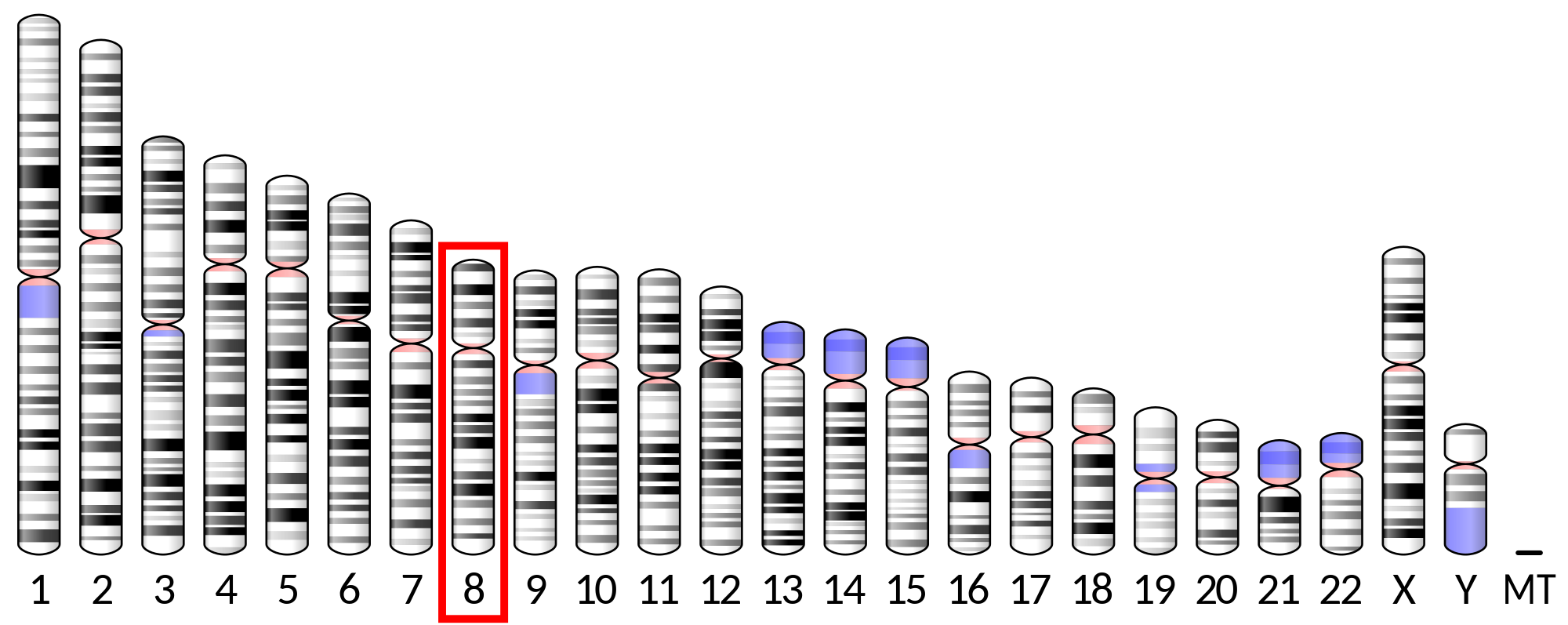
Lipoprotein lipase
Lipoprotein lipase (LPL) (EC 3.1.1.34, systematic name triacylglycerol acylhydrolase (lipoprotein-dependent)) is a member of the lipase gene family, which includes pancreatic lipase, hepatic lipase, and endothelial lipase. It is a water-soluble enzyme that hydrolyzes triglycerides in lipoproteins, such as those found in chylomicrons and very low-density lipoproteins (VLDL), into two free fatty acids and one monoacylglycerol molecule:
It is also involved in promoting the cellular uptake of chylomicron remnants, cholesterol-rich lipoproteins, and free fatty acids. LPL requires ApoC-II as a cofactor.
LPL is attached to the luminal surface of endothelial cells in capillaries by the protein glycosylphosphatidylinositol HDL-binding protein 1 (GPIHBP1) and by heparan sulfated peptidoglycans. It is most widely distributed in adipose, heart, and skeletal muscle tissue, as well as in lactating mammary glands.
In brief, LPL is secreted from heart, muscle and adipose parenchymal cells as a glycosylated homodimer, after which it is translocated through the extracellular matrix and across endothelial cells to the capillary lumen. After translation, the newly synthesized protein is glycosylated in the endoplasmic reticulum. The glycosylation sites of LPL are Asn-43, Asn-257, and Asn-359. Glucosidases then remove terminal glucose residues; it was once believed that this glucose trimming is responsible for the conformational change needed for LPL to form homodimers and become catalytically active. In the Golgi apparatus, the oligosaccharides are further altered to result in either two complex chains, or two complex and one high-mannose chain. In the final protein, carbohydrates account for about 12% of the molecular mass (55-58 kDa).
Homodimerization is required before LPL can be secreted from cells. After secretion, LPL is carried across endothelial cells and presented into the capillary lumen by the protein glycosylphosphatidylinositol-anchored high-density lipoprotein-binding protein 1.
Crystal structures of LPL complexed with GPIHBP1 have been reported. LPL is composed of two distinct regions: the larger N-terminus domain that contains the lipolytic active site, and the smaller C-terminus domain. These two regions are attached by a peptide linker. The N-terminus domain has an α/β hydrolase fold, which is a globular structure containing a central β sheet surrounded by α helices. The C-terminus domain is a β sandwich formed by two β sheet layers, and resembles an elongated cylinder.
The active site of LPL is composed of the conserved Ser-132, Asp-156, and His-241 triad. Other important regions of the N-terminal domain for catalysis includes an oxyanion hole (Trp-55, Leu-133), a lid region (residues 216-239), as well as a β5 loop (residues 54-64). The ApoC-II binding site is currently unknown, but it is predicted that residues on both N-and C-terminal domains are necessary for this interaction to occur. The C-terminal domain appears to confer LPL's substrate specificity; it has a higher affinity for large triacylglyceride-rich lipoproteins than cholesterol-rich lipoproteins. The C-terminal domain is also important for binding to LDL's receptors. Both the N-and C-terminal domains contain heparin binding sites distal to the lipid binding sites; LPL therefore serves as a bridge between the cell surface and lipoproteins. Importantly, LPL binding to the cell surface or receptors is not dependent on its catalytic activity.
The LPL non-covalent homodimer has a head-to-tail arrangement of the monomers. The Ser/Asp/His triad is in a hydrophobic groove that is blocked from solvent by the lid. Upon binding to ApoC-II and lipid in the lipoprotein, the C-terminal domain presents the lipid substrate to the lid region. The lipid interacts with both the lid region and the hydrophobic groove at the active site; this causes the lid to move, providing access to the active site. The β5 loop folds back into the protein core, bringing one of the electrophiles of the oxyanion hole into position for lipolysis. The glycerol backbone of the lipid is then able to enter the active site and is hydrolyzed.
Hub AI
Lipoprotein lipase AI simulator
(@Lipoprotein lipase_simulator)
Lipoprotein lipase
Lipoprotein lipase (LPL) (EC 3.1.1.34, systematic name triacylglycerol acylhydrolase (lipoprotein-dependent)) is a member of the lipase gene family, which includes pancreatic lipase, hepatic lipase, and endothelial lipase. It is a water-soluble enzyme that hydrolyzes triglycerides in lipoproteins, such as those found in chylomicrons and very low-density lipoproteins (VLDL), into two free fatty acids and one monoacylglycerol molecule:
It is also involved in promoting the cellular uptake of chylomicron remnants, cholesterol-rich lipoproteins, and free fatty acids. LPL requires ApoC-II as a cofactor.
LPL is attached to the luminal surface of endothelial cells in capillaries by the protein glycosylphosphatidylinositol HDL-binding protein 1 (GPIHBP1) and by heparan sulfated peptidoglycans. It is most widely distributed in adipose, heart, and skeletal muscle tissue, as well as in lactating mammary glands.
In brief, LPL is secreted from heart, muscle and adipose parenchymal cells as a glycosylated homodimer, after which it is translocated through the extracellular matrix and across endothelial cells to the capillary lumen. After translation, the newly synthesized protein is glycosylated in the endoplasmic reticulum. The glycosylation sites of LPL are Asn-43, Asn-257, and Asn-359. Glucosidases then remove terminal glucose residues; it was once believed that this glucose trimming is responsible for the conformational change needed for LPL to form homodimers and become catalytically active. In the Golgi apparatus, the oligosaccharides are further altered to result in either two complex chains, or two complex and one high-mannose chain. In the final protein, carbohydrates account for about 12% of the molecular mass (55-58 kDa).
Homodimerization is required before LPL can be secreted from cells. After secretion, LPL is carried across endothelial cells and presented into the capillary lumen by the protein glycosylphosphatidylinositol-anchored high-density lipoprotein-binding protein 1.
Crystal structures of LPL complexed with GPIHBP1 have been reported. LPL is composed of two distinct regions: the larger N-terminus domain that contains the lipolytic active site, and the smaller C-terminus domain. These two regions are attached by a peptide linker. The N-terminus domain has an α/β hydrolase fold, which is a globular structure containing a central β sheet surrounded by α helices. The C-terminus domain is a β sandwich formed by two β sheet layers, and resembles an elongated cylinder.
The active site of LPL is composed of the conserved Ser-132, Asp-156, and His-241 triad. Other important regions of the N-terminal domain for catalysis includes an oxyanion hole (Trp-55, Leu-133), a lid region (residues 216-239), as well as a β5 loop (residues 54-64). The ApoC-II binding site is currently unknown, but it is predicted that residues on both N-and C-terminal domains are necessary for this interaction to occur. The C-terminal domain appears to confer LPL's substrate specificity; it has a higher affinity for large triacylglyceride-rich lipoproteins than cholesterol-rich lipoproteins. The C-terminal domain is also important for binding to LDL's receptors. Both the N-and C-terminal domains contain heparin binding sites distal to the lipid binding sites; LPL therefore serves as a bridge between the cell surface and lipoproteins. Importantly, LPL binding to the cell surface or receptors is not dependent on its catalytic activity.
The LPL non-covalent homodimer has a head-to-tail arrangement of the monomers. The Ser/Asp/His triad is in a hydrophobic groove that is blocked from solvent by the lid. Upon binding to ApoC-II and lipid in the lipoprotein, the C-terminal domain presents the lipid substrate to the lid region. The lipid interacts with both the lid region and the hydrophobic groove at the active site; this causes the lid to move, providing access to the active site. The β5 loop folds back into the protein core, bringing one of the electrophiles of the oxyanion hole into position for lipolysis. The glycerol backbone of the lipid is then able to enter the active site and is hydrolyzed.