Community hub
Recent from talks
Knowledge base stats:
Talk channels stats:
Members stats:
Lobes of the brain
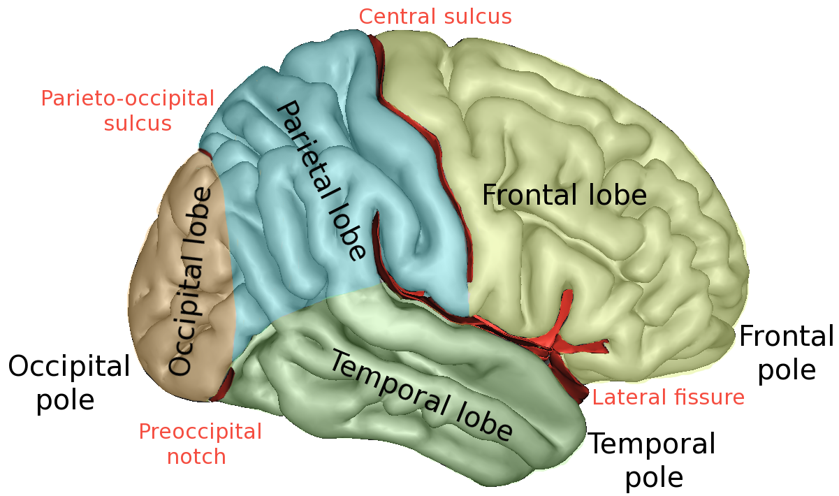
The lobes of the brain are the four major identifiable regions of the human cerebral cortex, and they comprise the surface of each hemisphere of the cerebrum. The two hemispheres are roughly symmetrical in structure, and are connected by the corpus callosum. Some sources include the insula and limbic lobe but the limbic lobe incorporates parts of the other lobes. The lobes are large areas that are anatomically distinguishable, and are also functionally distinct. Each lobe of the brain has numerous ridges, or gyri, and furrows, sulci that constitute further subzones of the cortex. The expression "lobes of the brain" usually refers only to those of the cerebrum, not to the distinct areas of the cerebellum.
The frontal lobe is located at the front of each cerebral hemisphere and positioned in front of the parietal lobe and above and in front of the temporal lobe. It is separated from the parietal lobe by a space between tissues called the central sulcus, and from the temporal lobe by a deep fold called the lateral sulcus, also called the Sylvian fissure. The precentral gyrus, which forms the posterior border of the frontal lobe, contains the primary motor cortex (area 4 under the Brodmann area architecture) which controls voluntary movements of specific body parts. The precentral region also contains the premotor cortex (Brodmann area 6).
The frontal lobe contains most of the dopamine-delicate neurons in the cerebral cortex. The dopamine system is associated with reward, attention, short-term memory tasks, planning, and motivation. Dopamine tends to limit and select sensory information arriving from the thalamus to the forebrain.[citation needed] A report from the National Institute of Mental Health says a gene variant that reduces dopamine activity in the prefrontal cortex is related to poorer performance and inefficient functioning of that brain region during working memory tasks, and to a slightly increased risk for schizophrenia.
The frontal lobe consists of the prefrontal cortex which is located in the most anterior (farthest away) section of the frontal lobe. It is critical for one's working memory and executive control which helps keep goals and complex tasks organized.
The divisions of the prefrontal cortex include orbital, medial, and lateral prefrontal cortex. Within the lateral prefrontal cortex there are two different divisions: the dorsolateral and ventrolateral prefrontal cortex. The dorsolateral prefrontal cortex is located on top of the ventrolateral prefrontal cortex and is mainly responsible for the executive control and manipulation of memories that are retrieved through episodic memory. The ventrolateral prefrontal cortex is important for the regulation of meaningful stimuli that a person experiences throughout their lifetime, such as images, letters, and names.
Damage to the prefrontal cortex can result in issues with one's long term and short-term memories, as well as create changes in people's behaviors and their abilities to plan and organize.
Damage can result from lesions or tumors that have been surgically removed, and traumatic brain injuries (TBI) experienced from a severe hit to the head causing damage to the brain from swelling. Most often a TBI is experienced within a person's childhood from playing competitive sports or an accident from normal play. Having a traumatic brain injury can increase your chances of developing neurological psychiatric problems and abusing substances, such as cannabis, is known to be a risk factor in developing symptoms associated with schizophrenia. A study found that schizophrenia symptoms (hearing voices, talking to people who were not there, etc.) worsened after the usage of cannabis, suggesting that a TBI from childhood can enhance a development of psychosis due to the changes seen in the white matter within the frontal-temporal areas.
The parietal lobe is positioned above the occipital lobe and behind the frontal lobe and central sulcus.
Hub AI
Lobes of the brain AI simulator
(@Lobes of the brain_simulator)
Lobes of the brain
The lobes of the brain are the four major identifiable regions of the human cerebral cortex, and they comprise the surface of each hemisphere of the cerebrum. The two hemispheres are roughly symmetrical in structure, and are connected by the corpus callosum. Some sources include the insula and limbic lobe but the limbic lobe incorporates parts of the other lobes. The lobes are large areas that are anatomically distinguishable, and are also functionally distinct. Each lobe of the brain has numerous ridges, or gyri, and furrows, sulci that constitute further subzones of the cortex. The expression "lobes of the brain" usually refers only to those of the cerebrum, not to the distinct areas of the cerebellum.
The frontal lobe is located at the front of each cerebral hemisphere and positioned in front of the parietal lobe and above and in front of the temporal lobe. It is separated from the parietal lobe by a space between tissues called the central sulcus, and from the temporal lobe by a deep fold called the lateral sulcus, also called the Sylvian fissure. The precentral gyrus, which forms the posterior border of the frontal lobe, contains the primary motor cortex (area 4 under the Brodmann area architecture) which controls voluntary movements of specific body parts. The precentral region also contains the premotor cortex (Brodmann area 6).
The frontal lobe contains most of the dopamine-delicate neurons in the cerebral cortex. The dopamine system is associated with reward, attention, short-term memory tasks, planning, and motivation. Dopamine tends to limit and select sensory information arriving from the thalamus to the forebrain.[citation needed] A report from the National Institute of Mental Health says a gene variant that reduces dopamine activity in the prefrontal cortex is related to poorer performance and inefficient functioning of that brain region during working memory tasks, and to a slightly increased risk for schizophrenia.
The frontal lobe consists of the prefrontal cortex which is located in the most anterior (farthest away) section of the frontal lobe. It is critical for one's working memory and executive control which helps keep goals and complex tasks organized.
The divisions of the prefrontal cortex include orbital, medial, and lateral prefrontal cortex. Within the lateral prefrontal cortex there are two different divisions: the dorsolateral and ventrolateral prefrontal cortex. The dorsolateral prefrontal cortex is located on top of the ventrolateral prefrontal cortex and is mainly responsible for the executive control and manipulation of memories that are retrieved through episodic memory. The ventrolateral prefrontal cortex is important for the regulation of meaningful stimuli that a person experiences throughout their lifetime, such as images, letters, and names.
Damage to the prefrontal cortex can result in issues with one's long term and short-term memories, as well as create changes in people's behaviors and their abilities to plan and organize.
Damage can result from lesions or tumors that have been surgically removed, and traumatic brain injuries (TBI) experienced from a severe hit to the head causing damage to the brain from swelling. Most often a TBI is experienced within a person's childhood from playing competitive sports or an accident from normal play. Having a traumatic brain injury can increase your chances of developing neurological psychiatric problems and abusing substances, such as cannabis, is known to be a risk factor in developing symptoms associated with schizophrenia. A study found that schizophrenia symptoms (hearing voices, talking to people who were not there, etc.) worsened after the usage of cannabis, suggesting that a TBI from childhood can enhance a development of psychosis due to the changes seen in the white matter within the frontal-temporal areas.
The parietal lobe is positioned above the occipital lobe and behind the frontal lobe and central sulcus.