
Recent from talks
Myxobolus cerebralis
Knowledge base stats:
Talk channels stats:
Members stats:
Myxobolus cerebralis
Myxobolus cerebralis is a myxosporean parasite of salmonids (salmon and trout species) that causes whirling disease in farmed salmon and trout and also in wild fish populations. It was first described in rainbow trout in Germany in 1893, but its range has spread and it has appeared in most of Europe (including Russia), the United States, South Africa, Canada and other countries from shipments of cultured and wild fish. In the 1980s, M. cerebralis was found to require a tubificid oligochaete (a kind of segmented worm) to complete its life cycle. The parasite infects its hosts with its cells after piercing them with polar filaments ejected from nematocyst-like capsules. This infects the cartilage and possibly the nervous tissue of salmonids, causing a potentially lethal infection in which the host develops a black tail, spinal deformities, and possibly more deformities in the anterior part of the fish.[citation needed]
Whirling disease affects juvenile fish (fingerlings and fry) and causes skeletal deformation and neurological damage. Fish "whirl" forward in an awkward, corkscrew-like pattern instead of swimming normally, find feeding difficult, and are more vulnerable to predation. The mortality rate is high for fingerlings, up to 90% of infected populations, and those that do survive are deformed by the parasites residing in their cartilage, bone, and neurological tissue. They act as a reservoir for the parasite, which is released into water following the fish's death. M. cerebralis is one of the most economically important myxozoans in fish, as well as one of the most pathogenic. It was the first myxosporean whose pathology and symptoms were described scientifically. The parasite is not transmissible to humans.
The taxonomy and naming of both M. cerebralis, and of myxozoans in general, have complicated histories. It was originally thought to infect fish brains (hence the specific epithet cerebralis) and nervous systems, though it soon was found to primarily infect cartilage, skeletal tissue, and nervous tissue. Attempts to change the name to Myxobolus chondrophagus, which would more accurately describe the organism, failed because of nomenclature rules[which?].[citation needed] Later, the organisms previously called Triactinomyxon dubium and T. gyrosalmo (class Actinosporea) were found to be, in fact, triactinomyxon stages of M. cerebralis, the life cycle of which was expanded to include the triactinomyxon stage. Similarly, other actinosporeans were folded into the life cycles of various myxosporeans.[citation needed]
M. cerebralis is one of the 1,350 known myxozoan parasites known to infect fish. Once thought to be a species of Protozoa, taxonomists noticed characteristics that more closely related M. cerebralis to the phylum Cnidaria. These features included cnidocysts, which are tentacles that are used to hold onto and prey upon the host. M. cerebralis has many diverse stages ranging from single cells to relatively large spores, not all of which have been studied in detail. This complex lifecycle involves two different hosts and numerous developmental stages. These stages happen through mitosis, endogeny, plasmotomy, or possibly meiosis. In the first part of its lifecycle, M. cerebralis is attached to its salmonid host externally. They then use their stinging tentacles to infect the host, causing the skeletal tissues and nervous system to become deformed.
Today, the myxozoans, previously thought to be multicellular protozoans, are considered animals by most scientists, though their status has not officially changed. Recent molecular studies suggest they are related to Bilateria or Cnidaria, with Cnidaria being closer morphologically because both groups have extrusive filaments. Bilateria were somewhat closer in some genetic studies, but those were found to have used samples that were contaminated by material from the host organism, and a 2015 study confirms they are cnidarians.[citation needed]
M. cerebralis has many diverse stages ranging from single cells to relatively large spores, not all of which have been studied in detail.
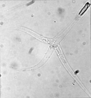
The stages that infect fish, called triactinomyxon spores, are made of a single style that is about 150 micrometers (μm) long and three processes or "tails", each about 200 micrometers long. These spores are typically oval shaped, and display asymmetrical symmetry. A sporoplasm packet at the end of the style contains 64 germ cells surrounded by a cellular envelope. There are also three polar capsules, each of which contains a coiled polar filament between 170 and 180 μm long, with about 5–6 coils in each filament. Polar filaments in both this stage and in the myxospore stage (see picture above) rapidly shoot into the body of the host, creating an opening through which the sporoplasm can enter. When it develops this polar filament it is able to attach to its host.
Upon contact with fish hosts and firing of the polar capsules, the sporoplasm contained within the central style of the triactinomyxon migrates into the epithelium or gut lining. Firstly, this sporoplasm undergoes mitosis to produce more amoeboid cells, which migrate into deeper tissue layers, to reach the cerebral cartilage.
Hub AI
Myxobolus cerebralis AI simulator
(@Myxobolus cerebralis_simulator)
Myxobolus cerebralis
Myxobolus cerebralis is a myxosporean parasite of salmonids (salmon and trout species) that causes whirling disease in farmed salmon and trout and also in wild fish populations. It was first described in rainbow trout in Germany in 1893, but its range has spread and it has appeared in most of Europe (including Russia), the United States, South Africa, Canada and other countries from shipments of cultured and wild fish. In the 1980s, M. cerebralis was found to require a tubificid oligochaete (a kind of segmented worm) to complete its life cycle. The parasite infects its hosts with its cells after piercing them with polar filaments ejected from nematocyst-like capsules. This infects the cartilage and possibly the nervous tissue of salmonids, causing a potentially lethal infection in which the host develops a black tail, spinal deformities, and possibly more deformities in the anterior part of the fish.[citation needed]
Whirling disease affects juvenile fish (fingerlings and fry) and causes skeletal deformation and neurological damage. Fish "whirl" forward in an awkward, corkscrew-like pattern instead of swimming normally, find feeding difficult, and are more vulnerable to predation. The mortality rate is high for fingerlings, up to 90% of infected populations, and those that do survive are deformed by the parasites residing in their cartilage, bone, and neurological tissue. They act as a reservoir for the parasite, which is released into water following the fish's death. M. cerebralis is one of the most economically important myxozoans in fish, as well as one of the most pathogenic. It was the first myxosporean whose pathology and symptoms were described scientifically. The parasite is not transmissible to humans.
The taxonomy and naming of both M. cerebralis, and of myxozoans in general, have complicated histories. It was originally thought to infect fish brains (hence the specific epithet cerebralis) and nervous systems, though it soon was found to primarily infect cartilage, skeletal tissue, and nervous tissue. Attempts to change the name to Myxobolus chondrophagus, which would more accurately describe the organism, failed because of nomenclature rules[which?].[citation needed] Later, the organisms previously called Triactinomyxon dubium and T. gyrosalmo (class Actinosporea) were found to be, in fact, triactinomyxon stages of M. cerebralis, the life cycle of which was expanded to include the triactinomyxon stage. Similarly, other actinosporeans were folded into the life cycles of various myxosporeans.[citation needed]
M. cerebralis is one of the 1,350 known myxozoan parasites known to infect fish. Once thought to be a species of Protozoa, taxonomists noticed characteristics that more closely related M. cerebralis to the phylum Cnidaria. These features included cnidocysts, which are tentacles that are used to hold onto and prey upon the host. M. cerebralis has many diverse stages ranging from single cells to relatively large spores, not all of which have been studied in detail. This complex lifecycle involves two different hosts and numerous developmental stages. These stages happen through mitosis, endogeny, plasmotomy, or possibly meiosis. In the first part of its lifecycle, M. cerebralis is attached to its salmonid host externally. They then use their stinging tentacles to infect the host, causing the skeletal tissues and nervous system to become deformed.
Today, the myxozoans, previously thought to be multicellular protozoans, are considered animals by most scientists, though their status has not officially changed. Recent molecular studies suggest they are related to Bilateria or Cnidaria, with Cnidaria being closer morphologically because both groups have extrusive filaments. Bilateria were somewhat closer in some genetic studies, but those were found to have used samples that were contaminated by material from the host organism, and a 2015 study confirms they are cnidarians.[citation needed]
M. cerebralis has many diverse stages ranging from single cells to relatively large spores, not all of which have been studied in detail.
The stages that infect fish, called triactinomyxon spores, are made of a single style that is about 150 micrometers (μm) long and three processes or "tails", each about 200 micrometers long. These spores are typically oval shaped, and display asymmetrical symmetry. A sporoplasm packet at the end of the style contains 64 germ cells surrounded by a cellular envelope. There are also three polar capsules, each of which contains a coiled polar filament between 170 and 180 μm long, with about 5–6 coils in each filament. Polar filaments in both this stage and in the myxospore stage (see picture above) rapidly shoot into the body of the host, creating an opening through which the sporoplasm can enter. When it develops this polar filament it is able to attach to its host.
Upon contact with fish hosts and firing of the polar capsules, the sporoplasm contained within the central style of the triactinomyxon migrates into the epithelium or gut lining. Firstly, this sporoplasm undergoes mitosis to produce more amoeboid cells, which migrate into deeper tissue layers, to reach the cerebral cartilage.
Recent media