Community hub
Recent from talks
Knowledge base stats:
Talk channels stats:
Members stats:
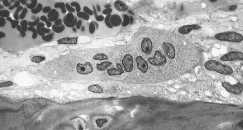
Osteoclast
An osteoclast (from Ancient Greek ὀστέον (osteon) 'bone' and κλαστός (clastos) 'broken') is a type of bone cell that removes bone tissue. This function is critical in the maintenance, repair, and remodeling of bones of the vertebral skeleton. The osteoclast disassembles and digests the composite of hydrated protein and mineral at a molecular level by secreting acid and a collagenase, a process known as bone resorption. This process also helps regulate the level of blood calcium.
Osteoclasts are found on those surfaces of bone that are undergoing resorption. On such surfaces, the osteoclasts are seen to be located in shallow depressions called resorption bays (Howship's lacunae). The resorption bays are created by the erosive action of osteoclasts on the underlying bone. The border of the lower part of an osteoclast exhibits finger-like processes due to the presence of deep infoldings of the cell membrane; this border is called ruffled border. The ruffled border lies in contact with the bone surface within a resorption bay. The periphery of the ruffled border is surrounded by a ring-like zone of cytoplasm, which is devoid of cell organelles but rich in actin filaments. This zone is called the clear zone or sealing zone. The actin filaments enable the cell membrane surrounding the sealing zone to be anchored firmly to the bony wall of Howship's lacunae. In this way, a closed subosteoclastic compartment is created between the ruffled border and the bone that is undergoing resorption. The osteoclasts secrete hydrogen ions, collagenase, cathepsin K and hydrolytic enzymes into this compartment. Resorption of bone matrix by the osteoclasts involves two steps: (1) dissolution of inorganic components (minerals), and (2) digestion of organic component of the bone matrix. The osteoclasts pump hydrogen ions into the subosteoclastic compartment and thus create an acidic microenvironment, which increases solubility of bone mineral, resulting in the release and re-entry of bone minerals into the cytoplasm of osteoclasts to be delivered to nearby capillaries. After the removal of minerals, collagenase and gelatinase are secreted into the subosteoclastic compartment. These enzymes digest and degrade collagen and other organic components of decalcified bone matrix. The degradation products are phagocytosed by osteoclasts at the ruffled border. Because of their phagocytic properties, osteoclasts are considered to be a component of the mononuclear phagocyte system (MPS). The activity of osteoclasts is controlled by hormones and cytokines. Calcitonin, a hormone of the thyroid gland, suppresses osteoclastic activity. Osteoclasts do not have receptors for parathyroid hormone (PTH). However, PTH stimulates osteoblasts to secrete a cytokine called osteoclast-stimulating factor, which is a potent stimulator of osteoclastic activity.
An odontoclast (/odon·to·clast/; o-don´to-klast) is an osteoclast associated with the absorption of the roots of deciduous teeth.
An osteoclast is a large multinucleated cell and human osteoclasts on bone typically have four nuclei and are 150–200 μm in diameter. When osteoclast-inducing cytokines are used to convert macrophages to osteoclasts, very large cells that may reach 100 μm in diameter occur. These may have dozens of nuclei, and typically express major osteoclast proteins but have significant differences from cells in living bone because of the not-natural substrate. The size of the multinucleated assembled osteoclast allows it to focus the ion transport, protein secretory and vesicular transport capabilities of many macrophages on a localized area of bone.
In bone, osteoclasts are found in pits in the bone surface which are called resorption bays, or Howship's lacunae. Osteoclasts are characterized by a cytoplasm with a homogeneous, "foamy" appearance. This appearance is due to a high concentration of vesicles and vacuoles. These vacuoles include lysosomes filled with acid phosphatase. This permits characterization of osteoclasts by their staining for high expression of tartrate resistant acid phosphatase (TRAP) and cathepsin K. Osteoclast rough endoplasmic reticulum is sparse, and the Golgi complex is extensive.
At a site of active bone resorption, the osteoclast forms a specialized cell membrane, the "ruffled border", that opposes the surface of the bone tissue. This extensively folded or ruffled border facilitates bone removal by dramatically increasing the cell surface for secretion and uptake of the resorption compartment contents and is a morphologic characteristic of an osteoclast that is actively resorbing bone.
Since their discovery in 1873 there has been considerable debate about their origin. Three theories were dominant: from 1949 to 1970 the connective tissue origin was popular, which stated that osteoclasts and osteoblasts are of the same lineage, and osteoblasts fuse together to form osteoclasts. After years of controversy it is now clear that these cells develop from the self fusion of macrophages. It was in the beginning of 1980 that the monocyte phagocytic system was recognized as precursor of osteoclasts. Osteoclast formation requires the presence of RANKL (receptor activator of nuclear factor κβ ligand) and M-CSF (Macrophage colony-stimulating factor). These membrane-bound proteins are produced by neighbouring stromal cells and osteoblasts, thus requiring direct contact between these cells and osteoclast precursors.
M-CSF acts through its receptor on the osteoclast, c-fms (colony-stimulating factor 1 receptor), a transmembrane tyrosine kinase-receptor, leading to secondary messenger activation of tyrosine kinase Src. Both of these molecules are necessary for osteoclastogenesis and are widely involved in the differentiation of monocyte/macrophage derived cells.
Hub AI
Osteoclast AI simulator
(@Osteoclast_simulator)
Osteoclast
An osteoclast (from Ancient Greek ὀστέον (osteon) 'bone' and κλαστός (clastos) 'broken') is a type of bone cell that removes bone tissue. This function is critical in the maintenance, repair, and remodeling of bones of the vertebral skeleton. The osteoclast disassembles and digests the composite of hydrated protein and mineral at a molecular level by secreting acid and a collagenase, a process known as bone resorption. This process also helps regulate the level of blood calcium.
Osteoclasts are found on those surfaces of bone that are undergoing resorption. On such surfaces, the osteoclasts are seen to be located in shallow depressions called resorption bays (Howship's lacunae). The resorption bays are created by the erosive action of osteoclasts on the underlying bone. The border of the lower part of an osteoclast exhibits finger-like processes due to the presence of deep infoldings of the cell membrane; this border is called ruffled border. The ruffled border lies in contact with the bone surface within a resorption bay. The periphery of the ruffled border is surrounded by a ring-like zone of cytoplasm, which is devoid of cell organelles but rich in actin filaments. This zone is called the clear zone or sealing zone. The actin filaments enable the cell membrane surrounding the sealing zone to be anchored firmly to the bony wall of Howship's lacunae. In this way, a closed subosteoclastic compartment is created between the ruffled border and the bone that is undergoing resorption. The osteoclasts secrete hydrogen ions, collagenase, cathepsin K and hydrolytic enzymes into this compartment. Resorption of bone matrix by the osteoclasts involves two steps: (1) dissolution of inorganic components (minerals), and (2) digestion of organic component of the bone matrix. The osteoclasts pump hydrogen ions into the subosteoclastic compartment and thus create an acidic microenvironment, which increases solubility of bone mineral, resulting in the release and re-entry of bone minerals into the cytoplasm of osteoclasts to be delivered to nearby capillaries. After the removal of minerals, collagenase and gelatinase are secreted into the subosteoclastic compartment. These enzymes digest and degrade collagen and other organic components of decalcified bone matrix. The degradation products are phagocytosed by osteoclasts at the ruffled border. Because of their phagocytic properties, osteoclasts are considered to be a component of the mononuclear phagocyte system (MPS). The activity of osteoclasts is controlled by hormones and cytokines. Calcitonin, a hormone of the thyroid gland, suppresses osteoclastic activity. Osteoclasts do not have receptors for parathyroid hormone (PTH). However, PTH stimulates osteoblasts to secrete a cytokine called osteoclast-stimulating factor, which is a potent stimulator of osteoclastic activity.
An odontoclast (/odon·to·clast/; o-don´to-klast) is an osteoclast associated with the absorption of the roots of deciduous teeth.
An osteoclast is a large multinucleated cell and human osteoclasts on bone typically have four nuclei and are 150–200 μm in diameter. When osteoclast-inducing cytokines are used to convert macrophages to osteoclasts, very large cells that may reach 100 μm in diameter occur. These may have dozens of nuclei, and typically express major osteoclast proteins but have significant differences from cells in living bone because of the not-natural substrate. The size of the multinucleated assembled osteoclast allows it to focus the ion transport, protein secretory and vesicular transport capabilities of many macrophages on a localized area of bone.
In bone, osteoclasts are found in pits in the bone surface which are called resorption bays, or Howship's lacunae. Osteoclasts are characterized by a cytoplasm with a homogeneous, "foamy" appearance. This appearance is due to a high concentration of vesicles and vacuoles. These vacuoles include lysosomes filled with acid phosphatase. This permits characterization of osteoclasts by their staining for high expression of tartrate resistant acid phosphatase (TRAP) and cathepsin K. Osteoclast rough endoplasmic reticulum is sparse, and the Golgi complex is extensive.
At a site of active bone resorption, the osteoclast forms a specialized cell membrane, the "ruffled border", that opposes the surface of the bone tissue. This extensively folded or ruffled border facilitates bone removal by dramatically increasing the cell surface for secretion and uptake of the resorption compartment contents and is a morphologic characteristic of an osteoclast that is actively resorbing bone.
Since their discovery in 1873 there has been considerable debate about their origin. Three theories were dominant: from 1949 to 1970 the connective tissue origin was popular, which stated that osteoclasts and osteoblasts are of the same lineage, and osteoblasts fuse together to form osteoclasts. After years of controversy it is now clear that these cells develop from the self fusion of macrophages. It was in the beginning of 1980 that the monocyte phagocytic system was recognized as precursor of osteoclasts. Osteoclast formation requires the presence of RANKL (receptor activator of nuclear factor κβ ligand) and M-CSF (Macrophage colony-stimulating factor). These membrane-bound proteins are produced by neighbouring stromal cells and osteoblasts, thus requiring direct contact between these cells and osteoclast precursors.
M-CSF acts through its receptor on the osteoclast, c-fms (colony-stimulating factor 1 receptor), a transmembrane tyrosine kinase-receptor, leading to secondary messenger activation of tyrosine kinase Src. Both of these molecules are necessary for osteoclastogenesis and are widely involved in the differentiation of monocyte/macrophage derived cells.