Hubbry Logo
search
logo
2221314

Reporter gene

logo
Community Hub0 Subscribers
Read side by side
from Wikipedia
A diagram of a how a reporter gene is used to study a regulatory sequence.

Reporter genes are molecular tools widely used in molecular biology, genetics, and biotechnology to study gene function, expression patterns, and regulatory mechanisms. These genes encode proteins that produce easily detectable signals, such as fluorescence, luminescence, or enzymatic activity, allowing researchers to monitor cellular processes in real-time. Reporter genes are often fused to regulatory sequences of genes of interest, enabling scientists to analyze promoter activity, transcriptional regulation, and signal transduction pathways. Common reporter gene systems include green fluorescent protein (GFP), β-galactosidase (lacZ), luciferase, and chloramphenicol acetyltransferase (CAT), each offering distinct advantages depending on the experimental application.[1] Their versatility makes reporter genes invaluable in fields such as drug discovery, gene therapy, and synthetic biology.[1]

Common reporters

[edit]

To introduce a reporter gene into an organism, scientists place the reporter gene and the gene of interest in the same DNA construct to be inserted into the cell or organism. For bacteria or prokaryotic cells in culture, this is usually in the form of a circular DNA molecule called a plasmid. For viruses, this is known as a viral vector. It is important to use a reporter gene that is not natively expressed in the cell or organism under study, since the expression of the reporter is being used as a marker for successful uptake of the gene of interest.[1]

Commonly used reporter genes that induce visually identifiable characteristics usually involve fluorescent and luminescent proteins. Examples include the gene that encodes jellyfish green fluorescent protein (GFP), which causes cells that express it to glow green under blue or ultraviolet light, the enzyme luciferase, which catalyzes a reaction with luciferin to produce light,[2] and the red fluorescent protein from the gene dsRed.[3][4][5][6][7] The GUS gene has been commonly used in plants, but luciferase and GFP are becoming more common.[8][9]

A common reporter in bacteria is the E. coli lacZ gene, which encodes the protein beta-galactosidase.[1] This enzyme causes bacteria expressing the gene to appear blue when grown on a medium that contains the substrate analog X-gal. An example of a selectable marker, which is also a reporter in bacteria, is the chloramphenicol acetyltransferase (CAT) gene, which confers resistance to the antibiotic chloramphenicol.[10]

History

[edit]

Reporters by discovery year

[edit]
Year Gene name Gene product Significance Assay Ref.
1961 lacZ β-galactosidase François Jacob and Jacques Monod were awarded a Nobel Prize in 1965 for their work. Enzyme assay, Histochemical (X-gal) [11][12]
1962 rfp Red fluorescent protein Microscopical, Spectrophotometry [13]
1979 cat Chloramphenicol acetyltransferase Used for measuring gene expression in eukaryotic cells. Chloramphenicol acetylation [10]
1985 luc Luciferase enzyme Provided a sensitive bioluminescent reporter for gene expression studies. Bioluminescence [4]
1987 gus B-Glucuronidase Became a widely used reporter gene in plant biology due to its high stability and easy detection in histochemical assays. Enabled visualization of gene expression patterns in plant tissues. Histochemical, Fluorometric [14]
1994 gfp Green fluorescent protein Enabled real-time visualization of gene expression in live cells. Fluorescence microscopy [3]

Transformation and transfection assays

[edit]

Many methods of transfection and transformation – two ways of expressing a foreign or modified gene in an organism – are effective in only a small percentage of a population subjected to the techniques. Thus, a method for identifying those few successful gene uptake events is necessary. Reporter genes used in this way are normally expressed under their own promoter (DNA regions that initiates gene transcription) independent from that of the introduced gene of interest; the reporter gene can be expressed constitutively ("always on") or inducibly. This independence is advantageous when the gene of interest is expressed under specific or hard-to-access conditions.[1]

Reporter genes employ diverse mechanisms to visualize or quantify gene activity:

  • Enzymatic reporters (e.g., LacZ) encode enzymes that catalyze reactions yielding a visible product. For example, β-galactosidase (encoded by LacZ) cleaves X-gal to produce a blue color, allowing easy identification of successful gene disruption (white colonies) versus intact genes (blue colonies).[7]
  • Bioluminescent reporters (e.g., luciferase) produce light via chemical reactions, enabling live-cell imaging and promoter studies without external light sources.[8]
  • Colorimetric reporters (e.g., CAT) generate detectable color changes when enzymes react with substrates, measurable via spectrophotometry or TLC.[9]
  • Selectable markers (e.g., Neo) confer antibiotic resistance (e.g., to G418), ensuring only transformed cells survive in selective media.[11][15]

In the case of selectable-marker reporters such as CAT, the transfected population can be grown on a chloramphenicol-containing substrate. Only cells with the CAT gene survive, confirming successful transformation.[10]

Gene expression assays

[edit]

Reporter genes can be used to assay for the expression of a gene of interest that is normally difficult to quantitatively assay.[1] Reporter genes can produce a protein that has little obvious or immediate effect on the cell culture or organism. They are ideally not present in the native genome to be able to isolate reporter gene expression as a result of the gene of interest's expression.[1][16]

To activate reporter genes, they can be expressed constitutively, where they are directly attached to the gene of interest to create a gene fusion.[17] This method is an example of using cis-acting elements where the two genes are under the same promoter elements and are transcribed into a single messenger RNA molecule. The mRNA is then translated into protein. It is important that both proteins be able to properly fold into their active conformations and interact with their substrates despite being fused. In building the DNA construct, a segment of DNA coding for a flexible polypeptide linker region is usually included so that the reporter and the gene product will only minimally interfere with one another.[18][19] Reporter genes can also be expressed by induction during growth. In these cases, trans-acting elements, such as transcription factors are used to express the reporter gene.[20][21]

Reporter gene assay have been increasingly used in high throughput screening (HTS) to identify small molecule inhibitors and activators of protein targets and pathways for drug discovery and chemical biology. Because the reporter enzymes themselves (e.g. firefly luciferase) can be direct targets of small molecules and confound the interpretation of HTS data, novel coincidence reporter designs incorporating artifact suppression have been developed.[22][23]
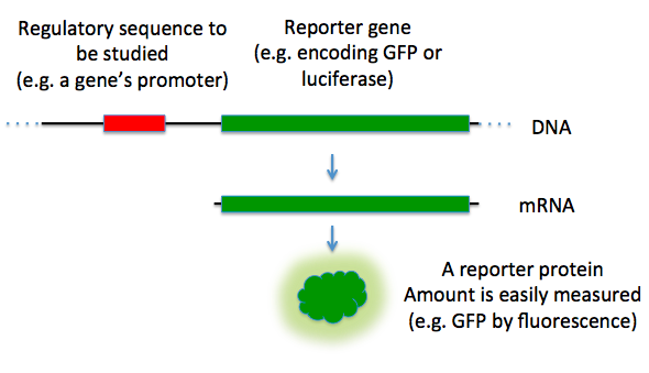
Promoter assays

[edit]

Reporter genes can be used to assay for the activity of a particular promoter in a cell or organism.[24] In this case there is no separate "gene of interest"; the reporter gene is simply placed under the control of the target promoter and the reporter gene product's activity is quantitatively measured. The results are normally reported relative to the activity under a "consensus" promoter known to induce strong gene expression.[25]

Limitations and advancements

[edit]

While reporter gene technology has become an essential component of molecular biology, its application still has limitations. One primary concern is the influence of genomic context on reporter expression. Reporter genes integrated into the genome can be subject to position-effect variegation, where the surrounding chromatin structure influences transcriptional activity. This can lead to inconsistent expression and complicate the interpretation of results, especially in stable cell lines and transgenic organisms.[26] Additionally, reporter expression may not always accurately reflect the activity of the endogenous gene of interest due to differences in post-transcriptional regulation, mRNA stability, or translational efficiency.[27]

Another common limitation is the cellular burden that reporter expression may impose. High levels of reporter protein production, such as fluorescent proteins or luciferases, can divert cellular resources, potentially impacting normal metabolism or physiology. This is particularly problematic in sensitive systems like stem cells or primary cell cultures, where even subtle changes in metabolism can influence cell behavior.[28] Additionally, some reporter systems, like luciferase assays, require the addition of exogenous substrates (e.g., luciferin), adds complexity and may reduce reproducibility, particularly in live animal models where substrate availability can vary.[28]

To address these challenges, several innovations have improved the reliability and flexibility of reporter gene technologies. One advancement involves the use of the 2A peptide, which allows the co-expression of multiple proteins from a single transcript without requiring a direct fusion. This approach enables the simultaneous expression of a gene of interest and a reporter while preserving the function of both.[29] Additionally, split-reporter systems, which produce a functional signal only when two proteins of interest interact, have become widely used in studies of protein–protein interactions due to their low background activity and high specificity.[30]

Applications

[edit]

Medical

[edit]

Tracking expression has allowed for multiple investigations into the progression of diseased cells.[31] Reporter genes have shown to provide critical insight into genes upregulated in cancer regulatory pathways as well as the identification into oncogenes and tumor suppressor genes. These have been used for further research into the development of therapeutics to stop further disease progression and metastasis.[31] Gene therapy has also been tracked through the use of reporter genes. This allows for the monitoring of gene therapy vectors to see if they are achieving intended results as well as to monitor patient safety for short and long term periods.[32] Therapeutics developed have benefited from the use of reporter genres such as a dual-reporter system developed for CRISPR/Cas9 models to monitor progression and success and benefits of being gene editing tools.[33]

Research

[edit]

The most commonly used application used for reporter genes has been for the identification of cis and trans acting elements. Through fusion to the promoter region of possible trans-cis acting elements, the change in fluorescence is measured and allows for tracking into transcriptional activity.[34] This provides useful information into understanding the pathways these elements are involved in and its regulatory uses for cell development and growth.[34] Immune responses are also a commonly used application of reporter genes and have benefited greatly through their use. They have allowed for further understanding in cell proliferation and differentiation into B-cells and T-cells during immune responses and have contributed to understanding activation through tracking cytokine signaling pathways.[35]

The development of reporter cell lines have also emerged with the discovery and use of reporter genes.[34] The cell lines are labelled with reporter genes to allow for fluorescent detection to help with identification into proteins used in cellular pathways and identification into protein localization.[30] This has allowed for a simple way to study protein progression that doesn't permit further experimentation for introduction and fusion of a reporter gene as the reporter gene is already present in the cell line.

A more complex use of reporter genes on a large scale is in two-hybrid screening, which aims to identify proteins that natively interact with one another in vivo. The yeast two-hybrid (Y2H) system, developed in the late 1980s and early 1990s, was an immense advancement in the use of reporter genes to study protein-protein interactions in vivo.[36]  This technique takes advantage of transcription factors' modular nature, which often consists of separate DNA-binding and activation domains. By genetically fusing two proteins of interest to these domains, researchers can detect physical interactions between them through the activation of a downstream reporter gene. Due to the simple genetic nature of the Y2H system, this technique significantly increased the accessibility of protein-protein interaction studies without the requirement of protein purification or complex biochemical assays. Experimental Y2H data have played a pivotal role in building large-scale synthetic human interactomes and in dissecting mechanisms in human disease.[37][38]

However, there are still some limitations. Y2H sometimes detects interactions that don't occur naturally or fails to detect weak or transient interactions. Due to its artificial setting, these failures could result from the absence of key factors such as post-translational modifications or compartmentalization. For example, Y2H has been shown to generate false positives due to indirect interactions mediated by host proteins, as demonstrated in studies of cyanobacterial PipX interactions where the self-interaction of PipX was found to be dependent on PII homologues from the host organism rather than a direct interaction.[39]

Massively parallel reporter assays (MPRAs) and machine learning are newer ways to study gene regulation with reporter genes. One major use is in synthetic biology and gene therapy, where researchers can design better regulatory elements to control gene expression.[40] For example, deep learning models trained on MPRA data have been used to optimize 5' untranslated regions (UTRs) for mRNA translation, enabling tailored designs that enhance gene-editing efficiency in the therapeutic context. This could make mRNA-based treatments more effective, as MPRAs also help identify how genetic variants affect gene expression, which is used in precision medicine and developing personalized treatments.[40]

Machine learning models trained on MPRA data can predict how different sequences impact gene activity, making it easier to design reporter genes that respond in specific ways. Combining MPRAs with next-gen sequencing also makes reporter gene experiments faster and more scalable. These advances could even improve mRNA-based vaccines and therapeutics by optimizing untranslated regions (UTRs) to boost stability and translation. For instance, modular MPRAs have uncovered context-specific regulatory sequences linked to type 2 diabetes, revealing enhancer-promoter interactions dependent on cell-specific transcription factors like HNF1.[41] Similarly, MPRA screens of cardiac enhancer variants have pinpointed functional noncoding sequences influencing QT interval variability, directly linking genetic variation to disease-associated gene dysregulation.[42]

See also

[edit]

References

[edit]
[edit]
Revisions and contributorsEdit on WikipediaRead on Wikipedia
from Grokipedia
A reporter gene is a gene that encodes a protein whose expression can be readily and specifically detected, often through enzymatic activity, fluorescence, or luminescence, allowing scientists to monitor the activity of promoters, enhancers, or other regulatory elements in molecular biology experiments.[1] These genes are typically fused to sequences of interest or used as selectable markers to track gene expression patterns, cellular transformations, and biological processes in living systems.[2] By producing distinguishable signals from endogenous proteins, reporter genes provide a sensitive and quantifiable readout of transcriptional regulation, making them indispensable tools in biotechnology and genetic research.[3] The concept of reporter genes emerged in the early 1980s with the adoption of the bacterial chloramphenicol acetyltransferase (CAT) gene to measure promoter activity in mammalian cells, marking a shift from indirect methods like Northern blotting to direct, enzyme-based assays.[4] This was followed by the widespread use of the Escherichia coli lacZ gene, encoding β-galactosidase, which produces a blue color upon substrate hydrolysis and has been a staple for visualizing gene expression in tissues and cells due to its stability and ease of detection.[1] The discovery and engineering of green fluorescent protein (GFP) from the jellyfish Aequorea victoria in the 1960s, with practical applications expanding in the 1990s, revolutionized non-invasive imaging by enabling real-time visualization of gene expression without substrates.[4] Other prominent examples include luciferase from fireflies or sea pansy, which catalyzes a bioluminescent reaction for high-sensitivity, low-background detection in live animals and high-throughput screens.[3] Reporter genes find broad applications in studying gene regulation, such as in promoter-reporter constructs where expression levels correlate with transcriptional activation or repression in response to stimuli like hormones or drugs.[3] In molecular imaging, they facilitate non-invasive tracking of cell migration, tumor growth, and gene therapy efficacy using techniques like fluorescence microscopy, bioluminescence imaging, or positron emission tomography (PET) when paired with complementary probes.[4] For instance, GFP variants and luciferase have been integrated into transgenic mouse models to trace developmental lineages or immune responses, while β-galactosidase remains favored for histological analysis in fixed tissues.[1] These tools also support drug discovery by evaluating bioactivity in reporter gene assays, such as measuring pathway activation in cell lines for biologics like monoclonal antibodies.[3] Despite their utility, challenges include potential immunogenicity of foreign proteins and the need for optimized delivery vectors to ensure accurate, cell-specific expression.[4]

Fundamentals

Definition and Purpose

A reporter gene is a non-endogenous gene that encodes a protein product easily distinguishable from endogenous proteins, allowing researchers to monitor the activity of associated regulatory elements such as promoters, enhancers, or other sequences controlling transcription, translation, or protein localization.[1] These genes are typically introduced into cells via genetic constructs where the reporter coding sequence is fused to the regulatory region of interest, providing a proxy for the behavior of the target gene without altering its native sequence.[5] The main purpose of reporter genes is to quantify gene expression levels and assess promoter activity indirectly, circumventing the difficulties of directly measuring many native gene products that may be present in low amounts or lack sensitive detection methods.[6] They also facilitate the study of cellular processes, including signal transduction pathways, and the validation of genetic engineering techniques by offering a measurable readout of experimental success.[7] This indirect approach enables non-invasive monitoring in living systems, supporting applications in molecular biology and biotechnology.[2] Reporter genes arose from the need to develop convenient, sensitive assays for gene expression in mammalian cells, where traditional methods often required cell lysis or were insufficiently quantitative, as demonstrated by the initial adaptation of bacterial chloramphenicol acetyltransferase for this role.[8] By providing a detectable signal tied to regulatory elements, they support high-throughput screening of gene function both in vitro and in vivo, accelerating research into gene regulation and cellular dynamics.[1] Common examples include genes encoding green fluorescent protein or luciferase, which yield visual or enzymatic outputs for tracking expression.[1]

Mechanisms of Detection

Reporter genes are detected through the biochemical activity of their encoded proteins, which convert substrates into quantifiable signals such as color, light, or fluorescence. Enzymatic reporters, such as β-galactosidase, catalyze the hydrolysis of chromogenic substrates like X-gal (5-bromo-4-chloro-3-indolyl-β-D-galactopyranoside), releasing an indolyl group that spontaneously dimerizes to form a blue insoluble product, allowing visual or spectrophotometric detection at approximately 620 nm.[9] Fluorescent reporters, like green fluorescent protein (GFP), generate signals via intrinsic fluorophores that absorb light at an excitation wavelength of 488 nm and emit green light at 509 nm, enabling non-invasive imaging without exogenous substrates.[10] Bioluminescent reporters, such as firefly luciferase, produce light through an ATP-dependent oxidation reaction where D-luciferin is converted to oxyluciferin, releasing photons in the process:
D-luciferin+O2+ATPfirefly luciferaseoxyluciferin+CO2+AMP+PPi+light (560 nm maximum) \text{D-luciferin} + \text{O}_2 + \text{ATP} \xrightarrow{\text{firefly luciferase}} \text{oxyluciferin} + \text{CO}_2 + \text{AMP} + \text{PP}_\text{i} + \text{light (560 nm maximum)}
This chemiluminescent reaction requires cofactors like Mg²⁺ and oxygen, yielding high sensitivity due to low background noise.[11] Signal quantification relies on specialized instrumentation tailored to the reporter type. For bioluminescent signals from luciferase, plate luminometers or imaging systems measure photon output in relative light units (RLU), capturing the flash or glow kinetics of the reaction.[12] Fluorescent signals from GFP are quantified using fluorescence microscopy for spatial resolution or flow cytometry/spectrofluorometry for population-level analysis, where intensity is proportional to protein concentration after correcting for excitation light scatter.[13] Enzymatic reporters like β-galactosidase are assessed via colorimetric assays, such as monitoring the absorbance of the blue product from X-gal hydrolysis or using chemiluminescent substrates like Galacton for enhanced sensitivity in microplate readers.[14] Detection sensitivity is influenced by several biophysical factors, including the reporter's quantum yield, substrate availability, and environmental conditions. Bioluminescent systems offer detection limits as low as 10⁻¹⁸ moles of enzyme due to their high signal-to-noise ratios (often >1000:1), but require cell-permeant substrates and can be quenched by tissue autofluorescence in vivo.[15] Fluorescent reporters provide real-time monitoring with signal-to-noise ratios around 10-100, limited by photobleaching and autofluorescence, while enzymatic assays achieve sensitivities of 10⁻¹⁵ to 10⁻¹⁶ moles but necessitate cell lysis and substrate addition, introducing variability from reaction kinetics.[9] Substrate requirements, such as ATP for luciferase or β-mercaptoethanol to stabilize β-galactosidase, further modulate detection thresholds.[16] To account for experimental variability, reporter signals are often normalized using co-transfected control reporters, such as Renilla luciferase, which provide an internal reference for transfection efficiency and cell viability. The normalized value, expressed as relative light units (RLU), is calculated as:
RLU=sample signalbackground signalcontrol reporter signal \text{RLU} = \frac{\text{sample signal} - \text{background signal}}{\text{control reporter signal}}
This approach mitigates differences in DNA uptake or cell number, ensuring reproducible quantification across replicates.[17]

Types of Reporter Genes

Enzymatic Reporters

Enzymatic reporters are genes that encode enzymes whose catalytic activity can be measured through the conversion of specific substrates into detectable products, such as colored, fluorescent, or radioactive compounds, allowing indirect quantification of gene expression levels. These reporters typically require the addition of exogenous substrates and often cell lysis for assaying, making them suitable for in vitro and ex vivo analyses where high sensitivity is needed to detect low levels of expression. Unlike non-enzymatic reporters, enzymatic ones amplify signals through catalytic turnover, providing robust detection in prokaryotic and eukaryotic systems, though they may introduce background noise from endogenous activities in certain organisms.[4] One of the earliest and most widely adopted enzymatic reporters is β-galactosidase, encoded by the lacZ gene from Escherichia coli. This enzyme hydrolyzes substrates like X-gal (5-bromo-4-chloro-3-indolyl-β-D-galactopyranoside) to produce a blue precipitate, enabling histochemical visualization, or chromogenic/fluorogenic substrates for quantitative assays via spectrophotometry or fluorometry. Introduced in the early 1980s through gene fusion techniques, lacZ became the first broadly used reporter for studying promoter activity and gene regulation in bacteria and yeast, with applications extending to mammalian cells despite challenges from endogenous β-galactosidase activity.[4] Another classic example is chloramphenicol acetyltransferase (CAT), encoded by the bacterial cat gene, which acetylates chloramphenicol using radiolabeled acetyl-CoA, detected via thin-layer chromatography and scintillation counting. Popular in the 1980s and pre-1990s for mammalian gene transfer studies due to its absence of endogenous counterparts in eukaryotes, CAT assays provided quantitative measures of transcriptional activity but were largely supplanted by safer alternatives owing to radioactivity requirements.[4] β-Glucuronidase (GUS), from the uidA gene of E. coli, cleaves substrates such as MUG (4-methylumbelliferyl-β-D-glucuronide) to yield the fluorescent product 4-methylumbelliferone or X-gluc for blue histochemical staining. Developed in the late 1980s, GUS excels in plant biology because of negligible endogenous activity in higher plants, facilitating precise localization of gene expression in tissues without interference, and its assays support both qualitative and quantitative readouts.[4] Luciferase reporters, such as firefly (Photinus pyralis luc gene) or Renilla (Renilla reniformis), catalyze the oxidation of luciferin or coelenterazine in an ATP-dependent manner to produce light, measurable by luminometry or bioluminescence imaging. First adapted as reporters in the late 1980s, these offer exceptional sensitivity for low-expression scenarios, with firefly luciferase enabling non-invasive whole-animal imaging after substrate injection, though Renilla variants provide dual-reporter normalization.[4] These enzymatic reporters share high sensitivity, often detecting expression at levels as low as a few molecules per cell due to catalytic amplification, making them ideal for quantitative analysis via spectrophotometry, fluorometry, or scintillation in low-expression contexts. They are particularly stable and non-toxic in prokaryotes, where bacterial origins minimize immunogenicity, but may exhibit potential toxicity or instability in eukaryotes from high expression or substrate metabolism. Advantages include cost-effective substrates for endpoint assays and versatility across organisms, yet disadvantages encompass the need for cell lysis in many protocols, which precludes real-time in vivo monitoring, and substrate costs that can limit scalability; additionally, endogenous enzyme activities (e.g., β-gal in mammals) necessitate controls for accuracy.[4]

Fluorescent and Bioluminescent Reporters

Fluorescent reporters, such as the green fluorescent protein (GFP) derived from the jellyfish Aequorea victoria, produce light through intrinsic fluorescence without requiring external substrates beyond excitation light.[18] GFP's chromophore forms via autocatalytic cyclization and oxidation of three internal amino acids (Ser-Tyr-Gly), enabling green emission at approximately 509 nm upon blue light excitation.[19] This property allows for non-destructive, real-time visualization of gene expression and protein localization in living cells and organisms.[20] Variants of GFP, including yellow fluorescent protein (YFP) and red fluorescent protein (RFP) such as DsRed from the coral Discosoma sp., expand the spectral range for multi-color imaging applications. YFP, engineered by introducing mutations like T203Y to shift emission to 527 nm, pairs effectively with cyan variants for fluorescence resonance energy transfer (FRET) studies of protein interactions.[21] RFP enables red-shifted emission around 583 nm, facilitating simultaneous tracking of multiple proteins without spectral overlap. In the 1990s, codon optimization of GFP for mammalian expression—replacing jellyfish codons with human-preferred synonyms—dramatically increased fluorescence yield in mammalian cells, making it a staple for live-cell imaging. Bioluminescent reporters, like Gaussia luciferase from the marine copepod Gaussia princeps, generate light through enzymatic oxidation of the substrate coelenterazine, producing blue emission at 480 nm without external excitation.[22] As a secreted protein, Gaussia luciferase allows non-invasive monitoring of gene expression via extracellular assays, ideal for in vivo studies in animals.[22] Both fluorescent and bioluminescent reporters support real-time, non-destructive tracking in intact cells and tissues, contrasting with substrate-dependent enzymatic methods by minimizing cell perturbation.[23] Key advantages of these reporters include precise visual localization of molecular events without cell lysis and compatibility with high-throughput live imaging.[24] However, fluorescent proteins suffer from autofluorescence interference in biological samples and photobleaching under prolonged excitation, limiting long-term observations.[25] Bioluminescent signals, while substrate-dependent, offer superior signal-to-noise ratios due to negligible background autofluorescence.[23] Recent advancements, such as near-infrared (NIR) fluorescent reporters developed in 2025, address tissue penetration limitations by shifting emission to 650–900 nm for deeper in vivo imaging with reduced scattering.[26] These chemogenetic NIR reporters, like nirFAST, enable tunable, high-contrast multiplexing with visible fluorophores, enhancing applications in whole-organism studies.[26]

Historical Development

Early Discoveries

The foundational work on reporter genes emerged from microbial genetics in the 1970s, where researchers sought safer and more precise methods to study gene regulation in operons, particularly the lac operon of Escherichia coli. Early techniques relied on radioisotopic labeling for measuring transcription, such as incorporating radiolabeled nucleotides into RNA, but these posed handling hazards and required destructive assays. To address this, enzymatic reporters like β-galactosidase (encoded by lacZ) were developed as non-radioactive alternatives, allowing quantification of promoter activity through colorimetric or fluorometric substrates.[4] A pivotal early application occurred in 1972, when lacZ assays were first used in bacteria to map promoter regions by fusing the gene to suspected regulatory sequences and measuring β-galactosidase activity, demonstrating that reporter expression quantitatively reflected target promoter strength. This proof-of-concept established that reporter activity could mirror the regulation of endogenous genes, such as inducible operons under varying nutrient conditions. By 1980, Casadaban and Cohen advanced the technique with in vitro lacZ fusion methods, enabling the cloning of exogenous promoters upstream of a functional lacZ fragment; these fusions were initially applied in bacteria but laid the groundwork for eukaryotic adaptations by facilitating precise transcriptional analysis without disrupting native gene function.[27] The chloramphenicol acetyltransferase (cat) gene, derived from bacterial antibiotic resistance plasmids, emerged as another enzymatic reporter in the early 1980s, with Gorman et al. demonstrating in 1982 its utility for assaying promoter activity in eukaryotic cells via thin-layer chromatography of acetylated products.[28] Initial challenges in eukaryotic expression, such as low lacZ translation due to codon bias and inefficient bacterial promoters, were overcome by pairing reporters with strong viral promoters like the Rous sarcoma virus long terminal repeat, enabling robust detection in mammalian systems. Additionally, early lacZ fusions in yeast during the 1980s, such as those transferring URA3-lacZ constructs from E. coli to Saccharomyces cerevisiae, served as precursors to interaction studies by validating heterologous expression and regulation in eukaryotes. These innovations by 1985, including the introduction of firefly luciferase by de Wet et al. for ultrasensitive bioluminescent detection in bacteria, solidified enzymatic reporters as versatile tools for dissecting gene control across organisms.[29]

Key Milestones and Evolutions

The cloning and expression of the green fluorescent protein (GFP) gene from the jellyfish Aequorea victoria in 1994 by Martin Chalfie, building on Douglas Prasher's 1992 cDNA isolation, marked a pivotal advancement in live-cell imaging, allowing non-invasive visualization of gene expression and protein localization in real time without exogenous substrates.[30] This eukaryotic adaptation shifted reporter systems toward dynamic, in vivo monitoring, contrasting with earlier prokaryotic enzymatic reporters. In the 1990s, the β-glucuronidase (GUS) reporter gained prominence in plant biology, facilitating histochemical detection of promoter activity in transgenic tissues and enabling large-scale screening of gene expression patterns during development and stress responses. The 2000s saw the rise of multimodal reporters, such as GFP-luciferase fusions, which combined fluorescence for cellular resolution with bioluminescence for whole-organism imaging, enhancing sensitivity in preclinical studies of gene therapy and tumor progression.[31] This era's innovations culminated in the 2008 Nobel Prize in Chemistry awarded to Osamu Shimomura, Martin Chalfie, and Roger Y. Tsien for the discovery and development of GFP, underscoring its transformative impact on biological imaging. Concurrently, secreted reporters like Gaussia luciferase emerged around 2009, enabling non-invasive monitoring through blood or urine sampling, which improved longitudinal tracking in animal models without tissue disruption.[12] In the 2020s, CRISPR-integrated reporters have facilitated precise tracking of genome editing outcomes, with systems incorporating fluorescent or luminescent markers at edit sites to quantify efficiency and off-target effects in real time.[32] These developments reflect a broader shift toward non-toxic, real-time reporters optimized for live imaging, including integrations with optogenetics for light-controlled gene expression and visualization in neural circuits.[33] By 2025, AI-driven design of spectral variants for fluorescent proteins has advanced multiplexed imaging, using neural networks to predict and engineer brighter, more orthogonal emitters for simultaneous tracking of multiple pathways in complex tissues.[34] That same year, secretory horseradish peroxidase (sHRP) was introduced as a cost-effective, non-lytic alternative to luciferase, offering stable, substrate-abundant detection for high-throughput promoter assays.[35] Throughout these evolutions, iterative engineering has focused on enhancing reporter stability, brightness, and spectral orthogonality, enabling their use in intricate biological systems like organoids and multicellular models while minimizing interference.[36]

Experimental Methods

Delivery Techniques

Delivery techniques for reporter genes involve methods to introduce reporter constructs into prokaryotic or eukaryotic cells, enabling subsequent monitoring of gene expression. In prokaryotes, such as bacteria, transformation is the primary approach, where exogenous DNA containing the reporter gene is taken up by competent cells. Common methods include electroporation, which uses electric pulses to create transient pores in the cell membrane for DNA entry; heat shock, involving a brief temperature elevation to facilitate DNA uptake; and conjugation, a natural process mediated by plasmids that transfer DNA between bacterial cells.[37][38] Stable integration of reporter genes in prokaryotes often occurs via plasmids, which replicate autonomously, or bacteriophages, which can integrate into the host genome as lysogens.[39] In eukaryotes, transfection methods are employed to deliver reporter constructs, distinguishing between transient expression, where the DNA remains episomal and is lost over cell divisions, and stable expression, where integration into the genome ensures long-term propagation. Chemical methods include lipofection, using cationic lipids to form complexes with DNA that fuse with the cell membrane, and calcium phosphate precipitation, which generates DNA-calcium phosphate co-precipitates for endocytosis. Physical techniques, such as electroporation, apply similar electric fields as in prokaryotes but optimized for eukaryotic cells. Biological approaches utilize viral vectors, like lentiviruses, which integrate reporter genes into the host genome for stable lines in dividing or non-dividing cells. As of 2025, CRISPR/Cas9 gene editing facilitates precise, site-specific integration of reporter genes into stable cell lines, enhancing reproducibility. Tools like STITCHR enable targeted insertion of reporter constructs for therapeutic applications.[40][41][42][43] To normalize transfection efficiency and select successfully transformed cells, selectable markers—such as antibiotic resistance genes—are co-delivered with reporter constructs, allowing growth only in cells that have incorporated the DNA. Transfection efficiency is calculated as the percentage of transfected cells relative to total cells, typically determined by flow cytometry detecting reporter signals like fluorescence from GFP. Challenges arise in hard-to-transfect cells, such as primary neurons, due to their post-mitotic state and robust membranes, often requiring specialized protocols like nucleofection to achieve viable delivery.[44][45][46] As of 2025, emerging trends in nanoparticle-based delivery, particularly lipid nanoparticles (LNPs) encapsulating reporter mRNA or DNA, are advancing in vivo applications by improving tissue targeting and reducing immunogenicity compared to traditional viral methods.[47][48]

Assay Protocols

Assay protocols for reporter genes typically begin after successful delivery of the reporter construct into cells, focusing on incubation to allow expression, optional treatments to induce activity, cell lysis or direct measurement, signal detection, and normalization to account for experimental variability. These procedures are standardized to ensure reproducibility and are commonly performed in mammalian cell lines such as HEK293 or HeLa. Incubation periods generally last 24–48 hours post-transfection to permit sufficient reporter protein accumulation, followed by treatments like inducers (e.g., doxycycline for Tet-inducible systems) if assessing regulatory elements. Controls for cell viability, such as the MTT assay, are integrated to verify that observed signals reflect gene activity rather than toxicity, where viable cells reduce MTT to formazan, measured spectrophotometrically at 570 nm.[49] For enzymatic reporters like firefly luciferase, the protocol involves lysing cells in a passive lysis buffer (e.g., 1X concentration prepared from 5X stock), adding substrate such as luciferin in assay reagent (e.g., LAR II), and measuring relative light units (RLU) via luminometry with a 2-second delay and 10-second integration. In high-throughput formats, 96-well plates are standard, enabling automation with plate luminometers for screening hundreds of samples; for instance, 20–50 µl lysate per well is mixed with 100 µl reagent for rapid readout. Dual-reporter setups, often pairing firefly luciferase with Renilla luciferase under a constitutive promoter (e.g., HSV-TK), enhance accuracy by normalizing the experimental signal to the control: normalized activity = firefly RLU / Renilla RLU, correcting for transfection efficiency and viability differences.[50][51][52] Fluorescent reporters such as green fluorescent protein (GFP) bypass lysis, allowing non-destructive measurement 24–72 hours post-transfection via flow cytometry for single-cell analysis (excitation 488 nm, emission 509 nm) or plate readers for bulk populations in 96-well formats. This enables real-time monitoring of expression dynamics, with sorting by fluorescence intensity to enrich for high expressors in functional studies. Bioluminescent variants like NanoLuc follow similar lysis and substrate addition steps as luciferase but offer brighter signals and longer half-lives (>2 hours), suitable for low-expression detection.[53][13][54] Data analysis emphasizes fold induction, calculated as treated RLU (or fluorescence intensity) divided by untreated control RLU, typically requiring >2-fold change for significance; experiments include at least three biological replicates with statistical tests like Student's t-test to assess variability (e.g., standard error of the mean). In dual setups, this ratio mitigates artifacts from uneven transfection. Recent advancements include automated, FACS-based protocols for CRISPR screens using dual-fluorescence reporters (e.g., GFP/mCherry readthrough systems), enabling genome-wide identification of regulators with high sensitivity in 2024 studies. As of 2025, massively parallel reporter assays (MPRA) enable high-throughput analysis of regulatory sequences by integrating thousands of constructs into safe harbor loci, combined with barcoded reporters for pooled screening. Additionally, protocols using CRISPR activation have been developed to rapidly verify silent gene reporter integration in human pluripotent stem cells.[55][56][57][58][59]

Applications in Research

Molecular Biology and Genetics

Reporter genes serve as essential tools in molecular biology and genetics for elucidating gene regulation and underlying genetic mechanisms. These genes, when fused to regulatory sequences such as promoters or enhancers, enable researchers to quantify transcriptional activity through easily detectable products like enzymatic reactions or fluorescence. In promoter assays, a typical construct comprises a promoter region ligated upstream of a reporter gene, such as lacZ encoding β-galactosidase or green fluorescent protein (GFP), where the reporter's expression level directly measures the promoter's regulatory strength in driving transcription. This fusion approach, often integrated into stable cell lines or transgenic models like those at the ROSA26 locus, facilitates precise mapping of how genetic elements control expression patterns.[1] Promoter and enhancer mapping relies on these fusion constructs to identify functional regulatory sequences. By cloning candidate DNA fragments upstream of a reporter like luciferase and transfecting them into appropriate cell types, scientists assess activity via luminescence, revealing active elements that influence gene expression. For instance, in comparative genomics, reporter assays have pinpointed cis-regulatory differences, such as specific nucleotides in the DDA3 promoter that diverge between humans and chimpanzees, leading to altered transcriptional output validated by mutagenesis. This method underscores the role of reporter genes in identifying cis-regulatory elements, which are non-coding sequences that modulate nearby gene activity without altering the protein-coding sequence.[1][60] Transcription factor binding assays further leverage reporter genes to study regulatory interactions. Constructs containing consensus binding sites for specific transcription factors (TFs), such as multiple repeats upstream of a luciferase reporter, are co-transfected with TF expression vectors; subsequent luciferase activity quantifies binding and activation efficiency. Luciferase-based systems are particularly valued for their sensitivity in detecting TF-mediated responses, as seen in assays monitoring interferon signaling or promoter activation by factors like FOXO. These assays provide insights into how TFs orchestrate genetic programs by binding responsive elements.[61] Split reporter systems extend reporter gene applications to protein-protein interactions (PPIs), a key aspect of genetic signaling networks. The reporter is fragmented into inactive halves, each fused to a query protein; interaction reconstitutes the reporter, producing a signal. The splitFAST system, derived from a 14 kDa fluorescent protein, offers rapid and reversible complementation for visualizing transient PPIs, such as those in the MAPK pathway (e.g., MEK1-ERK2), in live cells without background interference. This enables dynamic studies of genetic interactions underlying cellular processes like apoptosis or signal transduction.[62] In epigenetics, methylation-sensitive reporter genes track how DNA modifications influence gene regulation. Constructs like the Reporter of Genomic Methylation (RGM) incorporate promoters from imprinted genes, such as Snrpn, that are hypersensitive to CpG methylation; hypermethylation silences the downstream fluorescent reporter (e.g., GFP or tdTomato), allowing single-cell visualization of epigenetic changes during stem cell differentiation or reprogramming. This approach reveals how methylation at cis-regulatory elements represses transcription, providing a window into epigenetic control of genetic expression.[63] High-throughput enhancer screens using massively parallel reporter assays (MPRA) represent a 2025 advancement in genome-wide genetic studies. MPRA barcodes thousands of candidate enhancers in reporter constructs, delivered via lentiviral vectors to cells like human neurons; next-generation sequencing (NGS) quantifies RNA:DNA ratios to score activity. A recent study tested over 50,000 sequences, identifying 1,474 functional neuronal enhancers and 769 activity-altering variants, with strong correlations to in vivo mouse transgenic validations. This NGS integration enables scalable dissection of cis-regulatory landscapes, bridging individual assays to comprehensive genomic analyses.[58][64]

Biotechnology and Medicine

Reporter genes play a pivotal role in high-throughput drug screening within biotechnology, enabling the rapid assessment of compound effects on cellular pathways. Luciferase-based reporter assays, for instance, are widely employed to monitor transcriptional activation in response to potential therapeutics, allowing researchers to evaluate thousands of compounds simultaneously for their impact on gene expression and signal transduction. These assays utilize firefly or Gaussia luciferase as reporters fused to promoters of interest, providing sensitive, quantifiable bioluminescent signals that correlate with pathway activity, such as in GPCR signaling or splicing modulation. In drug discovery pipelines, dual-luciferase systems further enhance accuracy by normalizing for transfection efficiency and non-specific effects, facilitating the identification of modulators for targets like ASGR1 in metabolic disorders. In medicine, reporter genes facilitate non-invasive in vivo imaging of pathological processes, particularly tumor progression and metastasis. Green fluorescent protein (GFP) and luciferase reporters enable real-time visualization of neoplastic disease in animal models, where tumor-specific promoters drive reporter expression to indicate gene activity or cell proliferation. For example, luciferase-expressing tumors can be detected as early as one day post-implantation via bioluminescence imaging, offering higher sensitivity than fluorescence-based GFP methods due to reduced tissue autofluorescence. Systemic delivery of reporter constructs via viral vectors supports whole-organism tracking, allowing longitudinal monitoring of tumor growth and response to therapies without invasive procedures. Viral vector tracking in gene therapy relies heavily on reporter genes to assess delivery efficiency and transgene expression in vivo. Constitutively expressed reporters like luciferase or HSV1-tk enable positron emission tomography (PET) imaging of vector biodistribution, ensuring precise evaluation of transduction in target tissues such as the liver or tumors. In oncolytic virotherapy, reporter imaging accelerates clinical translation by quantifying viral replication and spread, while in CAR-T cell therapies, PET-based reporters like eDHFR or human-derived systems track cell persistence and tumor infiltration with high sensitivity—detecting as few as 11,000 cells per mm³. Recent 2025 advancements include hapten-based reporters for enhanced CAR-T monitoring via PET/CT, providing insights into therapeutic dynamics and optimizing personalized dosing strategies. Reporter cell lines engineered with stable reporter genes have advanced biologics potency assays, particularly for monoclonal antibodies and other therapeutics. These lines, incorporating luciferase or GFP under pathway-specific promoters, measure bioactivity through quantifiable signals like NFAT activation in response to antibody binding, supporting quality control in manufacturing. By 2025, innovations in CRISPR-knockin technology have produced stable reporter lines for cytokines, hormones, and mAbs, improving assay robustness and regulatory compliance for products like ADCs, where dual assessment of binding and cytotoxicity is required. Emerging biosensors integrate CRISPR-reporter fusions to detect environmental toxins, bridging biotechnology and medical diagnostics. Cas12a-based systems, coupled with fluorescent reporters, enable sensitive detection of contaminants like heavy metals or pesticides in water samples, achieving limits of detection in the nanomolar range via collateral cleavage-activated signals. These portable platforms, enhanced by machine learning for smartphone readout, support real-time environmental monitoring and inform personalized medicine by identifying toxin exposures linked to disease risk. Multimodal imaging combining PET and fluorescence reporters enhances in vivo applications, providing complementary anatomical and functional data. Human-derived dual reporters like TfR (transferrin receptor) allow simultaneous PET visualization of gene expression and fluorescence confirmation of cell localization, as demonstrated in stem cell tracking up to 60 days post-engraftment. In personalized medicine, such strategies monitor patient-specific responses to gene therapies, enabling tailored interventions based on real-time biodistribution and efficacy data.

Challenges and Innovations

Limitations

Reporter gene systems, while valuable for monitoring gene expression, face several biological limitations that can compromise their accuracy and applicability, particularly in vivo. Endogenous interference, such as autofluorescence from cellular components, often masks signals from fluorescent reporters like GFP, reducing detection sensitivity in complex tissues.[65] This issue is exacerbated in environments with high autofluorescence, such as metabolically active tissues, where background signals further diminish the signal-to-noise ratio.[66] Additionally, many reporter proteins, including GFP and HSV1-tk, exhibit immunogenicity, eliciting immune responses that limit long-term persistence and suitability for repeated or chronic studies.[67] Potential toxicity from reporters like GFP or tyrosinase can also alter cell behavior, introducing off-target effects such as reactive oxygen species production or disrupted cellular processes.[67][65] Technical constraints further hinder the reliability of reporter gene assays. Variable expression levels arise from position effects, where the genomic integration site influences chromatin context, leading to up to 1000-fold differences in mean expression and substantial noise in reporter output across cell populations.[68] Background noise in fluorescence-based assays, stemming from autofluorescence or leaky promoter activity, can generate false positives by mimicking induced expression, particularly with unstable or weakly controlled reporters.[66][69] Enzymatic reporters, such as luciferases, suffer from substrate instability; for instance, coelenterazine in Renilla luciferase systems degrades rapidly, causing signal decay and inconsistent quantification over time.[70] Multiplexing multiple reporters remains challenging due to spectral overlap in fluorescent systems, where emission spectra of variants like GFP-based probes blend, complicating simultaneous signal deconvolution and reducing the number of distinguishable channels to typically 3-4.[71] In heterogeneous cell populations, quantification errors are prevalent, as position variegation and epigenetic factors introduce variability that skews average expression measurements.[68] Long-term studies reveal additional issues, with reporter silencing—often via epigenetic mechanisms—leading to near-complete loss of expression (e.g., >55,000-fold RNA reduction over two years for GFP in primate liver), limiting applications in durable gene tracking.[72]

Recent Advancements

Recent advancements in reporter gene technology have focused on enhancing multimodal imaging capabilities, enabling simultaneous visualization through complementary modalities such as optical and positron emission tomography (PET). For instance, chimeric reporter genes combining NanoLuc luciferase with transmembrane domains allow for stable expression and dual optical-radiolabeling, facilitating real-time tracking of cells in vivo with improved sensitivity and depth penetration compared to single-modality systems.[73] Similarly, dual GFP-luciferase reporters have been integrated into cell lines for non-invasive fluorescence and bioluminescence imaging in tumor models, supporting longitudinal studies of tumor progression and therapeutic response.[74] Artificial intelligence has accelerated the design of brighter and more efficient fluorescent proteins, addressing limitations in signal intensity for deep-tissue imaging. The ESM3 AI model, for example, generated esmGFP, a novel fluorescent protein with enhanced brightness and photostability, simulating evolutionary processes to produce variants superior to traditional GFP derivatives.[75] These AI-optimized fluorophores exhibit quantum yields up to 0.8 and improved folding efficiency, enabling clearer visualization in live-cell applications without spectral overlap.[76] CRISPR/Cas9-mediated stable knock-in strategies have improved the reproducibility of reporter assays by generating homozygous fluorescent reporter cell lines with precise genomic integration. Optimized protocols now achieve over 90% efficiency in creating knock-in pools, minimizing variability in expression levels and supporting high-throughput screening in neuronal and stem cell models.[77] This approach counters transient transfection inconsistencies, providing long-term, heritable reporters for dynamic process monitoring. In 2025, genetically engineered secretory horseradish peroxidase (secHRP) emerged as a low-cost, stable alternative to traditional intracellular reporters, offering non-lytic, real-time promoter activity measurement with sensitivity comparable to luciferase while reducing assay costs by up to 80%.[35] SecHRP's extracellular secretion allows for simple, substrate-based detection without cell lysis, enhancing throughput in high-density cultures. Nano-reporters, such as NanoLuc-based systems, have advanced targeted delivery for precise spatiotemporal control. Integration of reporter genes with single-cell RNA sequencing (scRNA-seq) has expanded multi-omics capabilities, allowing simultaneous transcriptomic and functional readouts. Dual RNA cassette reporters, for example, decouple detection from quantification in multiplex assays, enabling >10-fold increase in cis-regulatory element profiling at single-cell resolution when combined with scRNA-seq data.[78] Designs for far-red, orange, and green orthogonal fluorophore-binding proteins enable 3-plex imaging with minimal crosstalk, allowing simultaneous tracking of multiple cellular events in complex tissues.[79] Transgenic "Tol" mice engineered for immunological tolerance to reporter proteins such as luciferase and GFP mitigate immunogenicity, enabling robust tumor growth and metastasis studies in immunocompetent models without rejection.[80] Bacterial biosensors incorporating CRISPR-activated reporters, such as Cas12a-coupled GFP expression, enable real-time pathogen detection with attomolar sensitivity in minutes, leveraging collateral cleavage for amplification-free diagnostics.[81] Advancements in non-viral vectors, including lipid nanoparticles, have improved reporter gene delivery efficiency to 60-80% in primary cells, offering safer alternatives to viral methods with reduced off-target effects and scalability for therapeutic applications.[82]

References

User Avatar
No comments yet.