Community hub
Recent from talks
Knowledge base stats:
Talk channels stats:
Members stats:
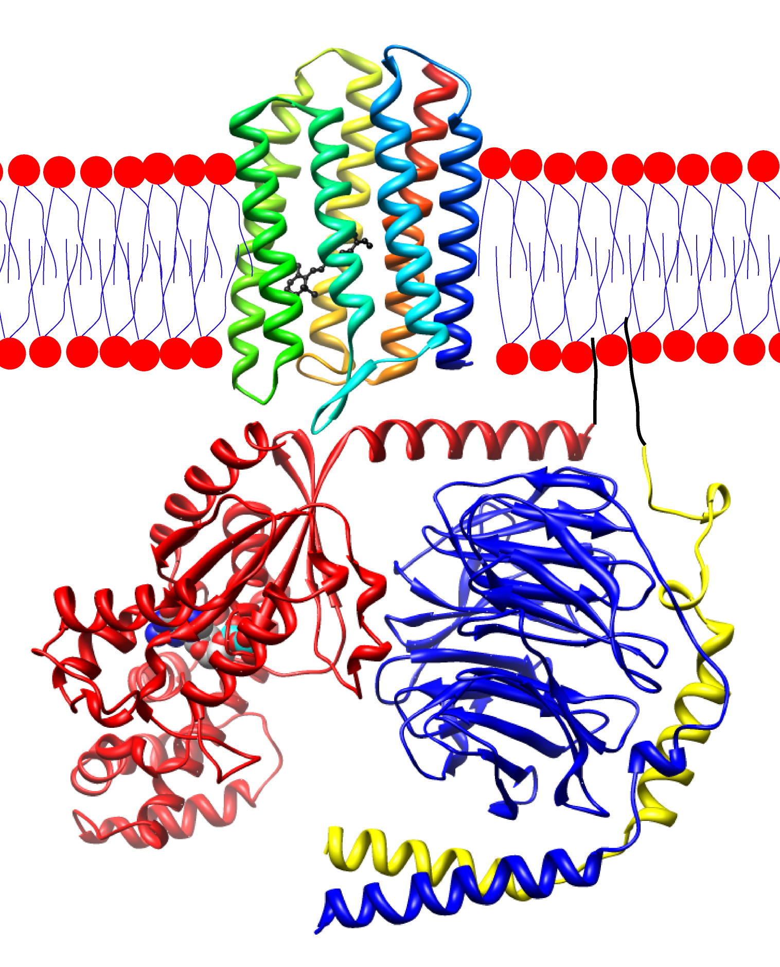
Transducin
Transducin (Gt) is a protein naturally expressed in vertebrate retina rods and cones and it is very important in vertebrate phototransduction. It is a type of heterotrimeric G-protein with different α subunits in rod and cone photoreceptors.
Light leads to conformational changes in the G protein–coupled receptor rhodopsin, which in turn leads to the activation of transducin. Transducin activates phosphodiesterase, which results in the breakdown of cyclic guanosine monophosphate (cGMP). The intensity of the flash response is directly proportional to the number of transducin activated.
Transducin is activated by metarhodopsin II, a conformational change in rhodopsin caused by the absorption of a photon by the rhodopsin moiety retinal. The light causes isomerization of retinal from 11-cis to all-trans. Isomerization causes a change in the opsin to become metarhodopsin II. When metarhodopsin activates transducin, the guanosine diphosphate (GDP) bound to the α subunit (Tα) is exchanged for guanosine triphosphate (GTP) from the cytoplasm. The α subunit dissociates from the βγ subunits (Tβγ). Activated transducin α-subunit activates cGMP phosphodiesterase. cGMP phosphodiesterase breaks down cGMP, an intracellular second messenger which opens cGMP-gated cation channels. Phosphodiesterase hydrolyzes cGMP to 5’-GMP. Decrease in cGMP concentration leads to decreased opening of cation channels and subsequently hyperpolarization of the membrane potential.
Transducin is deactivated when the α-subunit-bound GTP is hydrolyzed to GDP. This process is accelerated by a complex containing an RGS (Regulator of G-protein Signaling)-protein and the gamma-subunit of the effector, cyclic GMP phosphodiesterase.
The Tα subunit of transducin contains three functional domains: one for rhodopsin/Tβγ interaction, one for GTP binding, and the last for activation of cGMP phosphodiesterase.
There are different isoforms of Tα, seen in rod and cone cells. However, the isoforms exhibit functional interchangeability in the phototransduction cascade and shouldn't solely account for differences in light sensitivity. Although the focus for phototransduction is on Tα, Tβγ is crucial for rhodopsin to bind to transducin. The rhodopsin/Tβγ binding domain contains the amino and carboxyl terminal of the Tα. The amino terminal is the site of interaction for rhodopsin while the carboxyl terminal is that for Tβγ binding. The amino terminal might be anchored or in close proximity to the carboxyl terminal for activation of the transducin molecule by rhodopsin.
Interaction with photolyzed rhodopsin opens up the GTP-binding site to allow for rapid exchange of GDP for GTP. The binding site is in the closed conformation in the absence of photolyzed rhodopsin. Normally in the closed conformation, an α-helix located near the binding site is in a position which hinders the GTP/GDP exchange. A conformational change of the Tα by photolyzed rhodopsin causes the tilting of the helix, opening the GTP-binding site.
Once GTP has been exchanged for GDP, the GTP-Tα complex undergoes two major changes: dissociation from photolyzed rhodopsin and the Tβγ subunit and exposure of the phosphodiesterase (PDE) binding site for interaction with latent PDE. The conformational changes initiated in the transducin by binding of GTP are transmitted to the PDE binding site and cause it to be exposed for binding to PDE. The GTP-induced conformational changes could also disrupt the rhodopsin/Tβγ binding site and lead to dissociation from the GTP-Tα complex.
Hub AI
Transducin AI simulator
(@Transducin_simulator)
Transducin
Transducin (Gt) is a protein naturally expressed in vertebrate retina rods and cones and it is very important in vertebrate phototransduction. It is a type of heterotrimeric G-protein with different α subunits in rod and cone photoreceptors.
Light leads to conformational changes in the G protein–coupled receptor rhodopsin, which in turn leads to the activation of transducin. Transducin activates phosphodiesterase, which results in the breakdown of cyclic guanosine monophosphate (cGMP). The intensity of the flash response is directly proportional to the number of transducin activated.
Transducin is activated by metarhodopsin II, a conformational change in rhodopsin caused by the absorption of a photon by the rhodopsin moiety retinal. The light causes isomerization of retinal from 11-cis to all-trans. Isomerization causes a change in the opsin to become metarhodopsin II. When metarhodopsin activates transducin, the guanosine diphosphate (GDP) bound to the α subunit (Tα) is exchanged for guanosine triphosphate (GTP) from the cytoplasm. The α subunit dissociates from the βγ subunits (Tβγ). Activated transducin α-subunit activates cGMP phosphodiesterase. cGMP phosphodiesterase breaks down cGMP, an intracellular second messenger which opens cGMP-gated cation channels. Phosphodiesterase hydrolyzes cGMP to 5’-GMP. Decrease in cGMP concentration leads to decreased opening of cation channels and subsequently hyperpolarization of the membrane potential.
Transducin is deactivated when the α-subunit-bound GTP is hydrolyzed to GDP. This process is accelerated by a complex containing an RGS (Regulator of G-protein Signaling)-protein and the gamma-subunit of the effector, cyclic GMP phosphodiesterase.
The Tα subunit of transducin contains three functional domains: one for rhodopsin/Tβγ interaction, one for GTP binding, and the last for activation of cGMP phosphodiesterase.
There are different isoforms of Tα, seen in rod and cone cells. However, the isoforms exhibit functional interchangeability in the phototransduction cascade and shouldn't solely account for differences in light sensitivity. Although the focus for phototransduction is on Tα, Tβγ is crucial for rhodopsin to bind to transducin. The rhodopsin/Tβγ binding domain contains the amino and carboxyl terminal of the Tα. The amino terminal is the site of interaction for rhodopsin while the carboxyl terminal is that for Tβγ binding. The amino terminal might be anchored or in close proximity to the carboxyl terminal for activation of the transducin molecule by rhodopsin.
Interaction with photolyzed rhodopsin opens up the GTP-binding site to allow for rapid exchange of GDP for GTP. The binding site is in the closed conformation in the absence of photolyzed rhodopsin. Normally in the closed conformation, an α-helix located near the binding site is in a position which hinders the GTP/GDP exchange. A conformational change of the Tα by photolyzed rhodopsin causes the tilting of the helix, opening the GTP-binding site.
Once GTP has been exchanged for GDP, the GTP-Tα complex undergoes two major changes: dissociation from photolyzed rhodopsin and the Tβγ subunit and exposure of the phosphodiesterase (PDE) binding site for interaction with latent PDE. The conformational changes initiated in the transducin by binding of GTP are transmitted to the PDE binding site and cause it to be exposed for binding to PDE. The GTP-induced conformational changes could also disrupt the rhodopsin/Tβγ binding site and lead to dissociation from the GTP-Tα complex.