
Community hub
Recent from talks
Contribute something to knowledge base
Content stats: 0 posts, 0 articles, 1 media, 0 notes
Members stats: 0 subscribers, 0 contributors, 0 moderators, 0 supporters
Subscribers
Supporters
Contributors
Moderators
Hub AI
Vestibulo-ocular reflex AI simulator
(@Vestibulo-ocular reflex_simulator)
Hub AI
Vestibulo-ocular reflex AI simulator
(@Vestibulo-ocular reflex_simulator)
Vestibulo-ocular reflex
The vestibulo-ocular reflex (VOR) is a reflex that acts to stabilize gaze during head movement, with eye movement due to activation of the vestibular system, it is also known as the cervico-ocular reflex. The reflex acts to stabilize images on the retinas of the eye during head movement. Gaze is held steadily on a location by producing eye movements in the direction opposite that of head movement. For example, when the head moves to the right, the eyes move to the left, meaning the image a person sees stays the same even though the head has turned. Since slight head movement is present all the time, VOR is necessary for stabilizing vision: people with an impaired reflex find it difficult to read using print, because the eyes do not stabilise during small head tremors, and also because damage to reflex can cause nystagmus.
The VOR does not depend on what is seen. It can also be activated by hot or cold stimulation of the inner ear, where the vestibular system sits, and works even in total darkness or when the eyes are closed. However, in the presence of light, the fixation reflex is also added to the movement. Most features of VOR are present in kittens raised in complete darkness.
In lower animals, the organs that coordinate balance and movement are not independent from eye movement. A fish, for instance, moves its eyes by reflex when its tail is moved. Humans have semicircular canals, neck muscle "stretch" receptors, and the utricle (gravity organ). Though the semicircular canals cause most of the reflexes which are responsive to acceleration, the maintaining of balance is mediated by the stretch of neck muscles and the pull of gravity on the utricle (otolith organ) of the inner ear.
The VOR has both rotational and translational aspects. When the head rotates about any axis (horizontal, vertical, or torsional) distant visual images are stabilized by rotating the eyes about the same axis, but in the opposite direction. When the head translates, for example during walking, the visual fixation point is maintained by rotating gaze direction in the opposite direction, by an amount that depends on distance.
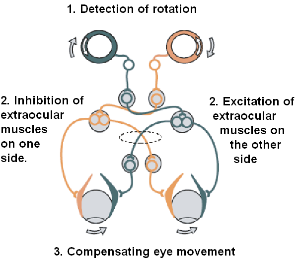
The vestibulo-ocular reflex is driven by signals arising from the vestibular system of the inner ear. The semicircular canals detect head rotation and provide the rotational component, whereas the otoliths detect head translation and drive the translational component. The signal for the horizontal rotational component travels via the vestibular nerve through the vestibular ganglion and end in the vestibular nuclei in the brainstem. From these nuclei, fibers cross to the abducens nucleus of the opposite side of the brain. Here, fibres synapse with 2 additional pathways. One pathway projects directly to the lateral rectus muscle of the eye via the abducens nerve. Another nerve tract projects from the abducens nucleus by the medial longitudinal fasciculus to the oculomotor nucleus of the opposite side, which contains motor neurons that drive eye muscle activity, specifically activating the medial rectus muscle of the eye through the oculomotor nerve.
Another pathway (not in picture) directly projects from the vestibular nucleus through the ascending tract of Deiter's to the medial rectus muscle motor neuron of the same side. In addition there are inhibitory vestibular pathways to the ipsilateral abducens nucleus. However no direct vestibular neuron to medial rectus motoneuron pathway exists.
Similar pathways exist for the vertical and torsional components of the VOR.
In addition to these direct pathways, which drive the velocity of eye rotation, there is an indirect pathway that builds up the position signal needed to prevent the eye from rolling back to center when the head stops moving. This pathway is particularly important when the head is moving slowly because here position signals dominate over velocity signals. David A. Robinson discovered that the eye muscles require this dual velocity-position drive, and also proposed that it must arise in the brain by mathematically integrating the velocity signal and then sending the resulting position signal to the motoneurons. Robinson was correct: the 'neural integrator' for horizontal eye position was found in the nucleus prepositus hypoglossi in the medulla, and the neural integrator for vertical and torsional eye positions was found in the interstitial nucleus of Cajal in the midbrain. The same neural integrators also generate eye position for other conjugate eye movements such as saccades and smooth pursuit.
Vestibulo-ocular reflex
The vestibulo-ocular reflex (VOR) is a reflex that acts to stabilize gaze during head movement, with eye movement due to activation of the vestibular system, it is also known as the cervico-ocular reflex. The reflex acts to stabilize images on the retinas of the eye during head movement. Gaze is held steadily on a location by producing eye movements in the direction opposite that of head movement. For example, when the head moves to the right, the eyes move to the left, meaning the image a person sees stays the same even though the head has turned. Since slight head movement is present all the time, VOR is necessary for stabilizing vision: people with an impaired reflex find it difficult to read using print, because the eyes do not stabilise during small head tremors, and also because damage to reflex can cause nystagmus.
The VOR does not depend on what is seen. It can also be activated by hot or cold stimulation of the inner ear, where the vestibular system sits, and works even in total darkness or when the eyes are closed. However, in the presence of light, the fixation reflex is also added to the movement. Most features of VOR are present in kittens raised in complete darkness.
In lower animals, the organs that coordinate balance and movement are not independent from eye movement. A fish, for instance, moves its eyes by reflex when its tail is moved. Humans have semicircular canals, neck muscle "stretch" receptors, and the utricle (gravity organ). Though the semicircular canals cause most of the reflexes which are responsive to acceleration, the maintaining of balance is mediated by the stretch of neck muscles and the pull of gravity on the utricle (otolith organ) of the inner ear.
The VOR has both rotational and translational aspects. When the head rotates about any axis (horizontal, vertical, or torsional) distant visual images are stabilized by rotating the eyes about the same axis, but in the opposite direction. When the head translates, for example during walking, the visual fixation point is maintained by rotating gaze direction in the opposite direction, by an amount that depends on distance.
The vestibulo-ocular reflex is driven by signals arising from the vestibular system of the inner ear. The semicircular canals detect head rotation and provide the rotational component, whereas the otoliths detect head translation and drive the translational component. The signal for the horizontal rotational component travels via the vestibular nerve through the vestibular ganglion and end in the vestibular nuclei in the brainstem. From these nuclei, fibers cross to the abducens nucleus of the opposite side of the brain. Here, fibres synapse with 2 additional pathways. One pathway projects directly to the lateral rectus muscle of the eye via the abducens nerve. Another nerve tract projects from the abducens nucleus by the medial longitudinal fasciculus to the oculomotor nucleus of the opposite side, which contains motor neurons that drive eye muscle activity, specifically activating the medial rectus muscle of the eye through the oculomotor nerve.
Another pathway (not in picture) directly projects from the vestibular nucleus through the ascending tract of Deiter's to the medial rectus muscle motor neuron of the same side. In addition there are inhibitory vestibular pathways to the ipsilateral abducens nucleus. However no direct vestibular neuron to medial rectus motoneuron pathway exists.
Similar pathways exist for the vertical and torsional components of the VOR.
In addition to these direct pathways, which drive the velocity of eye rotation, there is an indirect pathway that builds up the position signal needed to prevent the eye from rolling back to center when the head stops moving. This pathway is particularly important when the head is moving slowly because here position signals dominate over velocity signals. David A. Robinson discovered that the eye muscles require this dual velocity-position drive, and also proposed that it must arise in the brain by mathematically integrating the velocity signal and then sending the resulting position signal to the motoneurons. Robinson was correct: the 'neural integrator' for horizontal eye position was found in the nucleus prepositus hypoglossi in the medulla, and the neural integrator for vertical and torsional eye positions was found in the interstitial nucleus of Cajal in the midbrain. The same neural integrators also generate eye position for other conjugate eye movements such as saccades and smooth pursuit.
Recent media
Recent media