Hubbry Logo
search
logo
1545264

Doublecortin

logo
Community Hub0 Subscribers
Read side by side
from Wikipedia
DCX
Available structures
PDBOrtholog search: PDBe RCSB
Identifiers
AliasesDCX, DBCN, DC, LISX, SCLH, XLIS, doublecortin
External IDsOMIM: 300121; MGI: 1277171; HomoloGene: 7683; GeneCards: DCX; OMA:DCX - orthologs
Orthologs
SpeciesHumanMouse
Entrez
Ensembl
UniProt
RefSeq (mRNA)

NM_001110222
NM_001110223
NM_001110224
NM_010025

RefSeq (protein)

NP_001103692
NP_001103693
NP_001103694
NP_034155

Location (UCSC)Chr X: 111.29 – 111.41 MbChr X: 142.64 – 142.72 Mb
PubMed search[3][4]
Wikidata
View/Edit HumanView/Edit Mouse

Neuronal migration protein doublecortin, also known as doublin or lissencephalin-X is a protein that in humans is encoded by the DCX gene.[5]

Function

[edit]
Doublecortin expression in the rat dentate gyrus, 21st postnatal day. Oomen et al., 2009.[6]

Doublecortin (DCX) is a microtubule-associated protein expressed by neuronal precursor cells and immature neurons in embryonic and adult cortical structures. Neuronal precursor cells begin to express DCX while actively dividing, and their neuronal daughter cells continue to express DCX for 2–3 weeks as the cells mature into neurons. Downregulation of DCX begins after 2 weeks, and occurs at the same time that these cells begin to express NeuN, a neuronal marker.[7]

Due to the nearly exclusive expression of DCX in developing neurons, this protein has been used increasingly as a marker for neurogenesis. Indeed, levels of DCX expression increase in response to exercise,[8] and that increase occurs in parallel with increased BrdU labeling, which is currently a "gold standard" in measuring neurogenesis.

Doublecortin was found to bind to the microtubule cytoskeleton. In vivo and in vitro assays show that Doublecortin stabilizes microtubules and causes bundling.[9] Doublecortin is a basic protein with an iso-electric point of 10 typical of microtubule-binding proteins.


Knock out mouse

[edit]
Double layer hippocampus seen in Doublecortin knock out mice (right panels) compared to the normal hippocampus in wild type mice (left panels). Figure extracted from the work of the laboratory of Fiona Francis

In mice where the Doublecortin gene has been knocked out, cortical layers are still correctly formed. However, the hippocampi of these mice show disorganisation in the CA3 region. The normally single layer of pyramidal cells in mutants is seen as a double layer. These mice also have different behavior than their wild type littermates and are epileptic.[10]

Structure

[edit]
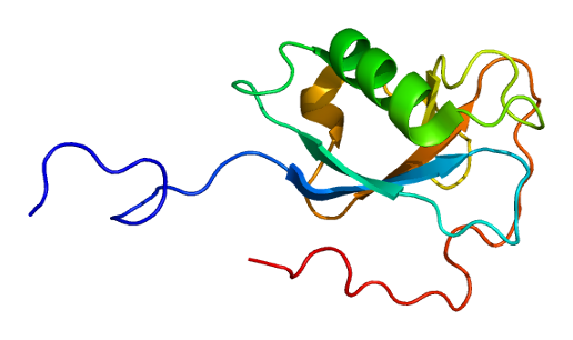
Doublecortin
solution structure of the N-terminal dcx domain of human doublecortin-like kinase
Identifiers
SymbolDCX
PfamPF03607
InterProIPR003533
SCOP21mfw / SCOPe / SUPFAM
CDDcd01617
Available protein structures:
Pfam  structures / ECOD  
PDBRCSB PDB; PDBe; PDBj
PDBsumstructure summary

The detailed sequence analysis of Doublecortin and Doublecortin-like proteins allowed the identification of a tandem repeat of evolutionarily conserved Doublecortin (DC) domains. These domains are found in the N terminus of proteins and consists of tandemly repeated copies of an around 80 amino acids region. It has been suggested that the first DC domain of Doublecortin binds tubulin and enhances microtubule polymerisation.[11]

Doublecortin has been shown to influence the structure of microtubules. Microtubule nucleated in vitro in the presence of Doublecortin have almost exclusively 13 protofilaments, whereas microtubule nucleated without Doublecortin are present in a range of different sizes.

Interactions

[edit]

Doublecortin has been shown to interact with PAFAH1B1.[12]

Clinical significance

[edit]

Doublecortin is mutated in X-linked lissencephaly and the double cortex syndrome, and the clinical manifestations are sex-linked. In males, X-linked lissencephaly produces a smooth brain due to lack of migration of immature neurons, which normally promote folding of the brain surface. Double cortex syndrome is characterized by abnormal migration of neural tissue during development which results in two bands of misplaced neurons within the subcortical white, generating two cortices, giving the name to the syndrome; this finding generally occurs in females.[13] The mutation was discovered by Joseph Gleeson and Christopher A. Walsh in Boston.[14][15] At least 49 disease-causing mutations in this gene have been discovered.[16]

See also

[edit]

References

[edit]

Further reading

[edit]
[edit]
Revisions and contributorsEdit on WikipediaRead on Wikipedia
from Grokipedia
Doublecortin (DCX) is a microtubule-associated protein encoded by the DCX gene on the X chromosome, essential for regulating neuronal migration and cytoskeletal organization during brain development.[1] It functions by binding to and stabilizing microtubules, which facilitates the movement of neurons to their proper positions in the developing cerebral cortex.[2] Mutations in DCX disrupt this process, leading to severe neurodevelopmental disorders such as lissencephaly in males and subcortical band heterotopia in females, often accompanied by epilepsy and intellectual disability.[1] The DCX gene produces a cytoplasmic protein containing two tandem doublecortin domains that interact with microtubules and other proteins like LIS1 to promote microtubule polymerization and stability.[3] Expressed primarily in the brain during fetal and early postnatal stages, DCX is critical in post-mitotic neurons undergoing migration and differentiation.[1] As part of a broader superfamily of DCX-like proteins, it shares conserved ubiquitin-like folds in its domains, which enable protein-protein interactions vital for cytoskeletal dynamics.[3] In addition to its core role in cortical development, DCX influences axonal growth and dendritic arborization in immature neurons, contributing to overall neural circuit formation.[2] Pathogenic variants in DCX typically result in a non-functional or unstable protein, impairing microtubule function and causing abnormal neuronal layering, as seen in the "smooth brain" appearance of lissencephaly.[4] Over 70 mutations are associated with isolated lissencephaly sequence, while more than 100 link to subcortical band heterotopia, highlighting DCX's dosage-sensitive nature due to its X-linked inheritance.[2]

Gene and Protein Basics

Gene Characteristics

The DCX gene, which encodes the doublecortin protein, is located on the long arm of the human X chromosome at cytogenetic band Xq23, specifically spanning genomic coordinates 111,293,779 to 111,412,192 (GRCh38.p14 assembly).[1] This locus covers approximately 118 kb of genomic DNA and consists of 9 exons, of which 6 are coding exons.[1][5] The DCX gene exhibits strong evolutionary conservation across mammalian species, reflecting its critical role in neuronal development. Orthologs include Dcx in the house mouse (Mus musculus, chromosome X), Norway rat (Rattus norvegicus), and other mammals such as chimpanzee and dog, with high sequence similarity in the doublecortin domains.[6][3] This conservation extends to vertebrates, underscoring the ancient origin of the DCX superfamily, which traces back to early metazoan lineages.[3] Alternative splicing of the DCX pre-mRNA generates multiple transcript variants in humans, leading to distinct protein isoforms. Eleven RefSeq mRNA isoforms have been annotated, including the full-length transcript NM_000555.3 (encoding a 365-amino-acid protein) and truncated forms such as those from NM_178152.3 and NM_178153.3, which lack portions of the C-terminal region.[1] These isoforms arise primarily from exon skipping and alternative polyadenylation sites, contributing to functional diversity in neuronal contexts.[1][7]

Protein Structure

The human doublecortin (DCX) protein is a microtubule-associated protein comprising 365 amino acids with a calculated molecular weight of approximately 40 kDa.[8] This compact structure enables its role in neuronal processes, with the protein encoded by the DCX gene on the X chromosome.[7] At its N-terminus, DCX contains two tandem doublecortin (DCX) domains, each approximately 80 amino acids in length, which serve as the primary sites for tubulin binding.[9] The N-terminal DCX domain spans residues 47–140, while the C-terminal DCX domain covers residues 170–260, sharing about 27% amino acid identity and exhibiting a conserved β-grasp fold with five β-strands surrounding a central α-helix.[9] Crystal structures of these domains, determined at resolutions around 1.8–2.5 Å (PDB IDs: 5IN7, 5IO9 for N-DCX; 5IP4 for C-DCX), reveal ubiquitin-like architectures that facilitate specific interactions with tubulin.[10] The domains are connected by a flexible linker, allowing independent or cooperative engagement with microtubule components.[10] The C-terminal region of DCX, spanning roughly residues 260–365, is a serine/proline-rich tail of about 100 amino acids that promotes protein self-association and contributes to microtubule bundling through domain-swapped dimerization.[10] Cryo-EM studies (e.g., EMD-2095) and integrative modeling of DCX-tubulin complexes demonstrate cooperative binding to the microtubule lattice, where the N-terminal domain contacts four α/β-tubulin dimers at inter-protofilament seams, stabilizing 13-protofilament microtubule structures.[11] This binding mode involves lattice-induced conformational changes in the C-terminal domain, enhancing overall affinity without altering protofilament number in standard assemblies.[11] DCX stability is modulated by post-translational modifications, particularly phosphorylation at multiple serine residues in the C-terminal region.[12] Key sites include Ser297, targeted by cyclin-dependent kinase 5 (CDK5), which alters the protein's conformation and binding properties, as well as other sites like Ser263 and Thr269 phosphorylated by kinases such as PKA and MAPK.[12] These modifications, identified through mass spectrometry and mutagenesis studies, influence DCX's structural integrity and association with microtubules without disrupting the core DCX domain folds.[12]

Biological Roles

Microtubule Regulation

Doublecortin (DCX) functions as a microtubule-associated protein that binds directly to the microtubule lattice with a stoichiometry of one DCX molecule per tubulin heterodimer, primarily interacting with polymerized tubulin rather than free heterodimers. This binding promotes microtubule nucleation by stabilizing early tubulin oligomers, leading to the preferential assembly of 13-protofilament microtubules, which constitute the predominant architecture in neuronal cells (70% with DCX versus 20% without). By facilitating nucleation, DCX lowers the critical tubulin concentration required for polymerization, thereby enhancing microtubule assembly rates in vitro.[13][14][15] DCX stabilizes microtubules against depolymerization by strengthening both longitudinal and lateral interactions between tubulin protofilaments, effectively suppressing dynamic instability. Specifically, it acts as an anti-catastrophe factor, reducing the frequency of depolymerization events to near zero at concentrations of 5 μM, and counteracts protofilament peeling by maintaining straight configurations. The binding affinity of DCX to microtubules can be modeled with a dissociation constant (Kd) of approximately 2 μM, reflecting its moderate strength interaction that allows dynamic regulation during neuronal development. This stabilization is particularly evident in assays where DCX-bound microtubules resist cold-induced disassembly.[15][13][14] In neuronal processes, DCX induces microtubule bundling at higher stoichiometric ratios (e.g., 3:2 DCX to tubulin), promoting parallel alignment and straight protofilament structures essential for cytoskeletal integrity. This bundling activity, mediated by cooperative binding involving the C-terminal domain, organizes microtubule arrays in growth cones and axons, supporting directed extension and advance in viscoelastic environments mimicking brain tissue. The N-terminal doublecortin domain primarily drives lattice stabilization in mature arrays, while the C-terminal region aids initial nucleation and bundling.[15][11][16][17]

Neuronal Migration and Differentiation

Doublecortin (DCX) plays a pivotal role in guiding radial and tangential neuronal migration during cortical development by stabilizing the leading processes of migrating neurons, which facilitates directed locomotion along radial glia or tangential pathways. In radial migration, DCX is expressed in projection neurons ascending from the ventricular zone to the cortical plate, where it concentrates in the distal leading processes to maintain cytoskeletal integrity and support nucleokinesis.[14] For tangential migration, DCX is enriched in interneurons originating from the ganglionic eminence, enabling their efficient traversal across the cortical intermediate zone by promoting microtubule polymerization and process extension.[18] This stabilization arises from DCX's microtubule-associated protein function, which bundles and stabilizes microtubules to counteract dynamic instability during migration.[19] Beyond migration, DCX contributes to neuronal differentiation by promoting dendritic branching and spine formation in immature neurons, essential for establishing synaptic connectivity. In cultured hippocampal neurons, varying DCX expression levels directly influence dendritic arborization: overexpression increases branch points and overall complexity, while reduction via RNAi leads to shorter, less branched dendrites with decreased total length. DCX achieves this by associating with actin-rich structures in growth cones and distal dendrites, facilitating branching while preventing excessive protrusions that could disrupt maturation.[19] For spine formation, DCX supports the maturation of dendritic protrusions into functional spines, as evidenced by altered spine density and morphology in neurons with modulated DCX levels, underscoring its role in transitioning from migratory to post-mitotic states. In adult neurogenesis, DCX is crucial for the integration of newly generated granule cells into the hippocampal dentate gyrus, where it sustains migration from the subgranular zone and promotes morphological maturation. DCX-positive immature neurons exhibit enhanced process extension and bipolar morphology, aiding their incorporation into existing circuits for functions like spatial learning.[20] This involves DCX-mediated microtubule stabilization that supports dendrite elongation and synapse formation in the adult context, distinct from embryonic roles but reliant on similar cytoskeletal mechanisms.[19] Experimental in vitro assays demonstrate that DCX overexpression accelerates neuronal migration speeds, as seen in cerebellar granule cells where transfected cells exhibit faster nucleokinesis and reduced branching pauses.[21] In slice cultures of cortical interneurons, reduction in DCX levels slows migration, with affected neurons covering shorter distances in longer times compared to controls, highlighting its dosage-dependent role in motility without altering directionality.[18] DCX integrates with the Reelin signaling pathway to ensure proper cortical layer formation, where Reelin from Cajal-Retzius cells guides DCX-expressing neurons to their laminar positions via shared regulation of cytoskeletal dynamics. This interaction occurs through downstream effectors like JNK-interacting protein 1 (JIP-1), linking Reelin's adhesive cues to DCX-dependent microtubule adjustments for precise layering.[19]

Expression and Regulation

Developmental Patterns

Doublecortin (DCX) exhibits high expression in the ventricular and subventricular zones of the developing brain, beginning at embryonic day 11 (E11) in mice, where scattered positive cells appear in the subventricular zone (SVZ) by E14, coinciding with the onset of neuronal migration.[22] In humans, DCX expression is elevated from mid-gestation, peaking in the neonatal period (0-0.24 years) within the SVZ and adjacent white matter of the prefrontal cortex, before a sharp decline during infancy.[23] Postnatally, in mice, robust DCX immunoreactivity persists in the SVZ through the first two weeks, supporting ongoing corticogenesis, while in humans, high levels extend into the first few postnatal weeks before diminishing significantly by toddlerhood (1.5-5 years).[22][23] The expression of DCX is transient in migrating neurons, reaching a peak during corticogenesis around E17 in mice within the intermediate zone and upper cortical plate, and gradually declining as neurons mature and integrate into the cortical layers.[22] In both rodents and primates, DCX levels drop markedly postnatally—by approximately 94% in humans and 77-86% in macaques from neonate to adult—reflecting the cessation of widespread neuronal migration and differentiation.[23] This pattern positions DCX as a commonly used marker for immature neurons during these phases, though it may also be expressed in other cell types.[24] However, the specificity of DCX as a marker has been debated, as it can be expressed in other cell types beyond immature neurons.[25] DCX protein localizes primarily to the soma periphery, dendrites, and axons of immature neurons, often aligning with microtubule networks to facilitate cytoskeletal dynamics during migration.[22] Its expression correlates with proliferation markers such as BrdU, as DCX-positive cells frequently include recently divided neuroblasts exiting the cell cycle, though DCX itself marks postmitotic stages. Environmental factors like exercise can upregulate DCX in developing and early postnatal brains, enhancing the density of positive neurons in neurogenic regions through promoted maturation.[26] Species-specific differences are evident in the duration of DCX expression; while it fades rapidly in the neocortex of both mice and humans by early postnatal stages, rodents display prolonged DCX immunoreactivity in the hippocampal dentate gyrus extending into adulthood, linked to sustained granule cell neurogenesis.[23] In contrast, human hippocampal expression declines exponentially with age, with low levels persisting into adulthood.[27]

Regulatory Mechanisms

The expression of doublecortin (DCX) is tightly controlled at the transcriptional level by key transcription factors that dictate its neuron-specific activation during development and neurogenesis. The repressor element 1-silencing transcription factor (REST), also known as NRSF, acts as a potent transcriptional repressor of DCX in non-neuronal cells and neural progenitors, binding to repressor element 1 (RE1) sites in the DCX promoter to suppress its expression; targeted downregulation of REST during neuronal differentiation relieves this repression, enabling DCX transcription in post-mitotic neurons.[28] Conversely, NeuroD1, a basic helix-loop-helix transcription factor, promotes DCX expression as part of its role in driving neuronal differentiation and maturation, with overexpression of NeuroD1 upregulating DCX alongside other neuronal markers in reprogramming protocols.[29] These opposing actions of REST and NeuroD1 ensure precise spatiotemporal control of DCX, aligning its expression with phases of neuronal commitment and migration. Post-transcriptional regulation further fine-tunes DCX levels through microRNAs (miRNAs) that influence mRNA stability and translation during neuronal maturation. For instance, miR-124, highly enriched in the brain, contributes to the transition from progenitor to immature neuron states by targeting non-neuronal transcripts, indirectly supporting DCX expression in early differentiation.[30] This miRNA-mediated control integrates with broader post-transcriptional networks to prevent ectopic DCX persistence in mature circuits. DCX protein activity and stability are modulated post-translationally, primarily through phosphorylation by multiple kinases that alter its binding affinity to microtubules. Cyclin-dependent kinase 5 (CDK5), in complex with p35, phosphorylates DCX at sites such as Ser297, reducing its microtubule-binding capacity and promoting dynamic cytoskeletal remodeling essential for neuronal migration; similarly, protein kinase A (PKA) phosphorylates DCX at Thr269/Ser265, further decreasing microtubule affinity and facilitating process extension at the leading edge.[31] These phosphorylation events are counterbalanced by phosphatases like PP1, maintaining a dynamic equilibrium that regulates DCX's role in microtubule stabilization. Environmental factors also influence DCX expression and activity, particularly in adult neurogenesis contexts where external stimuli drive adaptive responses. Environmental enrichment, characterized by increased sensory, social, and physical stimulation, upregulates DCX in the hippocampal dentate gyrus, enhancing the proliferation and differentiation of neural progenitors into migrating neuroblasts.[32] Likewise, brain injury, such as traumatic brain injury, induces DCX upregulation in the subgranular zone, promoting neurogenesis as a reparative mechanism despite potential trade-offs in neuronal survival.[33] These influences highlight DCX's responsiveness to experiential and pathological cues in the adult brain. Feedback loops between DCX and microtubule dynamics provide an additional layer of autoregulation, linking cytoskeletal state to DCX turnover. DCX binding stabilizes microtubules, but conversely, stable microtubules enhance DCX retention via reduced phosphorylation turnover. This bidirectional interplay, involving kinases like doublecortin-like kinase (DCLK) that phosphorylate DCX in a microtubule-dependent manner, ensures coordinated regulation of neuronal motility and polarity.[34]

Experimental Models

Knockout Mouse Phenotypes

Knockout mice deficient in doublecortin (Dcx) display normal lamination and layering in the neocortex, consistent with the protein's non-essential role in cortical neuronal migration in this species. In contrast, the hippocampus shows prominent abnormalities, particularly in the CA3 region, where pyramidal neurons fail to form a single compact layer and instead organize into a disrupted double-layered structure, with cells abnormally dispersed between the stratum pyramidale and stratum oriens. This lamination defect arises during postnatal development and persists into adulthood, accompanied by simplified dendritic arbors and altered mossy fiber projections in CA3 pyramidal cells, which contribute to increased neuronal excitability in the region.[35][36][37] These hippocampal alterations lead to enhanced seizure susceptibility in Dcx knockout mice, with rare spontaneous convulsive seizures observed via video-EEG monitoring and significantly increased sensitivity to convulsant agents such as pentylenetetrazol and kainic acid compared to wild-type littermates. Synaptic plasticity is disrupted, evidenced by a higher frequency of sharp wave-like events in CA3 slices and a lower threshold for inducing epileptiform activity with bicuculline, reflecting hyperexcitability and reduced inhibitory tone. Behaviorally, knockout mice exhibit hyperactivity in novel environments, impaired social interactions, reduced aggression following social deprivation, and a loss of behavioral lateralization, though hippocampal-dependent memory tasks such as spatial learning and fear conditioning remain largely intact.[38][39][37] Adult hippocampal neurogenesis in the dentate gyrus appears unaffected in Dcx knockout mice, with normal rates of progenitor proliferation, newborn neuron survival, and differentiation into mature granule cells, as assessed by BrdU labeling and NeuN expression. However, the lamination defects extend to subtle disorganization in the dentate gyrus granule cell layer in some cases, and while overall neurogenesis proceeds normally, the simplified dendritic morphology observed in hippocampal pyramidal neurons suggests potential impacts on integration of new neurons. Rescue experiments using Dcx re-expression via viral overexpression in neuronal cultures or electroporation have demonstrated partial restoration of migration and layering defects in hippocampal interneurons and pyramidal cells, confirming the specificity of the phenotype to Dcx loss.[36][40][41] The hippocampal phenotypes in Dcx knockout mice, particularly the double-layered organization of CA3 pyramidal cells, serve as an experimental analog to subcortical band heterotopia in humans, where DCX mutations cause ectopic neuronal bands and migration defects, though the mouse model lacks the severe cortical malformations seen in patients due to species-specific differences in DCX dependency.[42][43]

Other Model Systems

In non-mammalian model systems, doublecortin (DCX) and its homologs have been investigated to elucidate conserved roles in neuronal migration and axon guidance. In zebrafish, knockdown of the doublecortin-like kinase (DCLK), a close relative of DCX, using morpholino antisense oligonucleotides results in abnormal brain development, with increased apoptotic cells in the central nervous system and morphologically abnormal embryos in a dose-dependent manner. [44] In vitro neuronal culture assays have demonstrated DCX's critical role in migration. Silencing DCX using RNAi in adult subventricular zone (SVZ) cells reduces cell-autonomous migration and alters the migration substrate provided by astrocytes, as shown in Boyden chamber assays where DCX-knockdown cells exhibit slowed migration rates and disrupted leading process extension. [45] These assays confirm that DCX is necessary for chain migration in neuronal precursors, with defects mimicking those seen in vivo. [41] Conditional knockout models using Cre-lox systems have revealed region-specific roles of DCX in the mouse brain. For example, using Emx1-Cre to target forebrain regions or Nex-Cre for postmitotic neurons, selective deletion of DCX leads to localized disruptions in cortical layering and hippocampal dendrite formation without global lethality, highlighting differential contributions to radial versus tangential migration in distinct areas. [43] Such approaches demonstrate that DCX functions vary by brain region, with forebrain-specific knockouts showing more pronounced lamination defects than hippocampal-targeted ones. Induced pluripotent stem cell (iPSC)-derived models from patients with DCX mutations have been used to study neurodevelopmental defects, revealing impairments in neuronal migration and differentiation that contribute to lissencephaly phenotypes.[46]

Molecular Interactions

Key Protein Partners

Doublecortin (DCX) primarily interacts with platelet-activating factor acetylhydrolase 1B regulatory subunit 1 (PAFAH1B1, also known as LIS1), as evidenced by yeast two-hybrid screening, pull-down experiments, co-immunoprecipitation (co-IP) assays in COS-7 cells, and direct binding in vitro.[47] The C-terminal domain of DCX binds the WD40 repeats of LIS1.[47] DCX and LIS1 form a complex with cytoplasmic dynein.[48] DCX binds directly to tubulin heterodimers and microtubules, promoting their polymerization and stabilization, as demonstrated by in vitro binding assays using taxol-stabilized microtubules and cosedimentation experiments.[49] Structural studies further reveal that DCX's two DC domains engage tubulin protofilaments at specific lattice sites, with higher affinity for curved tubulin structures.[50] DCX associates with other microtubule-associated proteins (MAPs) such as MAP1B and tau, contributing to synergistic microtubule bundling through co-localization and functional overlap in neuronal processes, though direct binding evidence is limited.[51] DCX interacts with kinases including glycogen synthase kinase 3β (GSK3β), which phosphorylates DCX at specific serine residues, as shown by in vitro kinase assays and co-IP in neuronal lysates.[52] These phosphorylation events modulate DCX's localization, with evidence from mass spectrometry identifying GSK3β as a direct regulator.[52]

Functional Consequences

The LIS1-DCX complex plays a critical role in regulating cytoplasmic dynein to facilitate nuclear translocation during neuronal migration. In migrating neurons, this complex mediates the coupling of the nucleus to the centrosome, enabling coordinated nucleokinesis. Specifically, DCX interacts directly with dynein heavy and intermediate chains, forming a complex that, together with LIS1, maintains the structural integrity required for dynein-driven nuclear movement along microtubules. Disruption of this interaction, as observed in LIS1-deficient neurons, increases the nucleus-centrosome distance by approximately 70%, impairing migration efficiency.[48] DCX exhibits synergistic effects with other microtubule-associated proteins (MAPs), such as doublecortin-like kinase (DCLK), to enhance microtubule stability particularly in axons. This cooperation promotes robust microtubule polymerization and bundling, supporting axonal elongation and wiring in the developing cerebral cortex. In double-null models lacking both DCX and DCLK, axons display significant shortening and disrupted transport, underscoring their combined contribution to cytoskeletal integrity beyond individual functions.[53] Disruption of DCX interactions leads to altered cargo transport and neuronal polarity. By binding microtubules and modulating motor proteins like kinesin-3, DCX ensures polarized transport of cargos, such as Golgi components, into dendrites while restricting inappropriate entry into axons. Loss of DCX impairs this compartmentalization, resulting in defective kinesin-3 motility and reduced cargo delivery, which compromises neuronal polarity establishment. In DCX-deficient systems, microtubule organization falters, leading to mislocalized cargos and polarity defects that hinder proper dendritic elaboration.[54][55] Evidence from interaction mutants demonstrates defective microtubule bundling in vitro. Patient-derived DCX mutants, such as R89G and R59H, fail to associate properly with microtubules, resulting in diminished polymerization and bundling capacity when expressed in non-neuronal cells. These alleles exhibit loss-of-function phenotypes, with R59H additionally forming cytoplasmic aggregates that further disrupt bundling, highlighting how specific interaction defects compromise DCX's stabilizing role.[56] DCX participates in signaling cascades that link to the Wnt pathway to influence neuronal differentiation. As a downstream target, DCX expression is transcriptionally activated by Wnt/β-catenin signaling via promoter elements responsive to this pathway, promoting the transition from proliferation to differentiation in neural progenitors. This integration supports microtubule-dependent processes essential for differentiating neurons, such as process extension and maturation.[57] Recent studies indicate that DCX regulates tubulin polyglutamylation, which modulates interactions with other MAPs like tau, affecting microtubule stability and neuronal branching as of February 2025.[58]

Clinical and Pathological Aspects

Genetic Mutations

The DCX gene, located on chromosome Xq23.2, is subject to a variety of pathogenic variants that disrupt its function as a microtubule-associated protein essential for neuronal migration. Over 130 distinct pathogenic variants have been reported as of 2025, with missense mutations comprising the majority (approximately 65%), followed by nonsense, frameshift, splice site alterations, and small insertions/deletions; larger exon or whole-gene deletions/duplications are rarer but also documented.[59][60] These variants are cataloged in databases such as OMIM entry #300121 and ClinVar, with ongoing additions from clinical sequencing efforts.[5][61] DCX mutations follow an X-linked inheritance pattern, characterized by hemizygous effects in males and heterozygous effects in females due to X-chromosome inactivation mosaicism. In males, pathogenic variants typically result in severe classical lissencephaly, while in females, they lead to subcortical band heterotopia, often with milder or variable expressivity depending on skewed X-inactivation.[61][62] At the molecular level, these mutations impair DCX's ability to bind and stabilize microtubules, thereby reducing microtubule polymerization rates and bundling efficiency, which are critical for cytoskeletal dynamics during neuronal migration. For instance, missense mutations within the DCX domains, such as p.Arg192Trp, disrupt tubulin interactions and lead to diminished microtubule affinity and altered polymerization kinetics in cellular assays.[5] Similarly, nonsense and frameshift variants often result in truncated proteins that fail to polymerize microtubules effectively, abolishing normal binding.[63] Genotype-phenotype correlations reveal that the location and type of mutation influence severity; for example, variants in the N-terminal DCX domains tend to cause more profound disruptions and severe phenotypes in affected males, whereas C-terminal mutations are associated with milder effects, potentially due to partial retention of microtubule-binding capacity.[62][64] Truncating mutations generally correlate with more severe outcomes than missense variants, though expressivity varies, particularly in females.

Associated Disorders

Mutations in the DCX gene, encoding doublecortin, are primarily associated with X-linked disorders of neuronal migration, manifesting as distinct clinical syndromes in males and females due to the gene's location on the X chromosome.[61] In hemizygous males, DCX mutations cause X-linked lissencephaly type 1, characterized by a smooth cerebral cortex (agyria-pachygyria), severe intellectual disability, early-onset epilepsy (occurring in approximately 83% of cases, with 49% intractable), and motor abnormalities including hypotonia or spasticity.[61] Neuroimaging via MRI typically reveals anterior-predominant thick lissencephaly with shallow sulci and a simplified gyral pattern.[65] Affected males often exhibit profound developmental delays, with limited language acquisition and dependency on lifelong care.[61] Heterozygous females with DCX mutations typically present with double cortex syndrome, or subcortical band heterotopia (SBH), featuring a bilateral band of gray matter beneath a relatively normal cortical surface, leading to milder cognitive impairment, seizures (in about 85% of cases, 78% intractable), and behavioral challenges such as attention deficits.[61] The phenotypic variability in females arises from random X-chromosome inactivation, resulting in somatic mosaicism that spares some neuronal populations.[61] MRI in these cases shows the characteristic "double cortex" appearance, with the heterotopic band thickness correlating with seizure severity.[66] Diagnosis of DCX-related disorders relies on molecular genetic testing to identify pathogenic variants in the DCX gene, often prompted by neuroimaging evidence of migration defects, alongside clinical evaluation of neurodevelopmental symptoms. Molecular genetic testing, including next-generation sequencing, detects pathogenic variants in 94%-96% of cases.[61] Prenatal or postnatal MRI is essential for visualizing cortical malformations, while genetic confirmation via sequencing (detecting variants in up to 90% of SBH cases) guides prognosis and family counseling.[59] These disorders are rare, with classical lissencephaly occurring in approximately 1:100,000 births and DCX mutations accounting for 20-40% of such cases; the X-linked nature imparts a strong sex bias, with males experiencing more severe outcomes than carrier females.[61]

References

User Avatar
No comments yet.