Hubbry Logo
search
logo
1969519

Dynamic compression plate

logo
Community Hub0 Subscribers
Read side by side
from Wikipedia
Dynamic compression plate

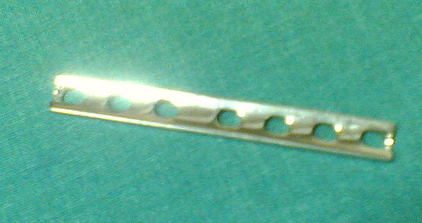
A dynamic compression plate (DCP) is a metallic plate used in orthopedics for internal fixation of bone,[1] typically after fractures. As the name implies, it is designed to exert dynamic pressure between the bone fragments to be transfixed. Dynamic compression is achieved either by attaching a tension device to a plate or by using a special dynamic compression plate. However, compression plating requires a longer surgical incision to allow insertion of the tension device and the possibility of refracture after the plate is removed. A neutralization plate is used to bridge a comminuted fracture; it also transmits bending or torsional forces from the proximal to the distal fragment. Plates used for buttressing prevent collapse by supporting an area of thin cortex or cancellous bone graft.[citation needed]

When plates are used, atrophy of the bone beneath the plate may predispose the bone to fracture. After removal of plates, fractures may occur through the screw holes.

Sliding screw plate

[edit]

The sliding screw plate (dynamic compression screw, dynamic hip screw) may be used to treat intertrochanteric fractures as well as other injuries. This device consists of a lag screw and a side plate with a barrel. The sliding screw telescopes and provides fixation while allowing impaction to occur at the fracture during healing and weight bearing. Among the complications of sliding screw plate fixation are "cutting out" of the nail, penetration of the screw into the joint, bending or breaking of the nail, and disengagement of the screw from the barrel and even protrusion of the screw into the vertebrae.[citation needed]

See also

[edit]

References

[edit]
Revisions and contributorsEdit on WikipediaRead on Wikipedia
from Grokipedia
The dynamic compression plate (DCP) is an orthopedic implant designed for the internal fixation of bone fractures, utilizing eccentric screw insertion to generate interfragmentary compression and achieve absolute stability, which promotes primary bone healing without callus formation.[1] Introduced in 1970 by Martin Allgöwer and colleagues as an advancement in plate technology, the DCP features specialized oval screw holes with an inclined or beveled floor that allows the screw head to slide and translate bone fragments laterally toward the fracture site upon tightening, thereby compressing the bone ends together.[2][3] To optimize compression, the plate is typically prebent or overbent by 1–2 mm over the fracture line, enabling it to function like a spring that presses both the near and far cortices into contact while minimizing gapping.[4] This design is particularly effective for transverse and oblique fractures in long bones, such as the femur, tibia, humerus, or radius, where it controls shear forces and micromotion to facilitate stable healing.[1][4] The DCP is often combined with lag screws for enhanced fixation in stable compression scenarios and is recognizable on radiographs by its distinctive oval holes.[3] A notable evolution of the DCP is the limited contact dynamic compression plate (LC-DCP), developed in the early 1990s to address concerns about vascular disruption and cortical bone porosity caused by extensive plate-to-bone contact in the original design.[5] The LC-DCP incorporates an undercut or grooved undersurface that reduces direct contact area by up to 50%, preserving periosteal blood supply, minimizing temporary osteoporosis under the plate, and allowing for better periosteal callus formation while retaining the core compression mechanism.[6] Both variants adhere to principles established by the Association for the Study of Internal Fixation (AO/ASIF), emphasizing precise application to avoid complications like screw loosening or delayed union.[4]

History and Development

Origins

The origins of compression plating trace back to early 20th-century efforts in internal fracture fixation, which initially relied on rigid plates without mechanisms for interfragmentary compression. In 1909, Belgian surgeon Albin Lambotte introduced one of the first practical metal plates for bone stabilization, featuring a thin, round design tapered at both ends to minimize soft tissue irritation; this plate used screws for attachment but provided absolute rigidity, limiting its ability to promote direct bone healing.[7] Similarly, in 1912, American surgeon William O. Sherman developed a plate system with self-tapping screws, emphasizing secure fragment alignment through rigid immobilization, though it too lacked dynamic features to apply controlled compression across the fracture site.[7] These innovations marked a shift from earlier wire-based methods but were constrained by material limitations and design simplicity. Early rigid plate designs encountered significant clinical challenges, including high rates of infection, delayed union, and non-union, primarily due to inadequate compression that failed to achieve stable interfragmentary contact. The absolute stability provided by these plates often resulted in excessive rigidity, leading to stress shielding, cortical porosis, and micromotion at the fracture interface, which compromised healing and increased susceptibility to postoperative infections from poor vascularity and biofilm formation on hardware.[8] Non-union rates were notably elevated in long bone fractures treated with such systems, as the lack of compression hindered primary bone healing and allowed for excessive strain that disrupted callus formation.[7] A pivotal advancement occurred in 1956 when George W. Bagby, working at the Mayo Clinic, modified the existing Collison plate by incorporating oval-shaped holes, enabling eccentric screw placement to generate dynamic interfragmentary compression during insertion. This design allowed the plate to slide slightly along the screw axis, applying controlled force to approximate fracture fragments without excessive rigidity, and was rigorously tested in canine femoral fracture models, where it demonstrated accelerated healing rates compared to non-compression controls.[9] Bagby's approach addressed prior deficiencies by promoting primary bone union through precise compression, reducing the shear forces that contributed to complications in earlier systems. Throughout the 1950s, several key publications and designs underscored the critical need for interfragmentary compression in fracture fixation, building on Bagby's prototype. Bagby and colleague J.M. Janes detailed the oval-hole mechanism in their 1956 work, highlighting its role in achieving compression via screw head eccentricity, which influenced subsequent engineering efforts.[10] These contributions emphasized that effective plating required not just stability but active fragment apposition to minimize healing time and complications, paving the way for formalized advancements in the following decade.[11]

AO Introduction and Evolution

The Association for the Study of Internal Fixation (AO), founded in 1958 by a group of Swiss surgeons led by Maurice E. Müller, aimed to standardize and advance fracture treatment through rigorous scientific research and education.[7] The organization's core principles emphasized anatomical reduction of fractures, stable internal fixation to promote primary bone healing without callus formation, preservation of vascular supply to the bone, and early functional rehabilitation to minimize complications like stiffness.[12] These tenets shifted the paradigm from conservative casting to operative intervention, focusing on rigid fixation that allows direct osteonal remodeling across the fracture gap under absolute stability. Building on these principles, the AO introduced the Dynamic Compression Plate (DCP) in 1969 as a major advancement in plating technology.[13] The DCP featured oval-shaped screw holes that enabled eccentric screw insertion to generate interfragmentary compression, paired with self-tapping cortical screws for secure anchorage without predrilling taps.[2] This design, inspired by earlier concepts like Bagby's 1956 oval hole plate for compression, allowed for precise control of fracture site stability while adhering to AO's rigid fixation goals.[7] In the 1990s, the AO evolved the DCP into the Limited Contact Dynamic Compression Plate (LC-DCP) to address concerns over periosteal blood supply disruption from broad plate-bone contact.[14] The LC-DCP incorporated undercuts along the plate's underside, reducing contact area to approximately 50% and minimizing vascular impairment and cortical atrophy.[13] Titanium alloys have been used for DCP and LC-DCP implants since the early 1970s alongside stainless steel, offering superior biocompatibility, a modulus of elasticity closer to bone, and reduced risk of allergic reactions.[15] Early clinical studies in the 1970s validated the DCP's efficacy, reporting primary bone healing rates of 80-90% in stable fractures treated with compression plating, with union times averaging 12-16 weeks and low complication rates when principles were followed.[2] These outcomes, documented in prospective series of long bone fractures, underscored the DCP's role in achieving direct healing without external support, influencing global adoption of AO techniques.[16]

Design Features

Plate Construction

The dynamic compression plate (DCP) is primarily constructed from biocompatible materials such as 316L stainless steel or titanium alloys, selected for their high corrosion resistance, biocompatibility, and mechanical strength suitable for load-bearing orthopedic applications.[17][18] These materials ensure durability in physiological environments while minimizing adverse tissue reactions.[19] DCP plates are available in narrow and broad profiles tailored to specific anatomical regions. The narrow profile, intended for the upper extremities and forearm, accommodates 3.5 mm screws and features a thickness of 2.0–3.3 mm and width of 10 mm.[20][21] In comparison, the broad profile, designed for the lower leg and thigh, uses 4.5 mm screws with a thickness of 4.8 mm and width of 16 mm to provide enhanced stability for larger bones.[22] Plates are produced in various lengths corresponding to 3–18 holes, enabling adaptation to different fracture spans.[23] Factory prebent options are available for certain DCP variants to approximate bone contours, such as a slight S-curve for femoral applications, though intraoperative contouring with bending tools is often required for precise fit.[24] Hole configurations consist of a combination of compression (oval-shaped) and round (neutral) holes, with center-to-center spacing typically ranging from 12–18 mm depending on plate size.[25][26] Plate thicknesses vary by system size, typically from ~2.0 mm for small fragment (3.5 mm screws) to ~5.0 mm for large fragment (4.5 mm screws), optimizing contact with bone while maintaining structural integrity.[26] An evolution to the limited-contact DCP (LC-DCP) incorporates undercuts in the plate undersurface to reduce bone contact area and preserve periosteal vascularity.[26]

Screw and Hole Mechanics

The holes in a dynamic compression plate (DCP) are oval-shaped, elongated along the longitudinal axis of the plate to accommodate eccentric screw placement that facilitates controlled fracture compression.[27] Each hole features a sloped edge, formed as part of an inclined cylinder on the side distant from the fracture, allowing the screw head to slide and generate axial compression as it tightens.[13] This design contrasts with round holes, which provide neutral fixation without such movement.[13] DCP systems utilize self-tapping cortical screws, typically 3.5 mm or 4.5 mm in diameter, with lengths ranging from 6 mm to 70 mm in 2 mm increments, and featuring hexagonal heads for precise torque application using a screwdriver.[26] These screws are designed for bicortical purchase, threading fully into the far cortex while the head seats against the plate hole.[27] In neutral position, screws are placed centrally in either oval or round holes, securing the plate to bone without inducing motion or compression.[13] For load or compression mode, eccentric placement in the oval hole positions the screw head against the sloped edge, causing the plate to glide relative to the bone fragment as the screw tightens, thereby drawing the fragments together.[27] This interaction relies on a specialized drill guide to offset the drill hole by about 1 mm from the center.[26] A key aspect of this mechanics is the recoil effect, where the plate's elastic deformation during screw tightening results in an additional 1-2 mm of fragment approximation after the screw head fully seats, enhancing interfragmentary contact.[1] The oval hole design also permits angulation tolerance of up to 20-25 degrees off-axis in the longitudinal direction and about 7 degrees transversely, allowing adaptive insertion while maintaining effective compression.[26]

Biomechanical Principles

Compression Mechanism

The dynamic compression plate (DCP) generates axial compression through eccentric loading of non-locking screws within its specialized oval holes. When a screw is inserted into the compression hole and positioned eccentrically toward the fracture site, tightening causes the screw head to engage the inclined surface of the hole, translating the plate laterally by up to 1 mm relative to the underlying bone fragment. This action induces a slight temporary bowing of the plate, followed by elastic recoil that draws the bone ends together, achieving interfragmentary compression across the fracture gap.[28][1] To optimize compression, particularly at the far cortex and prevent gapping at the near cortex, surgeons employ a pre-bending technique by contouring the plate 1-2 mm away from the bone surface over the fracture line prior to fixation. Upon screw insertion and tightening, the plate flattens against the bone, leveraging its elastic properties to produce additional compressive force through recoil. This method shifts pressure distribution toward the far cortex and enhances overall stability at the fracture interface.[28][1] The resulting interfragmentary pressure at the fracture site provides the rigid environment necessary for primary bone healing without callus formation. Pre-bending improves pressure distribution across a larger contact area.[1] The dynamic nature of the DCP allows limited interfragmentary micromotion following implantation, which supports bone remodeling under physiological loads while minimizing shear at the fracture site. This controlled motion arises from the plate's elastic deformation limits and the non-locking screw interface, contrasting with fully rigid systems.[28][1] The underlying mathematical basis for compression relies on a simplified application of Hooke's law, where the compressive force $ F $ is given by
F=kδ, F = k \cdot \delta,
with $ k $ representing the plate's axial stiffness (typically 100-150 N/mm for standard DCP constructs) and $ \delta $ the deflection from pre-bending or recoil. This model approximates the spring-like behavior during flattening, with forces of approximately 160-520 N under clinical torque levels up to 4 Nm.[1][29]

Load Distribution and Stability

The dynamic compression plate (DCP) functions primarily as a load-sharing device, distributing a significant portion of axial loads to the underlying bone after fracture reduction, which contrasts with fully rigid load-bearing plates that assume nearly all forces. This load-sharing supports primary bone healing by maintaining absolute stability while allowing the bone to experience physiological stresses, reducing the risk of stress shielding.[29][30] In terms of rigidity, DCP constructs achieve approximately 50-85% of intact bone stiffness in axial compression and bending, providing robust resistance to longitudinal forces, with comparable performance in torsion.[31] This differential stiffness ensures absolute stability for primary bone healing in transverse or short oblique fractures by limiting interfragmentary strain, a threshold that prevents fibrous tissue interposition and fosters direct osteonal remodeling.[28] The compression achieved through eccentric screw loading during implantation serves as the foundational mechanism for this strain control, enabling the plate to counteract shear and gap formation under load.[1] Stability in DCP fixation is influenced by key factors, including a minimum of six cortices engaged per main fragment (typically three bicortical screws on each side) to optimize frictional hold and prevent slippage, and a plate length spanning at least two times the fracture gap to distribute forces evenly and minimize localized stress concentrations.[32] Inadequate screw purchase or short plate working lengths can reduce torsional resistance, compromising long-term fixation.[33] Additionally, DCPs exhibit fatigue resistance under simulated physiological conditions, supporting early weight-bearing and healing.[34]

Surgical Applications

Indications

The dynamic compression plate (DCP) is primarily indicated for the treatment of transverse, short oblique, and simple multifragmentary fractures located in the diaphyseal regions of long bones, where interfragmentary compression can achieve absolute stability and promote primary bone healing.[35][36] These fracture patterns align with AO/OTA classification types A (simple) and B (wedge or multifragmentary diaphyseal), particularly when there is greater than 50% cortical contact to support load-sharing across the fracture site.[28][37] Common anatomical applications include midshaft fractures of the humerus, both-bone fractures of the radius and ulna, subtrochanteric and diaphyseal fractures of the femur, shaft fractures of the tibia and fibula, and midshaft clavicle fractures.[35][38][39] The DCP's design facilitates effective axial and interfragmentary compression in these locations, enhancing stability without excessive rigidity. DCP is contraindicated in highly comminuted fractures involving more than three fragments, as well as in osteoporotic bone, where locking compression plates provide superior angular stability and resistance to screw pullout.[40][41][30] In specific scenarios, such as atrophic nonunions or malunions necessitating corrective osteotomy, the DCP is indicated to apply controlled compression and stimulate union through direct cortical apposition.[42][43] The plate's load-sharing biomechanics are particularly advantageous in these cases, allowing preserved vascularity while achieving rigid fixation.[1]

Operative Technique

Preoperative planning for dynamic compression plate (DCP) implantation begins with thorough imaging, including anteroposterior and lateral X-rays or computed tomography scans, to assess the fracture pattern, bone quality, and soft tissue status.[28][44] Plate selection is based on the bone size and fracture location, with options such as 3.5 mm or 4.5 mm narrow or broad DCPs chosen for long bones like the humerus, radius, femur, or tibia.[28][45] The surgical approach involves an open reduction, typically through a standard incision that provides access to the fracture site while minimizing soft tissue disruption, such as the Thompson dorsal approach for forearm fractures.[28][45] Anatomical alignment is achieved using reduction forceps or clamps to appose fracture fragments, followed by provisional fixation with Kirschner wires (K-wires) to maintain length, rotation, and alignment.[28][44] Plate application requires contouring the DCP to match the bone's anatomy, with slight overbending (1-2 mm) over the fracture line to ensure even compression without gapping at the far cortex.[28][44] The plate is positioned spanning the fracture, centered over the site, and secured initially with a cortical screw inserted in neutral mode—perpendicular to the oval hole—into the proximal or distal fragment to stabilize the plate without inducing compression.[46][47] Compression is achieved by eccentric placement of subsequent screws in the dynamic compression holes. Using the AO drill guide oriented at an angle away from the fracture, a pilot hole is drilled eccentrically in the oval portion of the hole on the opposite fragment, followed by tapping and screw insertion.[27][47] As the screw tightens, its head slides down the inclined ramp of the hole, pulling the plate and compressing the fracture fragments to create interfragmentary lag effect; screws are tightened sequentially from both sides for balanced compression.[28][44] For greater control, an articulated tension device may be applied to the plate ends to generate up to 2 mm of additional compression before final screw placement.[28] Wound closure is performed in layers, with meticulous attention to soft tissue handling and protection of neurovascular structures, such as retracting the posterior interosseous nerve in forearm procedures.[45] Additional neutral mode screws are inserted proximally and distally to enhance stability, ensuring at least three cortices are engaged on each side of the fracture.[28] Postoperative management includes immobilization if needed and weight-bearing restrictions, such as non-weight-bearing for 6 weeks in tibial fractures to promote healing.[45] Specific instrumentation from the AO system is essential, including the dynamic compression drill guide for eccentric drilling, countersinks for screw head seating, and depth gauges for precise screw length measurement, alongside the tension device for controlled axial compression.[28][44]

Advantages and Complications

Benefits

The dynamic compression plate (DCP) promotes primary bone healing by achieving direct osteonal remodeling without callus formation, typically in the majority of appropriately stabilized cases.[32] This process involves the suppression of interfragmentary motion at the fracture site, enabling autogenuous welding of bone ends through haversian systems.[7] Clinical outcomes demonstrate high union rates with DCP fixation, achieving approximately 95% union for diaphyseal fractures by 12-16 weeks post-operatively.[48] For instance, in forearm diaphyseal fractures, union rates exceed 96% within 6-9 weeks.[7] The system's versatility allows application across various long bones, including the radius, ulna, humerus, and tibia, supporting early mobilization of adjacent joints within 2-6 weeks while maintaining stability.[7] DCP fixation is cost-effective due to reusable instrumentation sets in standard orthopedic systems, reducing overall procedural expenses compared to single-use alternatives.[49] Additionally, it exhibits lower infection rates of 2-5% versus 20-30% for external fixation methods, attributed to the internal nature of the implant avoiding pin tract complications.[50][51] Biologically, the compression mechanism of the DCP stabilizes the fracture hematoma, preserving the local biological environment and enhancing periosteal vascularity relative to non-compressive plate designs.[7] This load-sharing approach further supports efficient healing by minimizing stress shielding effects.[7]

Potential Issues

While dynamic compression plates (DCPs) provide reliable fixation for many fractures, several potential issues can arise, often linked to surgical technique, patient factors, or implant characteristics. Inadequate plate-to-bone contact during screw insertion can compromise the compression mechanism, leading to instability and increased risk of nonunion, particularly in diaphyseal fractures where absolute stability is required.[52] Malpositioning of the plate or screws, such as eccentric placement outside the oval hole's compression axis, may fail to achieve interfragmentary compression, resulting in delayed healing or malunion.[53] Additionally, undersized plates or insufficient screw numbers relative to fracture complexity can overload the implant, promoting hardware failure like screw loosening or plate bending under cyclic loading.[53] Infection remains a notable concern, with deep surgical site infections occurring in approximately 2-5% of cases, often necessitating implant removal, debridement, and antibiotics; risk factors include prolonged operative time and soft tissue stripping during plate application.[54] Hardware prominence or irritation is common in subcutaneous locations, such as the forearm or clavicle, leading to chronic pain, skin breakdown, or the need for elective removal in up to 20-30% of patients post-healing.[54] Nerve injuries, including radial nerve palsy in humeral fixations, can occur from direct trauma during drilling or indirect compression by the plate, with incidences reported around 5-10% in upper extremity applications.[55] Long-term complications include refracture after plate removal, attributed to temporary stress shielding that weakens the underlying bone, though DCPs mitigate this better than rigid plates due to their dynamic design; rates are higher if removal occurs before full remodeling (typically 12-18 months).[56] In osteoporotic bone, screw purchase may be insufficient, increasing pull-out risk and necessitating augmentation techniques like locking screws or bone grafting for gaps.[53] Mechanical failure, such as plate breakage, is rare (less than 2%) but more likely in high-energy fractures or noncompliant patients, often visualized radiographically as fracture lines at stress risers near screw holes.[57] Overall, these issues underscore the importance of meticulous preoperative planning and intraoperative adherence to AO principles to minimize adverse outcomes.[53]

References

User Avatar
No comments yet.