Hubbry Logo
MetalloproteinMetalloproteinMain
Open search
Metalloprotein
Community hub
Metalloprotein
logo
8 pages, 0 posts
0 subscribers
Be the first to start a discussion here.
Metalloprotein
Metalloprotein
from Wikipedia

The structure of hemoglobin. The heme cofactor, containing the metal iron, shown in green.

Metalloprotein is a generic term for a protein that contains a metal ion cofactor.[1][2] A large proportion of all proteins are part of this category. For instance, at least 1000 human proteins (out of ~20,000) contain zinc-binding protein domains[3] although there may be up to 3000 human zinc metalloproteins.[4]

Abundance

[edit]

It is estimated that approximately half of all proteins contain a metal.[5] In another estimate, about one quarter to one third of all proteins are proposed to require metals to carry out their functions.[6] Thus, metalloproteins have many different functions in cells, such as storage and transport of proteins, enzymes and signal transduction proteins, or infectious diseases.[7]

Most metals in the human body are bound to proteins. For instance, the relatively high concentration of iron in the human body is mostly due to the iron in hemoglobin.

Metal concentrations in humans organs (ppm = μg/g ash)[8]
Liver Kidney Lung Heart Brain Muscle
Mn (manganese) 138 79 29 27 22 <4-40
Fe (iron) 16,769 7,168 24,967 5,530 4,100 3,500
Co (cobalt) <2-13 <2 <2-8 --- <2 150 (?)
Ni (nickel) <5 <5-12 <5 <5 <5 <15
Cu (copper) 882 379 220 350 401 85-305
Zn (zinc) 5,543 5,018 1,470 2,772 915 4,688

Coordination chemistry principles

[edit]

In metalloproteins, metal ions are usually coordinated by nitrogen, oxygen or sulfur centers belonging to amino acid residues of the protein. These donor groups are often provided by side-chains on the amino acid residues. Especially important are the imidazole substituent in histidine residues, thiolate substituents in cysteine residues, and carboxylate groups provided by aspartate. Given the diversity of the metalloproteome, virtually all amino acid residues have been shown to bind metal centers. The peptide backbone also provides donor groups; these include deprotonated amides and the amide carbonyl oxygen centers. Lead(II) binding in natural and artificial proteins has been reviewed.[9]

In addition to donor groups that are provided by amino acid residues, many organic cofactors function as ligands. Perhaps most famous are the tetradentate N4 macrocyclic ligands incorporated into the heme protein. Inorganic ligands such as sulfide and oxide are also common.

Storage and transport metalloproteins

[edit]

These are the second stage product of protein hydrolysis obtained by treatment with slightly stronger acids and alkalies.

Oxygen carriers

[edit]

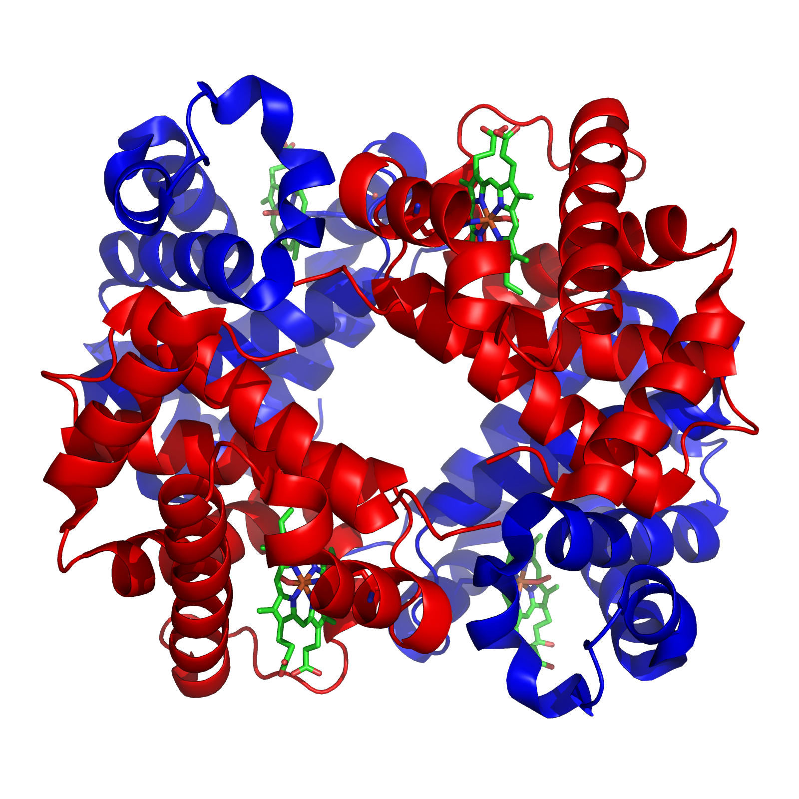
Hemoglobin, which is the principal oxygen-carrier in humans, has four subunits in which the iron(II) ion is coordinated by the planar macrocyclic ligand protoporphyrin IX (PIX) and the imidazole nitrogen atom of a histidine residue. The sixth coordination site contains a water molecule or a dioxygen molecule. By contrast the protein myoglobin, found in muscle cells, has only one such unit. The active site is located in a hydrophobic pocket. This is important as without it the iron(II) would be irreversibly oxidized to iron(III). The equilibrium constant for the formation of HbO2 is such that oxygen is taken up or released depending on the partial pressure of oxygen in the lungs or in muscle. In hemoglobin the four subunits show a cooperativity effect that allows for easy oxygen transfer from hemoglobin to myoglobin.[10]

In both hemoglobin and myoglobin it is sometimes incorrectly stated that the oxygenated species contains iron(III). It is now known that the diamagnetic nature of these species is because the iron(II) atom is in the low-spin state. In oxyhemoglobin the iron atom is located in the plane of the porphyrin ring, but in the paramagnetic deoxyhemoglobin the iron atom lies above the plane of the ring.[10] This change in spin state is a cooperative effect due to the higher crystal field splitting and smaller ionic radius of Fe2+ in the oxyhemoglobin moiety.

Hemerythrin is another iron-containing oxygen carrier. The oxygen binding site is a binuclear iron center. The iron atoms are coordinated to the protein through the carboxylate side chains of a glutamate and aspartate and five histidine residues. The uptake of O2 by hemerythrin is accompanied by two-electron oxidation of the reduced binuclear center to produce bound peroxide (OOH). The mechanism of oxygen uptake and release have been worked out in detail.[11][12]

Hemocyanins carry oxygen in the blood of most mollusks, and some arthropods such as the horseshoe crab. They are second only to hemoglobin in biological popularity of use in oxygen transport. On oxygenation the two copper(I) atoms at the active site are oxidized to copper(II) and the dioxygen molecules are reduced to peroxide, O2−
2
.[13][14]

Chlorocruorin (as the larger carrier erythrocruorin) is an oxygen-binding hemeprotein present in the blood plasma of many annelids, particularly certain marine polychaetes.

Cytochromes

[edit]

Oxidation and reduction reactions are not common in organic chemistry as few organic molecules can act as oxidizing or reducing agents. Iron(II), on the other hand, can easily be oxidized to iron(III). This functionality is used in cytochromes, which function as electron-transfer vectors. The presence of the metal ion allows metalloenzymes to perform functions such as redox reactions that cannot easily be performed by the limited set of functional groups found in amino acids.[15] The iron atom in most cytochromes is contained in a heme group. The differences between those cytochromes lies in the different side-chains. For instance cytochrome a has a heme a prosthetic group and cytochrome b has a heme b prosthetic group. These differences result in different Fe2+/Fe3+ redox potentials such that various cytochromes are involved in the mitochondrial electron transport chain.[16]

Cytochrome P450 enzymes perform the function of inserting an oxygen atom into a C−H bond, an oxidation reaction.[17][18]

Rubredoxin

[edit]
Rubredoxin active site.

Rubredoxin is an electron-carrier found in sulfur-metabolizing bacteria and archaea. The active site contains an iron ion coordinated by the sulfur atoms of four cysteine residues forming an almost regular tetrahedron. Rubredoxins perform one-electron transfer processes. The oxidation state of the iron atom changes between the +2 and +3 states. In both oxidation states the metal is high spin, which helps to minimize structural changes.

Plastocyanin

[edit]
The copper site in plastocyanin

Plastocyanin is one of the family of blue copper proteins that are involved in electron transfer reactions. The copper-binding site is described as distorted trigonal pyramidal.[19] The trigonal plane of the pyramidal base is composed of two nitrogen atoms (N1 and N2) from separate histidines and a sulfur (S1) from a cysteine. Sulfur (S2) from an axial methionine forms the apex. The distortion occurs in the bond lengths between the copper and sulfur ligands. The Cu−S1 contact is shorter (207 pm) than Cu−S2 (282 pm). The elongated Cu−S2 bonding destabilizes the Cu(II) form and increases the redox potential of the protein. The blue color (597 nm peak absorption) is due to the Cu−S1 bond where S(pπ) to Cu(dx2y2) charge transfer occurs.[20]

In the reduced form of plastocyanin, His-87 will become protonated with a pKa of 4.4. Protonation prevents it acting as a ligand and the copper site geometry becomes trigonal planar.

Metal-ion storage and transfer

[edit]

Iron

[edit]

Iron is stored as iron(III) in ferritin. The exact nature of the binding site has not yet been determined. The iron appears to be present as a hydrolysis product such as FeO(OH). Iron is transported by transferrin whose binding site consists of two tyrosines, one aspartic acid and one histidine.[21] The human body has no controlled mechanism for excretion of iron.[22] This can lead to iron overload problems in patients treated with blood transfusions, as, for instance, with β-thalassemia. Iron is actually excreted in urine[23] and is also concentrated in bile[24] which is excreted in feces.[25]

Copper

[edit]

Ceruloplasmin is the major copper-carrying protein in the blood. Ceruloplasmin exhibits oxidase activity, which is associated with possible oxidation of Fe(II) into Fe(III), therefore assisting in its transport in the blood plasma in association with transferrin, which can carry iron only in the Fe(III) state.

Calcium

[edit]

Osteopontin is involved in mineralization in the extracellular matrices of bones and teeth.

Metalloenzymes

[edit]

Metalloenzymes all have one feature in common, namely that the metal ion is bound to the protein with one labile coordination site. As with all enzymes, the shape of the active site is crucial. The metal ion is usually located in a pocket whose shape fits the substrate. The metal ion catalyzes reactions that are difficult to achieve in organic chemistry.

Carbonic anhydrase

[edit]
Active site of carbonic anhydrase. The three coordinating histidine residues are shown in green, hydroxide in red and white, and the zinc in gray.

In aqueous solution, carbon dioxide forms carbonic acid

CO2 + H2O ⇌ H2CO3

This reaction is very slow in the absence of a catalyst, but quite fast in the presence of the hydroxide ion

CO2 + OHHCO
3

A reaction similar to this is almost instantaneous with carbonic anhydrase. The structure of the active site in carbonic anhydrases is well known from a number of crystal structures. It consists of a zinc ion coordinated by three imidazole nitrogen atoms from three histidine units. The fourth coordination site is occupied by a water molecule. The coordination sphere of the zinc ion is approximately tetrahedral. The positively-charged zinc ion polarizes the coordinated water molecule, and nucleophilic attack by the negatively-charged hydroxide portion on carbon dioxide proceeds rapidly. The catalytic cycle produces the bicarbonate ion and the hydrogen ion[2] as the equilibrium:

H2CO3HCO
3
+ H+

favouring dissociation of carbonic acid at biological pH values.[26]

Vitamin B12-dependent enzymes

[edit]

The cobalt-containing Vitamin B12 (also known as cobalamin) catalyzes the transfer of methyl (−CH3) groups between two molecules, which involves the breaking of C−C bonds, a process that is energetically expensive in organic reactions. The metal ion lowers the activation energy for the process by forming a transient Co−CH3 bond.[27] The structure of the coenzyme was famously determined by Dorothy Hodgkin and co-workers, for which she received a Nobel Prize in Chemistry.[28] It consists of a cobalt(II) ion coordinated to four nitrogen atoms of a corrin ring and a fifth nitrogen atom from an imidazole group. In the resting state there is a Co−C sigma bond with the 5′ carbon atom of adenosine.[29] This is a naturally occurring organometallic compound, which explains its function in trans-methylation reactions, such as the reaction carried out by methionine synthase.

Nitrogenase (nitrogen fixation)

[edit]

The fixation of atmospheric nitrogen is an energy-intensive process, as it involves breaking the very stable triple bond between the nitrogen atoms. The nitrogenases catalyze the process. One such enzyme occurs in Rhizobium bacteria. There are three components to its action: a molybdenum atom at the active site, iron–sulfur clusters that are involved in transporting the electrons needed to reduce the nitrogen, and an abundant energy source in the form of magnesium ATP. This last is provided by a mutualistic symbiosis between the bacteria and a host plant, often a legume. The reaction may be written symbolically as

N2 + 16 MgATP + 8 e → 2 NH3 + 16 MgADP +16 Pi + H2

where Pi stands for inorganic phosphate. The precise structure of the active site has been difficult to determine. It appears to contain a MoFe7S8 cluster that is able to bind the dinitrogen molecule and, presumably, enable the reduction process to begin.[30] Some species of bacteria and archaea have also been shown to have Vanadium nitrogenases, which contain a VFe3S4 cluster and allows for an alternative pathway of nitrogen fixation in Molybdenum-deficient conditions.[31] The electrons are transported by the associated "P" cluster, which contains two cubical Fe4S4 clusters joined by sulfur bridges.[32]

Superoxide dismutase

[edit]
Structure of a human superoxide dismutase 2 tetramer

The superoxide ion, O
2
is generated in biological systems by reduction of molecular oxygen. It has an unpaired electron, so it behaves as a free radical. It is a powerful oxidizing agent. These properties render the superoxide ion very toxic and are deployed to advantage by phagocytes to kill invading microorganisms. Otherwise, the superoxide ion must be destroyed before it does unwanted damage in a cell. The superoxide dismutase enzymes perform this function very efficiently.[33]

The formal oxidation state of the oxygen atoms is −12. In solutions at neutral pH, the superoxide ion disproportionates to molecular oxygen and hydrogen peroxide.

O
2
+ 2 H+ → O2 + H2O2

In biology this type of reaction is called a dismutation reaction. It involves both oxidation and reduction of superoxide ions. The superoxide dismutase (SOD) group of enzymes increase the rate of reaction to near the diffusion-limited rate.[34] The key to the action of these enzymes is a metal ion with variable oxidation state that can act either as an oxidizing agent or as a reducing agent.

Oxidation: M(n+1)+ + O
2
→ Mn+ + O2
Reduction: Mn+ + O
2
+ 2 H+ → M(n+1)+ + H2O2.

In human SOD, the active metal is copper, as Cu(II) or Cu(I), coordinated tetrahedrally by four histidine residues. This enzyme also contains zinc ions for stabilization and is activated by copper chaperone for superoxide dismutase (CCS). Other isozymes may contain iron, manganese or nickel. The activity of Ni-SOD involves nickel(III), an unusual oxidation state for this element. The active site nickel geometry cycles from square planar Ni(II), with thiolate (Cys2 and Cys6) and backbone nitrogen (His1 and Cys2) ligands, to square pyramidal Ni(III) with an added axial His1 side chain ligand.[35]

Chlorophyll-containing proteins

[edit]
Hemoglobin (left) and chlorophyll (right), two extremely different molecules when it comes to function, are quite similar when it comes to its atomic shape. There are only three major structural differences; a magnesium atom (Mg) in chlorophyll, as opposed to iron (Fe) in hemoglobin. Additionally, chlorophyll has an extended isoprenoid tail and an additional aliphatic cyclic structure off the macrocycle.

Chlorophyll plays a crucial role in photosynthesis. It contains a magnesium enclosed in a chlorin ring. However, the magnesium ion is not directly involved in the photosynthetic function and can be replaced by other divalent ions with little loss of activity. Rather, the photon is absorbed by the chlorin ring, whose electronic structure is well-adapted for this purpose.

Initially, the absorption of a photon causes an electron to be excited into a singlet state of the Q band. The excited state undergoes an intersystem crossing from the singlet state to a triplet state in which there are two electrons with parallel spin. This species is, in effect, a free radical, and is very reactive and allows an electron to be transferred to acceptors that are adjacent to the chlorophyll in the chloroplast. In the process chlorophyll is oxidized. Later in the photosynthetic cycle, chlorophyll is reduced back again. This reduction ultimately draws electrons from water, yielding molecular oxygen as a final oxidation product.

Hydrogenase

[edit]

Hydrogenases are subclassified into three different types based on the active site metal content: iron–iron hydrogenase, nickel–iron hydrogenase, and iron hydrogenase.[36] All hydrogenases catalyze reversible H2 uptake, but while the [FeFe] and [NiFe] hydrogenases are true redox catalysts, driving H2 oxidation and H+ reduction

H2 ⇌ 2 H+ + 2 e

the [Fe] hydrogenases catalyze the reversible heterolytic cleavage of H2.

H2 ⇌ H+ + H
The active site structures of the three types of hydrogenase enzymes.

Ribozyme and deoxyribozyme

[edit]

Since discovery of ribozymes by Thomas Cech and Sidney Altman in the early 1980s, ribozymes have been shown to be a distinct class of metalloenzymes.[37] Many ribozymes require metal ions in their active sites for chemical catalysis; hence they are called metalloenzymes. Additionally, metal ions are essential for structural stabilization of ribozymes. Group I intron is the most studied ribozyme which has three metals participating in catalysis.[38] Other known ribozymes include group II intron, RNase P, and several small viral ribozymes (such as hammerhead, hairpin, HDV, and VS) and the large subunit of ribosomes. Several classes of ribozymes have been described.[39]

Deoxyribozymes, also called DNAzymes or catalytic DNA, are artificial DNA-based catalysts that were first produced in 1994.[40] Almost all DNAzymes require metal ions. Although ribozymes mostly catalyze cleavage of RNA substrates, a variety of reactions can be catalyzed by DNAzymes including RNA/DNA cleavage, RNA/DNA ligation, amino acid phosphorylation and dephosphorylation, and carbon–carbon bond formation.[41] Yet, DNAzymes that catalyze RNA cleavage reaction are the most extensively explored ones. 10-23 DNAzyme, discovered in 1997, is one of the most studied catalytic DNAs with clinical applications as a therapeutic agent.[42] Several metal-specific DNAzymes have been reported including the GR-5 DNAzyme (lead-specific),[43] the CA1-3 DNAzymes (copper-specific), the 39E DNAzyme (uranyl-specific)[44] and the NaA43 DNAzyme (sodium-specific).[45]

Signal-transduction metalloproteins

[edit]

Calmodulin

[edit]
EF-hand motif

Calmodulin is an example of a signal-transduction protein. It is a small protein that contains four EF-hand motifs, each of which is able to bind a Ca2+ ion.

In an EF-hand loop protein domain, the calcium ion is coordinated in a pentagonal bipyramidal configuration. Six glutamic acid and aspartic acid residues involved in the binding are in positions 1, 3, 5, 7 and 9 of the polypeptide chain. At position 12, there is a glutamate or aspartate ligand that behaves as a bidentate ligand, providing two oxygen atoms. The ninth residue in the loop is necessarily glycine due to the conformational requirements of the backbone. The coordination sphere of the calcium ion contains only carboxylate oxygen atoms and no nitrogen atoms. This is consistent with the hard nature of the calcium ion.

The protein has two approximately symmetrical domains, separated by a flexible "hinge" region. Binding of calcium causes a conformational change to occur in the protein. Calmodulin participates in an intracellular signaling system by acting as a diffusible second messenger to the initial stimuli.[46][47]

Troponin

[edit]

In both cardiac and skeletal muscles, muscular force production is controlled primarily by changes in the intracellular calcium concentration. In general, when calcium rises, the muscles contract and, when calcium falls, the muscles relax. Troponin, along with actin and tropomyosin, is the protein complex to which calcium binds to trigger the production of muscular force.

Transcription factors

[edit]
Zinc finger. The zinc ion (green) is coordinated by two histidine residues and two cysteine residues.

Many transcription factors contain a structure known as a zinc finger, a structural module in which a region of protein folds around a zinc ion. The zinc does not directly contact the DNA that these proteins bind to. Instead, the cofactor is essential for the stability of the tightly folded protein chain.[48] In these proteins, the zinc ion is usually coordinated by pairs of cysteine and histidine side-chains.

Other metalloenzymes

[edit]

See also

[edit]

References

[edit]
[edit]
Revisions and contributorsEdit on WikipediaRead on Wikipedia
from Grokipedia
A metalloprotein is a protein that binds one or more metal ions as cofactors through coordination bonds, typically involving donor atoms from amino acid side chains such as , , aspartate, or glutamate, or from exogenous ligands. These metal centers, often featuring transition metals like iron, , , or , are integral to the protein's structure and function, enabling roles that organic molecules alone cannot achieve. Metalloproteins comprise approximately one-third of all proteins in biological systems, underscoring their ubiquity across all domains of life. The structural diversity of metalloproteins arises from varied protein scaffolds, including α-helical bundles, β-sheets, and mixed folds, which position metal-binding sites with precise geometries to optimize reactivity. The coordination environment is defined by a primary of direct ligands and a secondary of nearby residues that fine-tune electronic properties, stability, and substrate specificity. For instance, groups in iron-containing metalloproteins feature rings coordinating the metal, while sites often involve tetrahedral arrangements with and donors. Functionally, metalloproteins catalyze essential reactions, with many acting as enzymes in classes like oxidoreductases, which facilitate electron transfer and redox processes critical for energy metabolism. Notable roles include oxygen transport in hemoglobin, nitrogen fixation in nitrogenase, and hydrolysis in carbonic anhydrase, where the metal ion lowers activation energies or stabilizes intermediates. Beyond catalysis, they support structural integrity, as in zinc fingers for DNA binding, and signaling pathways, highlighting their indispensability in cellular homeostasis and adaptation. The study of metalloproteins, rooted in , reveals evolutionary conservation of metal sites across protein families, often independent of overall fold, which informs targeting dysregulated forms in diseases like cancer and neurodegeneration. Advances in techniques such as and neutron diffraction continue to elucidate their mechanisms, emphasizing the metal's role in achieving high selectivity and efficiency in biological processes.

Fundamentals

Definition and Classification

Metalloproteins are proteins that contain one or more metal ions or metal clusters essential for their biological function, structure, or regulation, with the metals typically bound through coordination to side chains or cofactors either covalently or non-covalently. These metals, which include transition and non-transition elements such as iron, , , calcium, and , serve as cofactors that enable diverse roles in cellular processes. In humans, metalloproteins constitute approximately 30-40% of the , highlighting their prevalence and critical importance in . The study of metalloproteins traces back to the 19th century, when was identified as an iron-containing protein responsible for oxygen transport in blood, marking one of the earliest recognized examples. Modern structural insights emerged in the 1950s through , with the first atomic-resolution structures of (1958) and (1960) revealing the precise coordination of heme-bound iron, which revolutionized understanding of metal-protein interactions. These milestones shifted the field from biochemical characterization to detailed mechanistic analysis. Metalloproteins are classified in multiple ways to reflect their diversity. By metal type, they are grouped according to the incorporated element, such as iron-based (e.g., proteins like or non-heme iron-sulfur clusters in ), zinc-based (e.g., transcription factors), copper-based (e.g., blue copper proteins like ), calcium-based (e.g., for signaling), and molybdenum-based (e.g., for ). By function, they fall into categories including catalytic (s like ), transport and storage (e.g., for iron), (e.g., ), structural (e.g., stabilizing protein folds), and regulatory (e.g., modulating activity). By binding site, classifications distinguish (porphyrin-coordinated metals, primarily iron) from non-heme sites, and mononuclear (single metal ion) from polynuclear clusters (e.g., Fe-S clusters with 2-4 irons linked by sulfides). These schemes underscore the adaptability of metalloproteins, with coordination geometries like octahedral or tetrahedral motifs briefly linking to broader chemical principles.

Coordination Chemistry Principles

Metalloproteins feature metal ions bound to protein scaffolds through coordination bonds, where the metal acts as a Lewis acid and protein-derived groups or exogenous molecules serve as Lewis bases. These interactions are governed by principles of coordination chemistry, including ligand donor atom identity, , and , which dictate the electronic and functional properties of the site. The stability of these complexes arises from electrostatic attractions, covalent contributions, and thermodynamic factors, enabling precise control over reactivity in biological contexts. Common ligands in metalloproteins include side chains from such as the imidazole nitrogen of , the thiolate sulfur of , and the carboxylate oxygen of aspartate or glutamate, which provide , , and oxygen donors, respectively. Non-protein ligands, like molecules or dioxygen, often occupy remaining coordination sites, particularly in dynamic environments. These ligands are selected based on their ability to match the metal's electronic requirements, with being prevalent due to its versatile imidazolate donor. Coordination geometries in metalloproteins vary with the metal ion and set, commonly adopting tetrahedral arrangements for sites, octahedral for iron or magnesium, and square planar for in certain motifs. These geometries are influenced by , which describes how ligands split the d-orbitals of transition metals, affecting electronic transitions and reactivity. For octahedral complexes, the crystal field stabilization energy (CFSE) quantifies this splitting: CFSE=[0.4nt+0.6ne]Δo\text{CFSE} = [-0.4 n_t + 0.6 n_e] \Delta_o where ntn_t is the number of electrons in the t2gt_{2g} orbitals, nen_e is the number in the ege_g orbitals, and Δo\Delta_o is the octahedral splitting parameter. This energy stabilization favors specific geometries and oxidation states, enhancing site functionality. The stability and selectivity of metal binding are largely explained by the hard-soft acid-base (HSAB) theory, which predicts preferential interactions between hard acids (e.g., high-charge-density ions like Zn²⁺) and hard bases (e.g., oxygen donors from carboxylates) versus soft acids (e.g., Cu⁺) and soft bases (e.g., sulfur from thiolates). Entropy effects further modulate binding, as chelation by multidentate ligands reduces translational and rotational freedom but releases solvent molecules, contributing favorably to the overall free energy. Coordination environment also tunes redox potentials; for instance, soft sulfur ligands stabilize lower oxidation states, shifting potentials to more negative values and facilitating electron transfer. Spectroscopic methods are essential for characterizing these coordination sites. Ultraviolet-visible (UV-Vis) spectroscopy probes d-d transitions and charge-transfer bands, revealing geometry and ligand field strength. Electron paramagnetic resonance (EPR) detects unpaired electrons in paramagnetic metals like Cu²⁺ or Fe³⁺, providing information on spin state and ligand symmetry. Extended X-ray absorption fine structure (EXAFS) determines metal-ligand distances and coordination numbers with atomic precision, even in non-crystalline samples. These techniques, often used in combination, offer complementary insights into site structure and dynamics.

Abundance and Distribution

Prevalence in Biological Systems

Metalloproteins are ubiquitous across all domains of life, constituting a significant portion of the in diverse organisms. Estimates indicate that approximately 30% of all proteins in biological systems bind metal ions, with nearly half of all enzymes classified as metalloenzymes that require metal cofactors for catalytic activity. In prokaryotes, the prevalence is particularly high, with bioinformatic analyses suggesting that 20-30% of the in model organisms like involves metal-binding proteins, including around 144 iron-sulfur cluster-containing proteins alone, representing over 3% of the total . This proportion underscores the essential role of metalloproteins in prokaryotic and survival. The distribution of metalloproteins varies by organism type and environmental niche. They are present in , , and eukaryotes, but adaptations reflect physiological demands; for instance, aerobic and eukaryotes often feature iron-rich metalloproteins such as heme-containing for oxygen handling, while nitrogen-fixing prokaryotes like Rhizobium species express molybdenum-dependent enzymes for N₂ reduction. In eukaryotes, zinc-binding proteins comprise about 9% of the , higher than the 5-6% in prokaryotes, highlighting evolutionary divergences in metal utilization. Within cells, metalloproteins localize to specific compartments based on function. Zinc enzymes, such as , predominate in the for pH regulation and CO₂ transport, while iron-sulfur clusters are enriched in mitochondria for in the respiratory chain. In plant chloroplasts, magnesium ions coordinate within proteins, enabling light harvesting and . Proteomic analyses, particularly those combining with (ICP-MS), have been instrumental in mapping these distributions and quantifying metal content, revealing previously underappreciated metalloproteins in viruses; for example, several proteins, including the papain-like protease, feature structural zinc-binding sites essential for . Environmental metal availability profoundly influences metalloprotein expression and adaptation. In iron-limited conditions, bacteria like E. coli upregulate biosynthesis, such as enterobactin, to scavenge Fe³⁺ for incorporation into iron-dependent proteins like . Similarly, molybdenum scarcity can repress activity in diazotrophs, demonstrating how metalloproteins enable dynamic responses to stress.

Evolutionary Origins

The evolutionary origins of metalloproteins are rooted in the geochemical conditions of , where anaerobic oceans approximately 4 billion years ago were rich in bioavailable iron and , facilitating the formation of simple iron-sulfur (Fe-S) clusters as primordial cofactors in prebiotic chemistry. These clusters likely emerged in environments, where iron monosulfide minerals promoted the assembly of Fe-S structures essential for early processes, predating cellular . In the (LUCA), estimated to have existed around 3.8–4.2 billion years ago, dedicated machineries for Fe-S cluster biosynthesis were already present, supporting metabolic functions in an anaerobic, acetogenic . Phylogenetic analyses indicate that these ancient Fe-S proteins, such as , were central to LUCA's , with evidence from showing their conservation across bacterial and archaeal lineages. A pivotal milestone in metalloprotein diversification occurred during the (GOE) around 2.4 billion years ago, when rising atmospheric oxygen levels oxidized soluble Fe²⁺ to insoluble Fe³⁺, reducing iron bioavailability while solubilizing previously inaccessible metals like (as Cu²⁺) and . This geochemical shift prompted evolutionary adaptations, enabling the rise of copper- and -dependent proteins; for instance, copper enzymes such as cytochrome c oxidases emerged in aerobic post-GOE, replacing less efficient iron-based alternatives for oxygen reduction. Similarly, zinc fingers and superoxide dismutases incorporating proliferated in eukaryotes, reflecting a transition from iron-centric to more diverse metal utilization in oxygenated environments. enzymes, integral to anaerobic metabolisms like , trace back to early prokaryotes, with phylogenomic studies post-2020 revealing their presence in LUCA and widespread in ancient anaerobes, underscoring molybdenum's role in pre-GOE biogeochemical cycles. Gene duplication and horizontal gene transfer (HGT) further drove the diversification of metalloprotein active sites, allowing adaptation to varying metal availabilities. For example, ancient underwent to form modular chains, evolving into complex systems like those in modern through symmetrical microenvironment pairings. HGT facilitated the spread of metalloprotein genes across domains; bacterial were transferred to and eukaryotes, enabling the integration of Fe-S clusters into diverse respiratory pathways. In molybdenum nitrogenases, HGT events dating to the early distributed genes among prokaryotes, promoting nitrogen metabolism in anaerobic niches. In contemporary contexts, evolutionary pressures from have spurred the development of resistance mechanisms, such as -binding metallothioneins, which evolved from ancient zinc-binding scaffolds to sequester toxic ions in polluted environments. These proteins, often arising via in and plants exposed to industrial , exemplify ongoing metalloprotein adaptation to anthropogenic , with enhanced binding affinities conferring survival advantages.

Storage and Transport Functions

Oxygen Carriers

Oxygen carriers are metalloproteins specialized for the reversible binding and transport of molecular oxygen (O₂) to tissues, primarily through coordination to transition metal ions within protein active sites. In vertebrates, the most prominent examples are heme-containing proteins, where iron serves as the central metal. Hemoglobin (Hb), a tetrameric protein consisting of two α and two β subunits, facilitates cooperative oxygen binding in red blood cells, enabling efficient uptake in the lungs and release in peripheral tissues. This cooperativity arises from allosteric transitions between tense (T) and relaxed (R) states, allowing the protein to adapt to varying physiological demands. Myoglobin (Mb), in contrast, is a monomeric protein found in muscle tissues, functioning primarily as an molecule to support sustained contraction during periods of high demand. Its higher oxygen affinity compared to ensures rapid release to mitochondria under hypoxic conditions. The three-dimensional structure of myoglobin, first elucidated by , revealed a compact globular fold with eight α-helices enclosing the prosthetic group. The core mechanism of oxygen binding in these heme proteins involves ferrous iron (Fe²⁺) coordinated within a ring. The iron is axially ligated by a proximal residue, leaving a sixth coordination site available for O₂. Upon binding, the Fe²⁺ shifts from high-spin to low-spin, triggering conformational changes that facilitate subsequent bindings in . The overall reaction for hemoglobin oxygenation is represented as: Hb+4O2Hb(O2)4\mathrm{Hb + 4O_2 \rightleftharpoons Hb(O_2)_4} This equilibrium is modulated by allosteric effectors such as 2,3-bisphosphoglycerate (2,3-BPG), which binds to the deoxyhemoglobin T-state in a central cavity, stabilizing it and reducing oxygen affinity to promote unloading in tissues. The tetrameric architecture of hemoglobin, resolved by , underscores how subunit interfaces transmit these allosteric signals. Beyond heme-based carriers, non-heme variants exist in . , a copper-containing , serves as the primary oxygen transporter in arthropods and mollusks, where it binds O₂ at binuclear Cu(I) sites, turning upon oxygenation due to charge-transfer transitions. Each functional unit reversibly binds one O₂ molecule, with assembly into large multi-subunit complexes (up to 48 subunits in some mollusks) enabling high-capacity transport. , found in marine worms such as sipunculids and priapulids, employs a non-heme diiron center for O₂ binding, forming a peroxo-bridged Fe(III)-Fe(III) complex in the oxygenated state without color change. Physiologically, these proteins ensure tissue oxygenation by exploiting gradients in partial pressure (pO₂) and environmental factors. In hemoglobin, the Bohr effect describes the pH-dependent decrease in oxygen affinity, where lower pH (from CO₂ and H⁺ accumulation in active tissues) protonates specific residues, favoring O₂ release. This heterotropic allostery enhances delivery efficiency, with up to 2.5 protons released per O₂ bound at physiological pH. Disruptions in hemoglobin function, such as the Glu6Val mutation in the β-globin chain causing sickle cell anemia, lead to polymerization of deoxyhemoglobin under low pO₂, resulting in distorted erythrocytes, vaso-occlusion, and hemolytic crises. This single amino acid substitution, identified by Vernon Ingram, exemplifies how point mutations can profoundly impact metalloprotein stability and function.

Electron Transfer Proteins

Electron transfer proteins are a vital class of metalloproteins that mediate the movement of electrons in biological processes such as , , and metabolic pathways, primarily through redox-active metal centers including iron, iron-sulfur clusters, and ions. These proteins enable efficient transduction by shuttling electrons between donors and acceptors, often over distances of 10-14 via quantum mechanical tunneling, with rates optimized by the protein environment to minimize loss. The potentials of these metal centers, typically ranging from -0.5 V to +0.5 V versus the , dictate the thermodynamic favorability of electron flow, while structural features like the secondary fine-tune kinetics and specificity. Cytochromes represent a major family of heme-containing electron transfer proteins, where the iron atom in the porphyrin ring cycles between Fe(II) and Fe(III) states. In mitochondrial respiration, cytochrome c serves as a soluble carrier in the electron transport chain (ETC), transferring electrons from complex III (cytochrome bc1) to complex IV (cytochrome c oxidase), with a standard redox potential of approximately +0.25 V that positions it ideally between ubiquinone (+0.06 V) and oxygen (+0.82 V). This protein exemplifies octahedral iron coordination, often with histidine and methionine axial ligands, which stabilizes the heme and modulates the potential by about 100-200 mV compared to bis-histidine coordination. In photosynthesis, cytochrome f in the b6f complex similarly facilitates electron transfer from photosystem II to plastocyanin. The efficiency of cytochrome-mediated transfer follows Marcus theory, which describes the rate constant for non-adiabatic electron tunneling as ket=2πV2(4πλkBT)1/2exp[(ΔG+λ)24λkBT]k_{et} = \frac{2\pi}{\hbar} |V|^2 (4\pi \lambda k_B T)^{-1/2} \exp\left[ -\frac{(\Delta G + \lambda)^2}{4\lambda k_B T} \right], where VV is the electronic coupling, λ\lambda the reorganization energy, and ΔG\Delta G the driving force; in cytochromes, low λ\lambda values (around 0.5-1.0 eV) enable rates up to 10^6 s^{-1}. Iron-sulfur proteins, including rubredoxins and , utilize Fe-S centers for , particularly in anaerobic and photosynthetic electron flow. Rubredoxins feature a mononuclear iron atom tetrahedrally coordinated by four sulfurs, with potentials between -0.1 V and +0.05 V, enabling roles in bacterial respiration such as alkane in oleovorans. , containing [2Fe-2S], [3Fe-4S], or [4Fe-4S] clusters, exhibit lower potentials (-0.4 V to -0.3 V for plant-type [2Fe-2S]), facilitating electron delivery from to NADP+ reductase in chloroplasts or in anaerobic pathways like . These clusters support delocalized with reorganization energies of 0.6-0.8 eV, contributing to the ETC's complex I () where multiple Fe-S centers bridge flavin to . Blue copper proteins, such as plastocyanins and azurins, employ type 1 copper sites for rapid electron transfer, characterized by a trigonal geometry with two histidines, a cysteine, and a weakly bound methionine. Plastocyanins, found in chloroplast thylakoids, shuttle electrons from cytochrome f to photosystem I with a redox potential of about +0.37 V, supporting photosynthetic charge separation. Azurins, in bacterial periplasm, perform analogous functions in denitrification with potentials around +0.30 V. The "entatic state" in these proteins enforces a strained Cu(II) geometry that resembles the reduced Cu(I) form, reducing reorganization energy to ~0.1-0.2 eV and accelerating self-exchange rates to 10^3-10^4 M^{-1} s^{-1}, far exceeding typical Cu^{2+/+} couples, as per Marcus theory predictions. This entatic control, induced by the protein scaffold, ensures minimal structural change during redox cycling, optimizing turnover in metabolic chains.

Metal Ion Storage and Transfer

Metalloproteins play a crucial role in the sequestration, storage, and controlled delivery of metal ions within biological systems, preventing from free ions while ensuring availability for essential processes. For iron , serves as the primary intracellular storage protein, forming a spherical 24-subunit nanocage approximately 12 nm in diameter that encapsulates up to 4,500 Fe³⁺ ions in the form of a mineral core, thereby maintaining iron in a non-toxic, bioavailable state. In contrast, functions as the main serum transport protein, a bilobal that reversibly binds two Fe³⁺ ions per molecule in synergy with anions, facilitating safe circulation and delivery to cells via . Copper management involves specialized proteins that handle its redox-active nature to avoid oxidative damage. , a multidomain containing six to seven atoms, exhibits ferroxidase activity by oxidizing Fe²⁺ to Fe³⁺, which promotes iron loading into and accounts for about 95% of circulating transport in plasma. Metallothioneins, small cysteine-rich proteins with up to 30 residues per subunit, bind Cu⁺ and Zn²⁺ ions through thiolate clusters, enabling intracellular storage, detoxification of excess metals, and regulation of their across tissues. For calcium, intracellular buffering is mediated by proteins like , which utilizes multiple EF-hand motifs—helical loops that coordinate Ca²⁺ with high affinity—to rapidly sequester ions and maintain cytosolic concentrations, thereby protecting against overload in neurons and other cells. Similarly, parvalbumin, abundant in fast-twitch fibers, employs EF-hand domains to bind Ca²⁺ transiently during contraction-relaxation cycles, accelerating relaxation by facilitating ion removal from . Key mechanisms in metal ion handling distinguish apo-forms (metal-free proteins) from holo-forms (metal-bound), where metal insertion often stabilizes structure and enables function, as seen in the conformational shifts of and upon iron binding. Dedicated chaperones ensure targeted delivery; for instance, the copper chaperone for (CCS), a dimeric protein with a dedicated copper-binding domain, specifically shuttles Cu⁺ to Cu/Zn- () via direct protein-protein interaction, inserting the ion into the enzyme's without releasing free . Dysregulation of these systems underlies disorders such as hereditary hemochromatosis, where mutations in iron regulators like HFE lead to overload and excessive hepatic iron accumulation, causing tissue damage and . Likewise, Wilson's disease results from ATP7B mutations impairing export and maturation, leading to hepatic and neurological mishandling, with metallothioneins overwhelmed and free levels rising toxically.

Catalytic Functions

Hydrolase Enzymes

Hydrolase enzymes represent a key class of that catalyze the cleavage of chemical bonds through , often utilizing ions to activate water molecules as . serves as a paradigmatic example, featuring a (II) ion at its coordinated by three imidazole nitrogen atoms from residues (His94, His96, and His119) in a tetrahedral geometry, which polarizes a bound water molecule to form a essential for . This coordination enhances the enzyme's ability to interconvert and , facilitating rapid physiological responses. The catalytic mechanism of carbonic anhydrase involves the nucleophilic attack of the zinc-bound hydroxide on CO₂, forming a Zn-bound bicarbonate intermediate (Zn²⁺-HCO₃⁻) that dissociates to yield HCO₃⁻ and Zn²⁺-H₂O, followed by proton transfer (often via His64) to regenerate the active species. The process can be summarized as: \ceCO2+Zn2+OH>Zn2+HCO3>Zn2+H2O+HCO3\ce{CO2 + Zn^{2+}-OH^- -> Zn^{2+}-HCO3^- -> Zn^{2+}-H2O + HCO3^-} This yields the overall reaction \ceCO2+H2OHCO3+H+\ce{CO2 + H2O \rightleftharpoons HCO3^- + H^+}, with the enzyme achieving diffusion-limited kinetics at a turnover rate of approximately 106s110^6 \, \mathrm{s^{-1}} for the human α-isoform (hCA II). Carbonic anhydrase exists in distinct isozyme classes: α-forms predominate in animals, β-forms in plants and some bacteria, and γ-forms primarily in bacteria, each adapted to specific cellular environments while sharing zinc-dependent catalysis. Recent structural studies, including those from the 2020s, have revealed dynamic fluctuations in the zinc coordination sphere and active-site waters, underscoring how solvent dynamics contribute to product release and high efficiency. Physiologically, plays critical roles in regulation and CO₂ transport, enabling efficient in tissues and blood by accelerating formation and buffering protons in erythrocytes and renal cells. Inhibitors such as , which bind the site and block hydroxide formation, are clinically used to reduce in treatment by decreasing aqueous humor production. Beyond , other zinc-dependent hydrolases include matrix metalloproteinases (MMPs), which feature a conserved for hydrolyzing bonds in components like , thereby driving tissue remodeling during development and . , incorporating two ions and one magnesium ion in its , catalyzes the of phosphate esters to release inorganic phosphate, supporting mineralization and recycling. These enzymes highlight the versatility of metal coordination in promoting nucleophilic activation for diverse hydrolytic functions.

Redox Enzymes

Redox enzymes are a class of metalloproteins that facilitate essential for cellular , production, and defense against . These enzymes employ metal centers, such as , iron, and , to mediate , enabling the of reactions involving (ROS) and other substrates. By cycling between oxidation states, these metal ions lower activation energies and achieve high catalytic efficiencies, often approaching diffusion-limited rates. A prominent example is (SOD), particularly the copper-zinc form (Cu/Zn-SOD), which catalyzes the dismutation of radicals (O₂⁻•) into (H₂O₂) and molecular oxygen (O₂). The reaction proceeds via a cycle at the center, where Cu²⁺ is reduced to Cu⁺ by the first anion, followed by reoxidation of Cu⁺ by a second , with the ion stabilizing the structure. This mechanism operates at a near-diffusion-controlled rate constant of approximately 10⁹ M⁻¹ s⁻¹, ensuring rapid detoxification of in aerobic organisms. The overall reaction is: 2O2+2H+H2O2+O22 \text{O}_2^{\bullet-} + 2 \text{H}^+ \rightarrow \text{H}_2\text{O}_2 + \text{O}_2 Cu/Zn-SOD is ubiquitously expressed in eukaryotic cells, contributing to the first line of antioxidant defense by preventing superoxide accumulation, which can otherwise lead to damaging chain reactions. Cytochrome P450 enzymes represent another key family of redox metalloproteins, utilizing a heme iron center to perform monooxygenation of substrates such as hydrocarbons, steroids, and xenobiotics. In this process, the iron cycles through Fe²⁺, Fe³⁺, and high-valent Fe(IV)=O states, activated by NADPH-derived electrons and molecular oxygen, to insert one oxygen atom into the substrate (RH) while reducing the other to water. The catalytic cycle involves hydrogen atom abstraction from the substrate by the Fe(IV)=O species, forming a substrate radical that rebounds to the iron-oxo complex, yielding the hydroxylated product (ROH). The simplified stoichiometry is: RH+O2+NADPH+H+ROH+H2O+NADP+\text{RH} + \text{O}_2 + \text{NADPH} + \text{H}^+ \rightarrow \text{ROH} + \text{H}_2\text{O} + \text{NADP}^+ This radical rebound mechanism enables the diverse metabolic functions of P450s, including drug detoxification and biosynthesis of hormones, with over 50 human isoforms exhibiting substrate specificity. Other notable redox enzymes include catalase, which employs a heme iron center to decompose hydrogen peroxide, and xanthine oxidase, featuring a molybdenum cofactor for purine oxidation. Catalase catalyzes the breakdown of H₂O₂ via a two-stage ping-pong mechanism: the first H₂O₂ oxidizes the Fe³⁺ heme to a Fe⁴⁺=O porphyrin cation radical (Compound I), which then reacts with a second H₂O₂ to regenerate the resting state while producing water and oxygen. The reaction is: 2H2O22H2O+O22 \text{H}_2\text{O}_2 \rightarrow 2 \text{H}_2\text{O} + \text{O}_2 This high-turnover process (up to 10⁷ s⁻¹) protects cells from H₂O₂-mediated damage in peroxisomes and mitochondria. In contrast, xanthine oxidase oxidizes hypoxanthine to xanthine and xanthine to uric acid at the molybdenum center, involving nucleophilic attack by a substrate enolate on an oxidized Mo(VI) followed by electron transfer to FAD and iron-sulfur clusters, generating superoxide as a byproduct. These enzymes collectively manage ROS levels, with SOD and catalase forming a coordinated antioxidant network that mitigates oxidative stress in biological systems.

Nitrogen-Fixing Enzymes

Nitrogenase is the primary metalloprotein complex responsible for biological , catalyzing the reduction of atmospheric dinitrogen (N₂) to (NH₃) in prokaryotes. This enzyme system consists of two main components: the Fe protein, a homodimer containing a single [4Fe-4S] cluster that serves as the , and the MoFe protein, an α₂β₂ heterotetramer housing two types of metalloclusters per αβ dimer—the P-cluster ([8Fe-7S]) for transient electron storage and transfer, and the iron-molybdenum cofactor (), the site of N₂ reduction. The is a complex [MoFe₇S₉C] cluster featuring a central (C⁴⁻) and coordinated by homocitrate, which stabilizes the structure and facilitates substrate binding within the α-subunit. The catalytic mechanism follows the Lowe-Thorneley kinetic scheme, involving eight successive electron/proton transfers (E₀ to E₈ states) powered by the of 16 ATP molecules per N₂ reduced. The overall reaction is N₂ + 8 H⁺ + 8 e⁻ + 16 MgATP → 2 NH₃ + H₂ + 16 MgADP + 16 Pᵢ, with obligatory H₂ production arising from of accumulated hydrides at the . N₂ binds after four reducing equivalents accumulate as two [Fe–H–Fe] bridges (Janus intermediate, E₄), triggering H₂ release and initiating stepwise reduction via the alternating (A) pathway, which proceeds through diazene (N₂H₂) and (N₂H₄) intermediates before N–N bond cleavage and release of two NH₃ molecules. Alternative nitrogenase variants exist in certain , adapting to molybdenum scarcity: V-nitrogenase replaces with an iron-vanadium cofactor (FeVco, [VFe₇S₉C-homocitrate]) in the VFe protein (α₂β₂δ₂ heterooctamer) paired with a homologous Fe protein, exhibiting lower N₂ reduction efficiency but broader substrate range; Fe-only nitrogenase uses an all-iron cofactor (FeFeco) in place of or V, found in organisms like Azotobacter vinelandii under metal limitation, and operates via a similar mechanism but with even higher H₂ output and reduced activity. In biological systems, nitrogenase functions in both free-living diazotrophs, such as species in aerobic soils, where it contributes modest fixed nitrogen yields (up to 20 kg N ha⁻¹ yr⁻¹), and symbiotic associations, notably bacteria in root nodules of like soybeans, enabling high-efficiency fixation (up to 600 kg N ha⁻¹ yr⁻¹) by leveraging plant-derived carbohydrates. The process demands substantial energy, equivalent to 16 ATP per N₂ molecule, often accounting for 20-25% of the host plant's photosynthate in symbiotic systems. A major challenge is nitrogenase's extreme sensitivity to oxygen, which irreversibly inactivates the Fe protein and through oxidative damage, necessitating anaerobic microenvironments in free-living via rapid respiration or in symbioses through legume nodule barriers and protection. Recent advances in the include synthetic mimics, such as trigonal prismatic [Fe₆C] clusters capped by Mo or W, which replicate key structural motifs and enable electrocatalytic N₂ reduction, paving the way for bio-inspired . Ongoing efforts integrate these mimics into protein scaffolds for enhanced stability and activity under ambient conditions.

Photosynthetic Proteins

Photosynthetic proteins are metalloproteins that integrate light absorption, energy transfer, and electron transport in the process of , primarily utilizing magnesium in chlorophylls and transition metals like , iron, and for catalytic and functions. These proteins enable the conversion of into , with key components including light-harvesting complexes and reaction centers that coordinate metal centers to achieve high efficiency. In oxygenic , as found in , , and , these metalloproteins facilitate the , where water is split to produce oxygen and reducing equivalents. Central to this process are the chlorophyll proteins in photosystems I and II (PSI and PSII), where magnesium-porphyrin complexes serve as the primary light-absorbing pigments. In PSII, the reaction center , a special pair of molecules coordinated by magnesium, absorbs light at approximately 680 nm and initiates charge separation, driving to the acceptor pheophytin. Coupled to this is the (OEC), a Mn4CaO5 cluster that catalyzes water oxidation via the Kok cycle, accumulating four oxidizing equivalents to produce dioxygen:
2H2OO2+4H++4e2\mathrm{H_2O} \to \mathrm{O_2} + 4\mathrm{H^+} + 4e^-
The manganese ions in the OEC adopt distorted octahedral coordination, enabling sequential changes from Mn(III) to Mn(IV) states during the S0 to S4 transitions. Structural studies have revealed the cubane-like arrangement of three Mn and one Ca ions bridged by oxo ligands, with the fourth Mn loosely bound, facilitating substrate binding and O-O bond formation. This cluster's efficiency stems from its ability to couple , minimizing energy loss.
The cytochrome b6f complex links PSII to in the photosynthetic electron transport chain, employing iron-sulfur clusters and groups for proton translocation. The Rieske Fe-S center, a [2Fe-2S] cluster with one histidine-ligated iron, accepts electrons from or cytochrome c6 and transfers them to the high-potential H via the Rieske protein's conformational mobility. This complex operates via a modified Q-cycle, where plastoquinol oxidation at the Qo site bifurcates electrons: one to the Rieske center and the other across the membrane to L, then bH, reducing at the Qi site while translocating protons. The hemes bL and bH, along with the c1 heme, coordinate iron in bis-histidine ligation, ensuring vectorial electron flow and contributing to the proton motive force. Light-harvesting complexes, such as LHCII, the major antenna in , bind multiple Mg-chlorophyll a and b molecules to capture a broad spectrum of light and funnel excitation energy to the reaction centers. LHCII, a trimeric protein rich in chlorophylls coordinated by axial ligands from and water, achieves efficient energy transfer through (FRET), where dipole-dipole interactions between pigments enable near-unity quantum efficiency over distances of 1-10 nm. Recent spectroscopic studies using two-dimensional electronic spectroscopy have refined models of this process, revealing coherent delocalization and vibrational assistance that enhance transfer rates beyond classical Förster predictions, with quantum efficiencies approaching 95% under optimal conditions. The metalloprotein architecture of photosynthetic proteins traces its evolutionary origins to anoxygenic bacterial systems, such as those in , where type II reaction centers with Fe-S clusters preceded the emergence of oxygenic around 2.7-3 billion years ago in ancient . This transition involved and fusion events that integrated the Mn4Ca cluster into a PSI-like , enabling as an and transforming Earth's atmosphere. Advances in 2020s , including time-resolved techniques, have updated quantum efficiency models by quantifying vibronic couplings and environmental decoherence, showing how protein scaffolds tune metal-pigment interactions for robust energy conversion even under fluctuating light conditions.

Other Specialized Enzymes

Hydrogenases represent a diverse family of metalloenzymes that catalyze the reversible interconversion of molecular with protons and electrons, according to the reaction \ceH22H++2e\ce{H2 ⇌ 2H+ + 2e-}. The two predominant classes are [NiFe]-hydrogenases and [FeFe]-hydrogenases, differentiated by their metal content and architecture. In [NiFe]-hydrogenases, the catalytic core is a binuclear Ni-Fe center bridged by sulfurs and coordinated by non-protein ligands including (CO) and (CN⁻), enabling efficient H₂ oxidation under aerobic or anaerobic conditions. [FeFe]-hydrogenases, in contrast, feature a diiron subsite within a [4Fe-4S] cluster, often called the H-cluster, which supports high rates of H₂ evolution with low overpotentials, making them suitable for proton reduction. These enzymes facilitate anaerobic by coupling H₂ production or consumption to pathways, such as in sulfate-reducing or fermentative organisms. Beyond basic , hydrogenases contribute to processes, where [FeFe]-hydrogenases excel in H₂ production from renewable substrates like biomass-derived electrons. In , post-2020 engineering efforts have integrated genes into microbial hosts to boost yields via photo or dark fermentation, with enhancements from immobilization improving stability and electron transfer rates. Vitamin B₁₂-dependent enzymes exemplify cobalt-centered catalysis in radical-based rearrangements. (MCM), a key example, relies on the within the ring of adenosylcobalamin (AdoCbl) as its cofactor. The mechanism initiates with homolytic cleavage of the Co-C bond in AdoCbl, generating a 5'-deoxyadenosyl radical that abstracts a hydrogen from the substrate, triggering a radical migration to rearrange (R)- to . This process supports and in mammals, with the 's ability to stabilize the organometallic bond enabling the radical initiation at physiological temperatures. Structural studies reveal that the enzyme's accommodates the ring, positioning the for efficient homolysis while protecting the fragile radical intermediate. Carbon monoxide dehydrogenases (CODHs) are nickel-iron enzymes that mediate the reversible conversion of CO₂ to , a critical step in the for anaerobic microbes. The , termed the C-cluster, comprises a [NiFe₄S₄] unit where coordinates CO or CO₂, facilitating two-electron reduction with minimal . In this cluster, the Ni-Fe interaction enables CO₂ binding and activation, with electrons transferred via proximal iron-sulfur clusters to external acceptors like . CODHs support acetogenesis and by funneling CO into biosynthetic pathways, demonstrating the enzyme's role in fixing one-carbon units under reducing conditions. Soluble (sMMO) utilizes a non-heme diiron center to catalyze the selective oxidation of (CH₄) to (CH₃OH), enabling methanotrophic to harness this potent as a carbon source. The diiron(II) site in the hydroxylase component activates O₂ to form a transient bis(μ-oxo)diiron(IV) "diamond-core" intermediate, known as compound Q, which abstracts a from CH₄ and rebounds the hydroxyl group. This mechanism achieves high regio- and stereoselectivity despite the inert C-H bond in , with the protein matrix tuning the Fe-Fe distance to optimize O-O bond cleavage. sMMO's underscores the potential of diiron clusters for activating small alkanes in applications.

Regulatory Functions

Signal Transduction Proteins

Signal transduction proteins are a class of metalloproteins that utilize metal ions, particularly calcium (Ca²⁺), to propagate cellular signals in response to external or internal stimuli. These proteins typically feature EF-hand motifs, which are helix-loop-helix structures that coordinate Ca²⁺ ions, enabling rapid conformational changes that activate downstream effectors such as kinases and ion channels. By binding Ca²⁺ with micromolar affinities, they decode transient Ca²⁺ elevations into specific physiological responses, including and synaptic transmission. Calmodulin (CaM) exemplifies this role as a ubiquitous Ca²⁺ sensor, containing four EF-hand motifs that bind Ca²⁺ at two sites in the N-terminal lobe and two in the C-terminal lobe, with apparent dissociation constants (K_d) around 10⁻⁶ M. Upon Ca²⁺ binding, CaM undergoes a conformational change from a compact, closed state to an extended structure, exposing hydrophobic surfaces that interact with target proteins. This activates enzymes like Ca²⁺/calmodulin-dependent protein kinase II (CaMKII), which phosphorylates substrates to amplify signaling cascades. In striated muscle, (TnC) serves as the primary Ca²⁺-binding component of the complex, featuring four EF-hand motifs where the N-terminal sites (I and II) selectively bind Ca²⁺ to initiate contraction. Ca²⁺ binding to these regulatory sites induces a conformational shift in TnC, releasing the inhibitory peptide from and allowing to reposition on filaments, thereby facilitating actin-myosin cross-bridge formation and force generation. This mechanism ensures precise control of muscle relaxation and contraction in response to neural impulses. The core mechanism of these proteins involves the EF-hand's helix-loop-helix architecture, where Ca²⁺ coordination by oxygen atoms in the loop stabilizes an open conformation that propagates signals. Downstream effects include neurotransmitter release at synapses, where Ca²⁺-bound CaM interacts with priming proteins like Munc13 to trigger vesicle fusion. Mutations in TnC, such as those in the TNNC1 gene, disrupt Ca²⁺ sensitivity and are linked to cardiac arrhythmias, including , by altering myofilament Ca²⁺ handling and increasing arrhythmia susceptibility. Broader examples include annexins, a family of Ca²⁺-binding proteins that exhibit Ca²⁺-dependent affinity for membranes, facilitating processes like membrane repair and vesicle trafficking. Annexins bind anionic at low Ca²⁺ concentrations (micromolar range) via their convex core domains, bridging in a reversible manner to support cellular .

Transcription Factors

Transcription factors are a class of metalloproteins that rely on metal ions to adopt conformations enabling specific DNA binding and regulation of gene expression. Zinc finger proteins, particularly the C2H2 type, represent the most prevalent family, where each finger motif features a zinc ion (Zn²⁺) tetrahedrally coordinated by two cysteine and two histidine residues, stabilizing an antiparallel ββα fold that inserts into the DNA major groove for sequence-specific recognition. This coordination is essential for the structural integrity and DNA affinity of the fingers, as demonstrated in seminal studies on their modular architecture. A prototypical example is transcription factor IIIA (TFIIIA), which contains nine C2H2 zinc fingers and binds both DNA and RNA to regulate 5S ribosomal RNA gene transcription in Xenopus. TFIIIA's zinc-dependent fingers facilitate ordered assembly of the transcription initiation complex on the internal control region of the 5S gene, highlighting the role of metalloproteins in ribosomal biogenesis. In mammals, the Sp1 transcription factor employs three C2H2 zinc fingers for binding GC-rich promoter elements, where zinc-induced folding is critical for high-affinity DNA interaction and activation of housekeeping genes. Beyond zinc, other metals enable analogous regulation; for instance, the yeast ACE1 protein, a copper-dependent transcription factor, binds Cu(I) via cysteine-rich motifs, inducing a conformational change that promotes DNA binding to upstream activation sequences of copper homeostasis genes like CUP1 encoding metallothionein. This metal activation ensures transcriptional responses to copper excess, preventing toxicity. Iron-sensing mechanisms also involve metalloproteins that feedback on , such as iron-responsive element-binding proteins (IRE-BPs, or IRPs), which detect intracellular Fe²⁺/Fe³⁺ levels through their [4Fe-4S] cluster status and modulate translation of iron-related mRNAs, indirectly influencing transcriptional networks for . In , the Aft1 exemplifies direct iron regulation, where low iron stabilizes its nuclear localization and binding to promoters of iron uptake genes like FET3, forming a repletion feedback loop that represses uptake upon iron sufficiency. These regulatory loops maintain metal balance by coupling sensor domains to transcriptional outputs, with deficiency triggering activation and excess promoting repression. Recent advances in engineering have enabled precise ; for example, nucleases (ZFNs) designed via structural modeling achieve targeted cuts with reduced off-target effects, as shown in 2024 studies optimizing their modularity for therapeutic applications. Such innovations underscore the evolving utility of metal-dependent motifs in .

Emerging and Miscellaneous Roles

Structural and Sensing Proteins

Metalloproteins play crucial roles in providing structural integrity to biological tissues and organelles. In , a major component, lysyl oxidase () facilitates cross-linking of collagen fibrils through oxidative of residues, enhancing tensile strength and stability. is a -dependent that requires Cu²⁺ as a cofactor at its to catalyze the formation of intermediates, which spontaneously condense to form covalent cross-links such as aldimine or aldol structures. This is essential for the mechanical support of connective tissues like , tendons, and vessels, where deficiencies in copper availability impair cross-linking and lead to tissue fragility. Beyond its primary iron storage function, ferritin also contributes to structural roles through its self-assembling nanocage architecture, which templates of minerals. The 24-subunit protein shell, approximately 12 nm in diameter, creates a confined microenvironment that directs the and growth of or magnetite-like cores, preventing uncontrolled precipitation and providing mechanical reinforcement in cellular compartments. In certain organisms, such as and diatoms, ferritin variants optimize iron oxidation kinetics to support biomineral formation, illustrating its dual role in storage and structural templating. This nanocage design ensures controlled mineralization, which can influence cellular rigidity and protect against from free iron. Metalloproteins also enable environmental sensing by detecting metal ions or gases through allosteric conformational changes, allowing organisms to adapt to fluctuating conditions. In bacteria, the Zur protein serves as a zinc sensor and transcriptional repressor, binding Zn²⁺ at its metal-binding sites to enhance DNA affinity and downregulate zinc uptake genes under replete conditions. This allosteric activation maintains intracellular zinc homeostasis, preventing toxicity while ensuring availability for essential enzymes. Similarly, the CooA protein in Rhodospirillum rubrum functions as a heme-based gas sensor for carbon monoxide (CO), where CO binding to the ferrous heme induces a conformational shift that activates DNA binding and transcription of CO oxidation genes. These sensing mechanisms exemplify how metalloproteins transduce environmental signals into adaptive gene expression responses. In plants, blue copper proteins contribute to stress perception and adaptation, particularly under abiotic challenges like or heavy metal exposure. For instance, the blue copper protein (CPC) homologs, such as LpCPC in pumilum, enhance tolerance to saline-alkali stress by modulating scavenging and homeostasis when overexpressed. These proteins, characterized by their type-1 copper center with a distorted tetrahedral involving and ligands, facilitate that supports defenses during . Additionally, the basic blue protein (BBP), targeted by miR408a, regulates homeostasis and biosynthesis under stress, linking metal sensing to metabolic adjustments for survival. Such allosteric responses to availability enable plants to fine-tune without catalytic turnover. Emerging examples of structural metalloproteins include in , which biomineralize chains of Fe₃O₄ () nanocrystals for geomagnetic navigation. These organelles are enveloped by a associated with magnetosome membrane proteins (Mam proteins), such as MamP, which coordinate iron transport and crystal formation to assemble linear arrays providing moments. The cores, stabilized by Mam proteins, confer mechanical alignment and directional motility in microaerobic or anaerobic environments, representing an evolutionary adaptation for habitat sensing.

Biomedical and Synthetic Aspects

Metalloproteins play a critical role in various diseases associated with metal dyshomeostasis, where imbalances in essential metal ions disrupt protein function and contribute to pathology. In Alzheimer's disease, elevated levels of copper and zinc in amyloid-beta plaques promote oxidative stress and aggregation, exacerbating neurodegeneration. Copper dyshomeostasis specifically impairs cellular processes in affected brain regions, leading to protein misfolding and inflammation. Similarly, Menkes syndrome arises from defects in the ATP7A copper-transporting ATPase, a metalloprotein that fails to deliver copper to cuproenzymes, resulting in severe neurological deficits and connective tissue abnormalities. In therapeutics, metal-based drugs leverage interactions with metalloproteins to target cancer and enhance imaging. Cisplatin, a platinum-based chemotherapeutic, induces resistance in tumors partly through upregulation of matrix metalloproteinases (MMPs), zinc-dependent enzymes that degrade extracellular matrix and promote invasion. This interaction highlights MMPs as key regulators of cisplatin efficacy, influencing tumor progression and metastasis. Gadolinium-based contrast agents (GBCAs) used in MRI enhance visualization of tissues through their paramagnetic properties. However, free gadolinium ions may mimic calcium ions in calcium-binding proteins, such as calmodulin, potentially altering signaling pathways; these agents can disrupt calcium homeostasis by substituting for Ca²⁺ in metalloprotein active sites, which may lead to transient functional changes but also potential toxicity if retained. Diagnostics benefit from metalloprotein biomarkers that reflect disease states through metal alterations. In prostate cancer, prostate-specific antigen (PSA), a serine protease associated with zinc homeostasis in the prostate, serves as a key biomarker, with reduced zinc levels correlating to tumor progression and aiding in early detection when combined with serum PSA measurements. Zinc dyshomeostasis in prostatic tissue further enhances PSA's diagnostic utility, as low zinc promotes epithelial-to-mesenchymal transition and malignancy. Synthetic biology has advanced the design of artificial metalloproteins, expanding their biomedical potential. Computational methods have enabled de novo creation of heme-binding proteins in the 2020s, such as β-sheet scaffolds that incorporate for peroxidase-like activity, mimicking oxygenases. These designs achieve precise metal coordination, with recent examples using to enhance catalytic efficiency. Artificial metalloenzymes, repurposed from existing scaffolds, serve as mimics for catalysts, facilitating small-molecule transformations like CO oxidation under mild conditions. Looking ahead, AI-driven design of metalloproteins post-2023 promises breakthroughs in precision medicine and environmental applications. Tools like Metal-Installer automate the creation of custom metalloproteins with targeted binding sites, achieving high accuracy in metal coordination for therapeutic enzymes. Generative AI frameworks, such as SuperMetal, predict metal locations in proteins with sub-angstrom precision, enabling rapid prototyping of novel binders for heavy metal remediation or . Future prospects include integrating designed metalloproteins into nanobots for targeted therapies and engineering CO₂-fixing enzymes, like iron-sulfur cluster variants, to convert CO₂ to hydrocarbons efficiently. These innovations address gaps in current capabilities, potentially revolutionizing carbon capture and personalized .

References

Add your contribution
Related Hubs
User Avatar
No comments yet.