Hubbry Logo
MEN1MEN1Main
Open search
MEN1
Community hub
MEN1
logo
8 pages, 0 posts
0 subscribers
Be the first to start a discussion here.
MEN1
MEN1
from Wikipedia

MEN1
Available structures
PDBOrtholog search: PDBe RCSB
Identifiers
AliasesMEN1, MEAI, SCG2, menin 1
External IDsOMIM: 613733; MGI: 1316736; HomoloGene: 7418; GeneCards: MEN1; OMA:MEN1 - orthologs
Orthologs
SpeciesHumanMouse
Entrez
Ensembl
UniProt
RefSeq (mRNA)

NM_001168488
NM_001168489
NM_001168490
NM_008583

RefSeq (protein)

NP_001161960
NP_001161961
NP_001161962
NP_032609

Location (UCSC)Chr 11: 64.8 – 64.81 MbChr 19: 6.39 – 6.39 Mb
PubMed search[3][4]
Wikidata
View/Edit HumanView/Edit Mouse

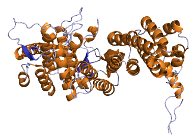
Menin is a protein that in humans is encoded by the MEN1 gene.[5] Menin is a putative tumor suppressor associated with multiple endocrine neoplasia type 1 (MEN-1 syndrome) and has autosomal dominant inheritance.[6] Variations in the MEN1 gene can cause pituitary adenomas, hyperparathyroidism, pancreatic neuroendocrine tumors, gastrinoma, and adrenocortical cancers.

In vitro studies have shown that menin is localized to the nucleus, possesses two functional nuclear localization signals, and inhibits transcriptional activation by JunD. However, the function of this protein is not known. Two messages have been detected on northern blots but the larger message has not been characterized. Two variants of the shorter transcript have been identified where alternative splicing affects the coding sequence. Five variants where alternative splicing takes place in the 5' UTR have also been identified.[5]

History

[edit]

In 1988, researchers at Uppsala University Hospital and the Karolinska Institute in Stockholm mapped the MEN1 gene to the long arm of chromosome 11.[7] The gene was finally cloned in 1997.[8]

Genomics

[edit]

The gene is located on long arm of chromosome 11 (11q13) between base pairs 64,570,985 and 64,578,765. It has 10 exons and encodes a 610-amino acid protein.

Over 1300 mutations have been reported to date (2010). The majority (>70%) of these are predicted to lead to truncated forms are scattered throughout the gene. Four - c.249_252delGTCT (deletion at codons 83-84), c.1546_1547insC (insertion at codon 516), c.1378C>T (Arg460Ter) and c.628_631delACAG (deletion at codons 210-211) have been reported to occur in 4.5%, 2.7%, 2.6% and 2.5% of families.[6]

Clinical implications

[edit]

The MEN1 phenotype is inherited via an autosomal-dominant pattern and is associated with neoplasms of the pituitary gland, the parathyroid gland, and the pancreas (the 3 "P"s). While these neoplasias are often benign (in contrast to tumours occurring in MEN2A), they are adenomas and, therefore, produce endocrine phenotypes. Pancreatic presentations of the MEN1 phenotype may manifest as Zollinger–Ellison syndrome.

MEN1 pituitary tumours are adenomas of anterior cells, typically prolactinomas or growth hormone-secreting. Pancreatic tumours involve the islet cells, giving rise to gastrinomas or insulinomas. In rare cases, adrenal cortex tumours are also seen.

Role in cancer

[edit]

Most germline or somatic mutations in the MEN1 gene predict truncation or absence of encoded menin resulting in the inability of MEN1 to act as a tumor suppressor gene.[9] Such mutations in MEN1 have been associated with defective binding of encoded menin to proteins implicated in genetic and epigenetic mechanisms.[10] Menin is a 621 amino acid protein associated with insulinomas[11] which acts as an adapter while also interacting with partner proteins involved in vital cell activities such as transcriptional regulation, cell division, cell proliferation, and genome stability. Insulinomas are neuroendocrine tumors of the pancreas with an incidence of 0.4 %[citation needed] which usually are benign solitary tumors but 5-12 % of cases have distant metastasis at diagnosis.[12] These familial MEN-1 and sporadic tumors may arise either due to loss of heterozygosity or the chromosome region 11q13 where MEN1 is located, or due to presence of mutations in the gene.[13][14]

MEN1 mutations comprise mostly frameshift deletions or insertions, followed by nonsense, missense, splice-site mutations and either part or complete gene deletions resulting in disease pathology.[15] Frameshift and nonsense mutations result in a supposed inactive and truncated menin protein while splice-site mutations result in incorrectly spliced mRNA. Missense mutations of MEN1 are especially important as they result in a change to crucial amino acids needed in order to bind and interact with other proteins and molecules. As menin is located predominantly in the nucleus,[16] these mutations can impact the stability of the cell and may further affect functional activity or expression levels of the protein. Studies have also shown that single amino acid changes in genes involved in oncogenic disorders may result in proteolytic degradation leading to loss of function and reduced stability of the mutant protein; a common mechanism for inactivating tumor suppressor gene products.[17][18] MEN1 gene mutations and deletions also play a role in the development of hereditary and a subgroup of sporadic pituitary adenomas and were detected in approximately 5% of sporadic pituitary adenomas.[19] Consequently, alterations of the gene represent a candidate pathogenetic mechanism of pituitary tumorigenesis especially when considered in terms of interactions with other proteins, growth factors, oncogenes play a rule in tumorigenesis.

Although the exact function of MEN1 is not known, the Knudson "two-hit" hypothesis provides strong evidence that it is a tumor suppressor gene. Familial loss of one copy of MEN1 is seen in association with MEN-1 syndrome. Tumor suppressor carcinogenesis follows Knudson's "two-hit" model.[20] The first hit is a heterozygous MEN1 germline mutation either developed in an early embryonic stage and consequently present in all cells at birth for the sporadic cases, or inherited from one parent in a familial case. The second hit is a MEN1 somatic mutation, oftentimes a large deletion occurring in the predisposed endocrine cell and providing cells with the survival advantaged needed for tumor development.[21] The MEN-1 syndrome often exhibits tumors of parathyroid glands, anterior pituitary, endocrine pancreas, and endocrine duodenum. Less frequently, neuroendocrine tumors of lung, thymus, and stomach or non-endocrine tumors such as lipomas, angiofibromas, and ependymomas are observed neoplasms.[22]

In a study of 12 sporadic carcinoid tumors of the lung, five cases involved inactivation of both copies of the MEN1 gene. Of the five carcinoids, three were atypical and two were typical. The two typical carcinoids were characterized by a rapid proliferative rate with a higher mitotic index and stronger Ki67 positivity than the other typical carcinoids in the study. Consequently, the carcinoid tumors with MEN1 gene inactivation in the study were considered to be characterized by more aggressive molecular and histopathological features than those without MEN1 gene alterations.[23]

Pharmaceuticals

[edit]

Anticancer drugs that exert their pharmacological effect by interfering with menin's interactions include the approved drug revumenib and the experimental drugs enzomenib, icovamenib, and ziftomenib.

Interactions

[edit]

MEN1 has been shown to interact with:

References

[edit]

Further reading

[edit]
[edit]
Revisions and contributorsEdit on WikipediaRead on Wikipedia
from Grokipedia
Multiple endocrine neoplasia type 1 (MEN1) is a rare inherited disorder caused by mutations in the MEN1 gene on chromosome 11q13, leading to the development of multiple tumors in endocrine glands, most commonly the parathyroid glands, the endocrine pancreas, and the gland. This autosomal dominant condition, also known as Wermer's syndrome, has a prevalence of approximately 1 in 30,000 individuals and affects males and females equally, with symptoms often first appearing in the second or third decade of life; approximately 10% of cases result from de novo mutations. The MEN1 gene encodes the protein menin, a tumor suppressor that regulates and division; inactivating mutations disrupt this function, resulting in tumor formation, though most tumors are benign but can become functionally active or, rarely, malignant. The hallmark of MEN1 is , occurring in over 90% of patients by age 50, often presenting as multiglandular parathyroid adenomas or that cause hypercalcemia, leading to symptoms such as fatigue, kidney stones, , and gastrointestinal issues. Enteropancreatic neuroendocrine tumors affect 30-80% of individuals, including gastrinomas (causing Zollinger-Ellison syndrome with peptic ulcers and diarrhea) and insulinomas (resulting in ), while pituitary adenomas occur in about 30-40% of cases, frequently prolactinomas that may cause , , or headaches. Less common manifestations include adrenal cortical tumors, thymic or bronchial carcinoids, and facial angiofibromas, with the syndrome's high —reaching 95% by age 40—necessitating lifelong surveillance. Diagnosis of MEN1 relies on clinical criteria, including the presence of two or more endocrine tumors associated with the syndrome, a family history of MEN1 in a first-degree relative, or identification of a MEN1 mutation through , which is recommended for at-risk individuals starting at age 5-10. Management focuses on early detection via regular biochemical screening (e.g., serum calcium, , ) and imaging (e.g., MRI, CT), with treatments such as surgical resection for symptomatic tumors, medical therapies for excess, and for families, as there is no cure; early detection and appropriate management have substantially improved , though it remains reduced compared to the general population, with recent studies showing that only about 28% of deaths in recognized and treated patients are directly related to MEN1.

Gene Overview

Location and Structure

The MEN1 gene is located on the long arm of chromosome 11 at the q13.1 band. Its genomic coordinates, according to the GRCh38 assembly, span from 64,803,516 to 64,811,294, encompassing approximately 8 kb of DNA. This positioning was confirmed through linkage studies and positional cloning efforts that mapped the gene to this region based on familial inheritance patterns of multiple endocrine neoplasia type 1. The gene consists of 10 s separated by 9 introns, with the overall genomic structure spanning about 9 kb, including regulatory elements. 1 is entirely untranslated, and the coding sequence begins in 2 with the initiation codon, extending through 2 to 10 to encode a 610-amino-acid protein known as menin; the 3' portion of 10 (approximately 832 ) serves as the . The intron- boundaries are precisely defined, with splice sites following the GT-AG consensus rule, facilitating accurate pre-mRNA processing. The promoter region lies upstream of exon 1 and includes Alu-type repetitive elements, which are short interspersed nuclear elements characteristic of primate genomes and contribute to transcriptional regulation. This promoter drives the expression of a major 2.8-kb mRNA transcript, with transcription initiation potentially occurring within exon 1. The Alu elements in the upstream regulatory region represent a human-specific , as they are absent in non-primate orthologs, though the core coding sequence shows high conservation. Evolutionarily, the MEN1 gene is highly conserved across , with orthologs identified in vertebrates such as mice (on ) and , as well as like fruit flies and snails, indicating an ancient role in cellular processes; however, it is absent in simpler organisms like and . The exon-intron organization, particularly exons 2 through 9, is well preserved between and murine genomes, underscoring functional importance.

Transcription and Regulation

The MEN1 undergoes primary transcript processing that includes , generating multiple isoforms with potential functional diversity. The predominant isoform, MENIN isoform 2, consists of 610 and is considered the reference transcript, while events occur across the entire , including a novel in 7 that is highly expressed in various tissues. These events produce isoforms featuring in-frame insertions, deletions, or unique amino termini, with at least four alternative splice variants identified through 5' RACE and RT-PCR analyses. Such isoform diversity may influence menin's interactions with partner proteins, though the functional implications in normal physiology remain under investigation. Regulatory elements controlling MEN1 transcription include a minimal promoter region spanning -135 to -36 base pairs upstream of exon 2, which contains initiator (Inr) elements and is modulated by upstream cis-regulatory sequences such as UR1-UR5 and smaller elements C1-C5 located between -325 and -107. Promoter activity exhibits cell-type specificity, with reduced function in endocrine-derived cells compared to non-endocrine lines, and is subject to negative feedback from menin itself, as overexpression in Men1-null fibroblasts dose-dependently suppresses promoter-driven reporter activity. Epigenetic modifications, including promoter hypermethylation, contribute to downregulated MEN1 expression, as observed in pancreatic ductal adenocarcinomas where methylation correlates with decreased menin levels in 5 of 24 tumors, and in pancreatic neuroendocrine tumors where it occurs frequently. Additionally, histone modifications play a role in regulation; for instance, menin interacts with SUV39H1 to promote H3K9 trimethylation at target loci, and ChIP-seq studies have mapped menin binding to distal enhancers that loop to promoters, influencing chromatin accessibility, though direct application to MEN1 autoregulation requires further validation. MEN1 exhibits ubiquitous basal expression across human tissues but shows elevated levels in endocrine organs, consistent with its role in neuroendocrine . reveals widespread MEN1 transcripts in adult tissues, with enhanced expression in proliferating cells entering , suggesting cell cycle-linked transcriptional control. Quantitative data from the GTEx consortium indicate median transcripts per million (TPM) values of approximately 1.8 in the , 1.3 in the and , and 0.85 in (GTEx Analysis Release V10, dbGaP Accession phs000424.v10.p2), underscoring higher expression in these tissues relative to non-endocrine organs like . This pattern aligns with the gene's association with endocrine tumorigenesis when dysregulated.

Menin Protein

Structure and Domains

Menin is a 610-amino acid nuclear scaffold protein encoded by the MEN1 gene, lacking any transmembrane domains and featuring intrinsically disordered regions (IDRs), including one flanked by two nuclear localization signals (NLSs) in the C-terminal half that facilitate its nuclear import. The protein's folded core adopts a distinctive curved left-hand , consisting of an N-terminal domain formed by a β-hairpin, a central palm domain with a deep binding pocket, and C-terminal finger-like extensions. This overall , resolved in crystal form at 2.1–2.9 resolution (e.g., PDB entry 3U84 for unbound menin), lacks homology to other known eukaryotic protein domains beyond the NLSs. Key structural features include the C-terminal NLSs, which ensure nuclear localization, and interaction domains within the palm that bind partners like the mixed-lineage leukemia (MLL) complex via conserved motifs such as FPX XP. structures of menin-MLL1 complexes (e.g., PDB 3U85) highlight atomic coordinates for critical residues, including Arg-392 and Gln-359 in the pocket, which stabilize MLL binding through hydrogen bonds and hydrophobic interactions. A cryo-electron microscopy structure at 3.2 Å resolution (PDB 8GPN) captures menin in complex with a nucleosome bearing histone H3 dimethylated at lysine 79 (H3K79me2), revealing multivalent contacts where the palm and finger domains engage the histone H3 tail via a π-cation interaction involving Arg-334 and the methylated lysine. Post-translational modifications of menin include phosphorylation primarily at serine/threonine residues, such as the constitutive site at Ser-543, Ser-583, and dynamic sites like Ser-394 (induced by ionizing radiation or UV) and Ser-487, which occur without altering core stability or localization. Ubiquitination involves polyubiquitin chains that target menin for proteasomal degradation, with patterns enhanced by MEN1 mutations disrupting stabilizing interactions.

Cellular Functions

Menin functions as a tumor suppressor by regulating through the transcriptional activation of inhibitors (CDKIs), particularly p27^{Kip1} and p18^{Ink4c}. These inhibitors enforce arrest at the [G1 phase](/page/G1 phase) by binding and inhibiting complexes, thereby preventing uncontrolled cellular growth. Menin achieves this regulation by recruiting the mixed-lineage leukemia (MLL) complex to the promoter and coding regions of the CDKN1B (encoding p27^{Kip1}) and CDKN2C (encoding p18^{Ink4c}) genes, facilitating their expression in a menin-dependent manner. Experimental evidence from Men1-null embryonic fibroblasts demonstrates that re-expression of wild-type menin increases p27^{Kip1} and p18^{Ink4c} transcription by 4- to 5-fold, as measured by reporter assays, while menin mutants associated with MEN1 syndrome (e.g., L22R, A242V) fail to do so due to impaired DNA binding. In hyperplastic parathyroid glands from MEN1 patients, the percentage of p27^{Kip1}-negative cells is increased (34.4% vs. 20% in normal tissues; p=0.11), underscoring menin's role in maintaining proliferative control. Beyond direct transcriptional effects, menin's tumor suppressive activity is reinforced by its epigenetic modifications that promote the expression of these CDKIs, linking proliferation control to . In mouse models, loss of menin leads to down-regulation of p27^{Kip1} and p18^{Ink4c}, resulting in deregulated , as observed in islet cell hyperplasia. This mechanism highlights menin's essential role in balancing cellular growth, with p18^{Ink4c} showing particularly strong collaboration with menin in suppressing neuroendocrine proliferation, as evidenced by accelerated tumorigenesis in combined Men1 and Cdkn2c knockout mice. Menin plays a critical epigenetic role by interacting with histone methyltransferases MLL1 and MLL2, enabling trimethylation of at 4 (H3K4me3), an active mark that promotes transcription. As a , menin stabilizes the MLL1/2 complexes at target promoters, facilitating H3K4 and subsequent opening for transcriptional machinery access. This process is vital for activating genes involved in development and differentiation, such as Hox cluster genes, where menin ensures stable long-term expression during embryogenesis. Genome-wide studies in menin-null mouse embryonic stem cells reveal that menin is required for H3K4me3 at nearly all Hox loci, with loss leading to profound reductions in and . The interaction is direct and specific to MLL1/2 among the MLL family, as demonstrated by co-immunoprecipitation assays showing menin's binding to the MLL1 SET domain, which catalyzes the reaction. This epigenetic function extends to broader activation, where menin-MLL complexes maintain H3K4me3 patterns at enhancers and promoters, supporting cellular identity and preventing oncogenic . In addition to transcriptional activation, menin's epigenetic contributions ensure precise control of landscapes, with levels directly correlating with menin occupancy in ChIP-seq analyses across multiple cell types. Disruption of menin-MLL binding, as modeled by small-molecule inhibitors, rapidly reduces at target loci, confirming the complex's necessity for methylation fidelity. These roles position menin as a key of epigenetic , influencing long-range interactions essential for normal cellular function. Menin contributes to and genome stability by stimulating (HR), a high-fidelity pathway for repairing double-strand breaks (DSBs). Menin accumulates at DSB sites in response to DNA damage, where it interacts with checkpoint Chk1 to enhance HR efficiency. In human cells engineered with the DR-GFP/I-SceI reporter system, overexpression of menin increases HR-mediated repair by promoting GFP expression in DSB-induced cells, as quantified by (significant increase in double-positive cells, p<0.05). This stimulation depends on menin's N-terminal domain, as deletion mutants (ΔNH₂) abolish the interaction with Chk1 and repair enhancement. and re-ChIP experiments further show menin and Chk1 co-localize at I-SceI-induced breaks, with menin facilitating the recruitment of HR factors. The process involves the ATM/ATR signaling pathway, as inhibition (an ATM/ATR blocker) reduces menin-stimulated repair by approximately 90%. These findings indicate menin's role in maintaining genomic integrity by prioritizing error-free HR over mutagenic alternatives like . Menin's involvement in HR extends to broader genome stability, where its absence sensitizes cells to DSB-inducing agents, as evidenced by increased γ-H2AX foci in menin-depleted lines. By modulating Chk1 activity at damage sites, menin ensures timely resolution of replication-associated breaks, preventing chromosomal aberrations. This function is conserved across cell types, with menin promoting HR in both cycling and quiescent cells. Menin participates in telomere maintenance by regulating telomerase activity, primarily through transcriptional repression of the human telomerase reverse transcriptase (hTERT) , which encodes the catalytic subunit of telomerase. Menin binds to the hTERT promoter in a sequence-independent manner, inhibiting its activity and thereby limiting telomere elongation in normal cells. reporter assays in various cell lines demonstrate that menin overexpression reduces hTERT promoter activity in a cell-type-specific , while siRNA-mediated knockdown of menin fails to significantly up-regulate hTERT mRNA, suggesting indirect or context-dependent regulation. In experimental models, menin knockdown in HEK293T cells promotes telomerase component (TERC) localization to s, slowing telomere erosion rates as measured by telomere restriction fragment analysis. This indicates that menin normally disrupts telomerase assembly or access, contributing to controlled telomere shortening. Evidence from Men1 models supports this, where homozygous null islets exhibit altered telomerase dynamics and mild telomere instability, though not leading to immediate length aberrations. Additionally, menin localizes to s during , as shown by in spermatocytes, where it associates with telomeric repeats without altering overall telomerase activity in overexpressing cells. These observations highlight menin's role in fine-tuning telomerase to prevent excessive telomere maintenance, which could otherwise promote replicative . Menin also regulates T cell differentiation and function, influencing immune responses through transcriptional control.

Role in Disease

Association with MEN1 Syndrome

Multiple endocrine neoplasia type 1 (MEN1) syndrome is a rare autosomal dominant hereditary disorder resulting from germline mutations in the MEN1 gene, which encodes the tumor suppressor protein menin. The syndrome exhibits near-complete , with over 95% of mutation carriers developing clinical manifestations by age 40 and approaching 100% by age 50. Its is estimated at approximately 1 in 30,000 individuals, though reported rates vary from 3 to 20 per 100,000 depending on population and diagnostic criteria. About 10% of cases arise from de novo mutations, meaning affected individuals have no family history of the syndrome, while the remainder are inherited from an affected parent with a 50% transmission risk per offspring. The hallmark features of MEN1 syndrome involve multifocal tumors primarily affecting the parathyroid glands, , and enteropancreatic neuroendocrine tissues, often leading to endocrine dysfunction. Parathyroid involvement manifests as in over 90% of cases by age 50, typically due to multiglandular or adenomas causing hypercalcemia. Pituitary adenomas occur in 30-40% of patients, commonly prolactinomas or non-functioning tumors that may result in hormonal excesses or mass effects. Enteropancreatic tumors, seen in 30-70% of cases, include gastrinomas (leading to Zollinger-Ellison syndrome with peptic ulcers) and insulinomas, among others, which can be functional or malignant. These manifestations usually emerge in adulthood, with parathyroid tumors appearing earliest (average onset in the 20s), followed by enteropancreatic and pituitary lesions. The pathogenesis of MEN1 syndrome adheres to Knudson's for tumor suppressor genes, where the inherited MEN1 represents the first hit, inactivating one in all cells. Tumor formation requires a second somatic hit, typically (LOH) at 11q13 encompassing the wild-type MEN1 , observed in over 90% of affected tumors. This biallelic inactivation disrupts menin function, promoting uncontrolled in endocrine tissues.

Involvement in Sporadic Cancers

Somatic inactivating in the MEN1 gene occur in 20-40% of sporadic parathyroid adenomas, contributing to tumorigenesis independent of hereditary syndromes. These often lead to loss of menin function, promoting uncontrolled in parathyroid chief cells. Similarly, approximately 30% of sporadic gastrinomas harbor MEN1 somatic , frequently accompanied by allelic deletions, which drive Zollinger-Ellison syndrome-like tumor development. In non-endocrine sporadic cancers, MEN1 alterations play emerging roles; for instance, reduced menin expression is observed in sporadic breast cancers, where it interacts with to influence tumor progression, though direct mutations are infrequent. In sporadic cancers, particularly castration-resistant forms, MEN1 mutations occur in about 17% of cases and may exert an oncogenic effect by enhancing signaling. Loss-of-heterozygosity (LOH) at the locus encompassing MEN1 is a common mechanism across various sporadic malignancies, including parathyroid adenomas (26-37% frequency), tumors, and carcinoids, facilitating biallelic inactivation. In sporadic neuroendocrine tumors (NETs), MEN1 somatic alterations provide prognostic insights; The Cancer Genome Atlas (TCGA) analyses of pancreatic NETs reveal MEN1 mutations in 37% of cases, often without direct survival correlation but associated with distinct molecular subtypes influencing therapeutic response. Specifically, in sporadic thymic carcinoids, MEN1 mutations and 11q losses are frequent, correlating with heightened malignant potential, local invasion, and early , as highlighted in 2025 molecular reviews.

Mutations and Variants

Types of Pathogenic Mutations

Pathogenic mutations in the MEN1 gene primarily lead to loss of function of the menin protein and are responsible for the majority of (MEN1) cases. As of 2025, more than 1,800 distinct have been reported in the literature, cataloged in specialized databases such as the Open Variation Database (LOVD) and the Universal Mutation Database (UMD-MEN1). These are evenly distributed across the 10 exons of the gene, with relative hotspots observed in exons 2 and 10. The spectrum of pathogenic mutations includes several major types, with frameshift insertions and deletions being the most prevalent at approximately 40-50%, followed by missense mutations (20-25%), nonsense mutations (10-20%), and splice-site alterations (7-11%). Frameshift and nonsense mutations typically result in premature termination codons and truncated proteins, while missense mutations involve single substitutions that may disrupt protein function. Splice-site mutations affect intron-exon boundaries, leading to aberrant mRNA processing. Large genomic deletions, encompassing one or more exons, represent about 5% of cases and are often identified through (MLPA) rather than standard sequencing. Variant classification follows the American College of Medical Genetics and (ACMG) guidelines, distinguishing pathogenic/likely pathogenic from benign/likely benign ones based on criteria such as population frequency, computational predictions, functional assays, and segregation data. Missense , in particular, pose classification challenges and frequently require additional evidence like tools or studies to confirm pathogenicity. Recent case reports continue to identify novel , including double-substitution that alter multiple adjacent and contribute to the expanding genetic landscape.

Genotype-Phenotype Correlations

Although no definitive genotype-phenotype correlations have been established for overall, certain patterns have emerged from large cohort studies, particularly linking type to disease severity and presentation. Truncating , such as nonsense and frameshift variants, are associated with earlier disease onset and greater tumor multiplicity compared to non-truncating , likely due to complete loss of menin function. In contrast, missense , especially those affecting specific domains like the nuclear localization signal (NLS), have been linked to atypical or less penetrant features, including isolated or unusual tumor types such as macro-prolactinomas. Epidemiological studies from diverse cohorts provide supporting evidence for these associations. For instance, in a multicenter of over 100 Korean patients, truncating mutations correlated with a significantly higher prevalence of pituitary neuroendocrine tumors (59.7% vs. 36.0% in non-truncating cases). Similarly, Italian cohort data indicate that and frameshift mutations are more strongly tied to aggressive gastroenteropancreatic neuroendocrine tumors (GEP-NETs) than missense variants. Disease and expression in MEN1 are also influenced by genetic modifiers, including loss of the wild-type (second hit) via mechanisms like , which is observed in nearly all MEN1-associated tumors and drives tumorigenesis. Environmental factors, such as dietary influences on , may interact with mutations to modulate , though evidence remains limited and indirect. Founder effects in specific populations can further shape phenotypic variability; for example, recurrent mutations in Italian cohorts have been associated with distinct patterns of tumor distribution and earlier onset in affected families, highlighting the role of population-specific .

Molecular Interactions

Protein-Protein Interactions

Menin, the protein encoded by the MEN1 gene, participates in a wide array of protein-protein interactions that underpin its function in cellular processes. High-throughput approaches such as two-hybrid (Y2H) screening and affinity purification-mass spectrometry (AP-MS) have mapped an extensive interactome, identifying over 50 potential binding partners, which underscore menin's integration into diverse multiprotein complexes. A primary set of interactors involves the mixed-lineage family proteins MLL1 and MLL2, which are methyltransferases central to H3K4 and epigenetic regulation. Menin binds directly to the N-terminal region of MLL1 via two distinct menin-binding motifs (MBM1 spanning residues 4-15 and MBM2 spanning residues 23-40), forming a high-affinity complex with a (Kd) of approximately 10 nM. This interaction has been robustly demonstrated through co-immunoprecipitation assays in and epithelial cell lines, where menin stabilizes MLL1 recruitment to promoters without altering its enzymatic activity. Similar binding occurs with MLL2, sharing the same structural motifs and affinity profile, facilitating cooperative epigenetic modification. Menin also forms a direct interaction with SMAD3, the receptor-regulated in the TGF-β signaling pathway. This association promotes SMAD3-dependent activation of growth-inhibitory genes and has been confirmed by co-immunoprecipitation in COS cells, specific to SMAD3 without involving SMAD2 or SMAD4. The binding requires the MH2 domain of SMAD3. In the context of , menin interacts with RPA2, the 32-kDa subunit of the heterotrimer, which coats single-stranded DNA during replication and repair processes. This partnership was first identified via Y2H screening reconstituting Ras signaling and subsequently validated by co-immunoprecipitation in human cell lysates, revealing menin's recruitment to DNA damage sites to support . Experimental studies indicate that RPA2 engages menin's N-terminal region (up to residue 286). These interactions display tissue-specific modulation, with enhanced binding affinities and stability observed in endocrine tissues such as the and pituitary, where menin’s associations with MLL1/2 and SMAD3 are amplified to enforce control and suppress proliferation.

Signaling Pathways

Menin, the protein encoded by the MEN1 gene, serves as a critical scaffold in multiple cellular signaling pathways, primarily exerting tumor-suppressive effects through and epigenetic modifications. As a component of the mixed-lineage leukemia (MLL) complex, menin facilitates lysine 4 trimethylation (), which activates in a context-dependent manner, while also repressing oncogenic signals in pathways such as Wnt/β-catenin and . Dysregulation of these pathways due to MEN1 loss promotes tumorigenesis, particularly in endocrine tissues, by altering proliferation, , and metabolic signaling. In the Wnt/β-catenin pathway, menin functions as a transcriptional repressor by directly interacting with β-catenin, reducing its nuclear accumulation and thereby inhibiting downstream target gene activation. This repression is evident in mouse embryonic fibroblasts and islet β-cells, where menin overexpression suppresses β-catenin-mediated transcription, limiting . Conversely, MEN1 deficiency in murine models leads to β-catenin stabilization and pathway hyperactivation, driving the expression of proliferative genes like Axin2 and contributing to formation. This repressive role underscores menin's integration into Wnt signaling to maintain cellular homeostasis. Menin also antagonizes signaling, a pathway involved in and oncogenesis, by epigenetically repressing key effectors such as Gli1. Through recruitment of the protein arginine methyltransferase PRMT5, menin dimethylates arginine 2 (H3R2me2), which competes with and silences Hedgehog target genes in and MEN1-associated tumors. Ablation of menin in murine models enhances Hedgehog pathway activity, promoting islet cell proliferation and tumor progression, independent of canonical ligand-receptor interactions. Regarding apoptosis regulation, menin interacts with the JunD to repress its transcriptional activity, thereby inhibiting cell growth and promoting . In murine embryonic fibroblasts, menin overexpression induces by blocking JunD-mediated anti-apoptotic , while menin-JunD binding prevents JunD by JNK . Loss of this interaction shifts JunD from a growth suppressor to a promoter, facilitating in MEN1-deficient cells. Pathway dysregulation in MEN1 loss is exemplified by elevated c-Myc expression in neuroendocrine tumors, where menin normally represses c-Myc transcription via modulation at its promoter. In pancreatic models, Men1 upregulates c-Myc mRNA, enhancing proliferation and neoplastic transformation. This effect integrates with cross-talk to the PI3K/AKT pathway, where menin inhibits AKT activation by regulating its cellular localization, suppressing AKT1-driven proliferation and anti-apoptotic signals in endocrine and non-endocrine cells. Consequently, MEN1 deficiency amplifies PI3K/AKT signaling, promoting tumor progression through enhanced survival and metabolic reprogramming. As of 2025, therapeutic targeting of menin protein-protein interactions, such as the menin-MLL interface with small-molecule inhibitors, has advanced understanding of these complexes in and endocrine tumors.

History and Research

Discovery and Cloning

The (MEN1) syndrome, also known as Wermer syndrome, was first linked to chromosome through genetic studies in affected families during the . In 1988, Larsson et al. demonstrated tight linkage between the MEN1 locus and the muscle gene (PYGM) on in a large Swedish pedigree, establishing the chromosomal location for further mapping efforts. Subsequent linkage analyses in additional families refined the candidate to approximately a 400-kilobase interval within , supported by (LOH) observations in MEN1-associated tumors indicating a tumor suppressor mechanism. Positional cloning of the MEN1 was achieved in 1997 by two independent groups using complementary strategies centered on LOH analysis and genomic sequencing within the refined interval. The U.S. (NIH) team, led by Chandrasekharappa et al., identified the by systematically sequencing candidate transcripts in the minimal LOH region, isolating a 2.8-kilobase mRNA expressed ubiquitously and encoding a 610-amino-acid protein named menin; they reported in 15 of 21 MEN1 families and somatic with LOH in 13 of 24 tumors. Concurrently, the European MEN1 Consortium, led by Lemmens et al., used trapping and cDNA selection from a chromosome 11-specific library to clone the same , confirming inactivating such as and frameshift alterations in affected individuals, which truncated the menin protein and supported its role as a tumor suppressor. These initial findings established MEN1 as a classic tumor suppressor gene, with biallelic inactivation via germline mutation plus somatic LOH or second-hit mutation mirroring Knudson's two-hit hypothesis in MEN1 tumorigenesis. The gene's discovery, published in April 1997, enabled the first genetic testing for MEN1 and shifted research toward understanding menin's function beyond its identification.

Recent Advances

Recent advances in MEN1 research since the 2020s have leveraged CRISPR-Cas9 technology to dissect menin's tumor-suppressive roles beyond endocrine tissues. Genome-wide CRISPR screens have identified menin as a key regulator in solid tumors, such as liver cancer, where its knockout promotes metastasis in vivo models, revealing a dual function in suppressing tumor growth and invasion in non-endocrine contexts. Similarly, isogenic cell lines generated via CRISPR editing of patient-derived iPSCs have enabled precise studies of MEN1 loss-of-function, highlighting menin's involvement in chromatin remodeling and gene expression in various cancers. These functional studies underscore menin's broader oncogenic implications, expanding from classical endocrine neoplasia to sporadic solid malignancies. Therapeutic developments targeting menin have progressed notably, with inhibitors like revumenib entering clinical trials primarily for harboring MLL (KMT2A) rearrangements, where menin-MLL disruption drives leukemogenesis. Preclinical extensions to MEN1 models demonstrate that menin inhibition can reverse aberrant in contexts, suggesting potential repurposing for MEN1-associated pancreatic and pituitary lesions, though clinical translation remains exploratory. Concurrently, the 2025 American Association of Clinical Endocrinology (AACE) consensus statement emphasizes expanded for MEN1, recommending it for individuals with suggestive clinical features or family history, alongside biochemical surveillance to enable early intervention. Emerging research highlights overlaps between MEN1 and MEN4, where mutations mimic MEN1 phenotypes, with menin regulating p27 ( product) transcription, leading to shared parathyroid and pituitary tumor risks in up to 3% of MEN1-negative cases. AI-driven tools for mutation prediction are gaining traction, integrating with genomic data to classify MEN1 variants' pathogenicity, improving diagnostic accuracy in ambiguous cases. In imaging, 2024-2025 studies validate 68Ga-DOTATATE PET/CT as superior for detecting MEN1-associated duodeno-pancreatic neuroendocrine tumors, outperforming conventional modalities with higher sensitivity (up to 87%) and guiding precise surgical management. These multimodal advances address persistent gaps in tumor localization and personalized therapy for MEN1 patients.

References

Add your contribution
Related Hubs
User Avatar
No comments yet.