Hubbry Logo
MYD88MYD88Main
Open search
MYD88
Community hub
MYD88
logo
8 pages, 0 posts
0 subscribers
Be the first to start a discussion here.
MYD88
MYD88
from Wikipedia

MYD88
Available structures
PDBOrtholog search: PDBe RCSB
Identifiers
AliasesMYD88, MYD88D, myeloid differentiation primary response 88, innate immune signal transduction adaptor, MYD88 innate immune signal transduction adaptor, IMD68
External IDsOMIM: 602170; MGI: 108005; HomoloGene: 1849; GeneCards: MYD88; OMA:MYD88 - orthologs
Orthologs
SpeciesHumanMouse
Entrez
Ensembl
UniProt
RefSeq (mRNA)

NM_010851

RefSeq (protein)

NP_034981

Location (UCSC)Chr 3: 38.14 – 38.14 MbChr 9: 119.17 – 119.17 Mb
PubMed search[3][4]
Wikidata
View/Edit HumanView/Edit Mouse

Myeloid differentiation primary response 88 (MYD88) is a protein that, in humans, is encoded by the MYD88 gene.[5][6] originally discovered in the laboratory of Dan A. Liebermann (Lord et al. Oncogene 1990) as a Myeloid differentiation primary response gene.

Function

[edit]

The MYD88 gene provides instructions for making a protein involved in signaling within immune cells. The MyD88 protein acts as an adapter, connecting proteins that receive signals from outside the cell to the proteins that relay signals inside the cell.

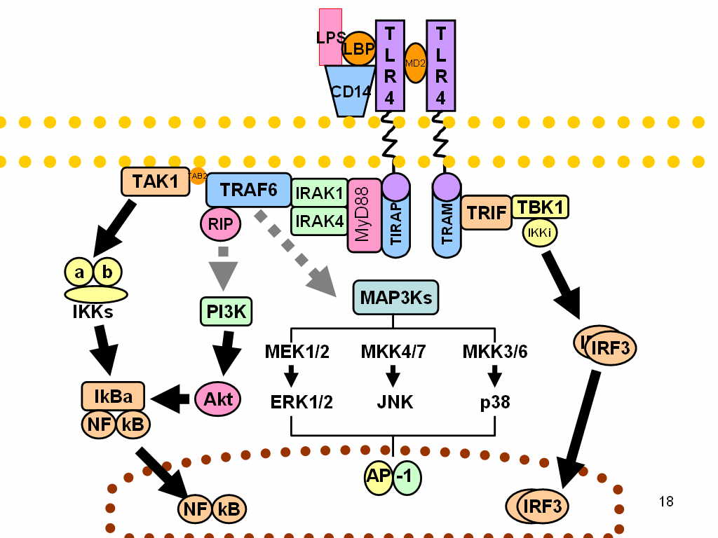
In innate immunity, the MyD88 plays a pivotal role in immune cell activation through Toll-like receptors (TLRs), which belong to large group of pattern recognition receptors (PRR). In general, these receptors sense common patterns which are shared by various pathogens – Pathogen-associated molecular pattern (PAMPs), or which are produced/released during cellular damage – damage-associated molecular patterns (DAMPs).[7]

TLRs are homologous to Toll receptors, which were first described in the ontogenesis of fruit flies Drosophila, being responsible for dorso-ventral development. Hence, TLRs have been proved in all animals from insects to mammals. TLRs are located either on the cellular surface (TLR1, TLR2, TLR4, TLR5, TLR6) or within endosomes (TLR3, TLR7, TLR8, TLR9) sensing extracellular or phagocytosed pathogens, respectively. TLRs are integral membrane glycoproteins with typical semicircular-shaped extracellular parts containing leucine-rich repeats responsible for ligand binding, and Intracellular parts containing Toll-Interleukin receptor (TIR) domain.[8]

After ligand binding, all TLRs, apart from TLR3, interact with adaptor protein MyD88. Another adaptor protein, which is activated by TLR3 and TLR4, is called TIR domain-containing adapter-inducing IFN-β (TRIF). Subsequently, these proteins activate two important transcription factors:

  • NF-κB is a dimeric protein responsible for expression of various inflammatory cytokines, chemokines and adhesion and costimulatory molecules, which in turn triggers acute inflammation and stimulation of adaptive immunity
  • IRFs is a group of proteins responsible for expression of type I interferons setting the so-called antiviral state of a cell.

TLR7 and TLR9 activate both NF-κB and IRF3 through MyD88-dependent and TRIF-independent pathway, respectively.[8]

The human ortholog MYD88 seems to function similarly to mice, since the immunological phenotype of human cells deficient in MYD88 is similar to cells from MyD88 deficient mice. However, available evidence suggests that MYD88 is dispensable for human resistance to common viral infections and to all but a few pyogenic bacterial infections, demonstrating a major difference between mouse and human immune responses.[9] Mutation in MYD88 at position 265 leading to a change from leucine to proline have been identified in many human lymphomas including ABC subtype of diffuse large B-cell lymphoma[10] and Waldenström's macroglobulinemia.[11]

Interactions

[edit]

Myd88 has been shown to interact with:

Gene polymorphisms

[edit]

Various single nucleotide polymorphisms (SNPs) of the MyD88 have been identified. and for some of them an association with susceptibility to various infectious diseases[22] and to some autoimmune diseases like ulcerative colitis was found.[23]

References

[edit]

Further reading

[edit]
[edit]
Revisions and contributorsEdit on WikipediaRead on Wikipedia
from Grokipedia
MYD88, or myeloid differentiation primary response 88, is a gene located on the short arm of chromosome 3 at position 3p22.2 that encodes a cytosolic adapter protein essential for innate immune signal transduction. The MyD88 protein serves as a key mediator in the signaling pathways activated by Toll-like receptors (TLRs) and the interleukin-1 receptor (IL-1R) family, linking extracellular pathogen detection to intracellular inflammatory responses. Expressed ubiquitously but at particularly high levels in immune tissues such as bone marrow and the appendix, MyD88 plays a central role in both innate and adaptive immunity by facilitating the activation of proinflammatory genes. Structurally, the MyD88 protein consists of an N-terminal death domain (DD) for interactions with downstream kinases like IRAK4 and IRAK1/2, a central intermediate domain, and a C-terminal Toll/IL-1R homology domain (TIR) that binds to the TIR domains of TLRs and IL-1Rs. Upon ligand binding to these receptors, MyD88 is recruited to form the myddosome complex—a oligomeric assembly of six MyD88 molecules with four each of IRAK4 and IRAK2/1—which propagates signals leading to the activation of (NF-κB), mitogen-activated protein kinases (MAPKs), and AP-1 transcription factors. This cascade results in the transcription of genes encoding cytokines such as tumor necrosis factor-alpha (TNF-α), interleukin-6 (IL-6), and interleukin-1 beta (IL-1β), crucial for mounting an effective immune defense against bacterial and viral pathogens. Dysfunction or dysregulation of MyD88 has profound implications for health, with germline mutations causing MyD88 deficiency—an autosomal recessive disorder characterized by recurrent pyogenic bacterial infections due to impaired TLR and IL-1R signaling. Conversely, somatic gain-of-function mutations, most notably the L265P variant in the TIR domain, drive constitutive activation and are found in over 90% of cases, as well as in subsets of other B-cell malignancies like (DLBCL) and IgM (IgM-MGUS). These mutations highlight MyD88's dual role in immune protection and oncogenesis, positioning it as a potential therapeutic target for inflammatory and neoplastic disorders.

Gene and Discovery

Discovery History

The MYD88 gene was first identified in 1990 during studies of in murine M1 myeloid leukemia cells induced to differentiate by interleukin-6 (IL-6), where it emerged as one of the primary response genes upregulated in this process, leading to its naming as "myeloid differentiation primary response 88." This initial characterization highlighted its potential role in myeloid cell maturation, though its function remained unclear at the time. Subsequent work in 1997 focused on the full-length cDNA and elucidating the gene's , revealing a protein with a Toll/interleukin-1 receptor (TIR) domain and a death domain homologous to those in IL-1 receptor-associated kinase (IRAK) proteins, suggesting involvement in IL-1 signaling pathways. Interspecific backcross mapping localized the murine Myd88 to the distal region of , while assigned the human MYD88 homolog to chromosome 3p21–3p22, positioning it within genomic regions linked to inflammatory responses. These findings established MYD88 as an evolutionarily conserved adaptor potentially bridging receptors to downstream effectors. Early functional characterization in 1997–1998 confirmed MyD88's adaptor role in IL-1 signaling through two-hybrid screens demonstrating direct interactions between its domain and IRAK, facilitating signal transduction from the IL-1 receptor. By 1998, similar assays extended this to Toll-like receptors (TLRs), identifying MyD88 as the key TIR domain-containing adaptor essential for human Toll (TLR4)-induced activation, thus linking it to innate immune recognition of pathogens. Pivotal evidence came from the generation of MyD88-deficient mice in 1998, which displayed profound defects in IL-1- and IL-18-mediated responses, including impaired T-cell proliferation and induction, underscoring its non-redundant role in innate immunity. Further studies in the early , including expanded phenotypes, reinforced MyD88's broad requirement across TLR pathways for production.

Genomic Location and Organization

The MYD88 gene is located on the short arm of human chromosome 3 at cytogenetic band 3p22.2, specifically spanning genomic coordinates 38,138,661–38,143,022 on reference assembly GRCh38.p14. This positions it within a region associated with immune response regulation, though the gene itself occupies approximately 4.4 kb of genomic DNA, including introns and regulatory elements. The gene structure comprises 5 exons, with exon 1 being entirely non-coding and contributing primarily to the (UTR) of the transcript. The complete coding sequence is distributed across exons 2–5, yielding a principal mRNA transcript (RefSeq NM_002468.5) of 2,667 nucleotides in length, which encodes a 297-amino-acid . Alternative splicing generates additional isoforms, but the canonical form supports the core adaptor function in innate immunity signaling. The promoter region in the 5' flanking sequence includes putative TATA and CAAT boxes, along with binding sites for transcription factors AP-1 and , facilitating inducible expression in response to inflammatory cues. MYD88 exhibits strong evolutionary conservation, with orthologs identified in a wide range of mammals and beyond, reflecting its fundamental role in pathways. Zoo-blot analyses and sequence comparisons demonstrate high similarity, particularly in the C-terminal Toll/interleukin-1 receptor (TIR) domain, which is preserved across vertebrates to maintain protein-protein interaction interfaces essential for signaling.

Protein Structure

Domains and Motifs

The MYD88 protein consists of 296 and has a of approximately 33 . It possesses a modular architecture with an N-terminal death domain (DD) spanning residues 1–109, which mediates homotypic interactions with death domains of downstream effectors, a central intermediate domain covering residues 110–160 that connects these domains and contributes to regulation of oligomerization and localization, and a C-terminal Toll/interleukin-1 receptor (TIR) domain spanning residues 161–296, which enables binding to TIR domains of upstream receptors. The TIR domain exhibits a conserved horseshoe-shaped fold typical of TIR family proteins, formed by a central five-stranded parallel β-sheet surrounded by α-helices and interconnecting loops. This structure is stabilized by five key subdomains: boxes 1–3, which contain conserved residues essential for overall folding and stability; the BB-loop, a surface-exposed region involved in dimerization interfaces; and patches A and B, which contribute to specificity in TIR-TIR interactions. These elements collectively enable the domain to facilitate signal propagation without possessing intrinsic enzymatic activity, functioning instead as a scaffolding adaptor to recruit and organize signaling complexes. The atomic structure of the MYD88 TIR domain (residues 157–296) was resolved by NMR spectroscopy in 2009, revealing dimerization interfaces primarily involving the BB-loop and αC helix, which bury significant surface area (~1,200 Ų) to promote stable oligomerization. This structural insight highlights how the domain's motifs enable higher-order assemblies critical for downstream signaling, while the absence of catalytic residues underscores MYD88's role as a non-enzymatic mediator in innate immune pathways.

Post-Translational Modifications

MYD88 undergoes several post-translational modifications that fine-tune its role as an adapter protein in (TLR) and interleukin-1 receptor (IL-1R) signaling, influencing protein stability, localization, and activation of downstream pathways such as . Phosphorylation at serine residues within the death domain, including Serine 34, has been identified as a potential regulatory site, with studies indicating that substitution at this position can impair activation without directly attributing the defect to loss of . Additional sites, such as Ser244, Tyr257, and Tyr276, have been mapped through proteomic analyses, potentially modulating MyD88 interactions with signaling partners, though specific kinases remain to be fully elucidated. Ubiquitination targets multiple residues on MYD88, including K95, K115, K119, K127, K190, K214, K231, K238, K250, K256, K262, K282, and K291, as determined by mass spectrometry-based . K63-linked polyubiquitination at these sites, facilitated by ligases like members of the Pellino family in conjunction with TRAF6 in the signaling complex, promotes MYD88 oligomerization and recruitment of effectors to enhance and MAPK activation. Conversely, K48-linked polyubiquitination directs MYD88 to proteasomal degradation, limiting prolonged signaling; for instance, the SPOP targets specific lysines (e.g., K231, K262) for this modification to suppress . Deubiquitinases like CYLD specifically remove K63-linked chains from MYD88 to inhibit excessive TLR responses. These modifications have been confirmed through studies integrated into databases like PhosphoSitePlus, with significant updates around 2015 incorporating large-scale proteomic data from immune cells to validate sites and their functional impacts.

Biological Function

Role in Innate Immunity

MyD88 serves as an essential adaptor protein in the , primarily by facilitating from most Toll-like receptors (TLRs), excluding TLR3, and members of the interleukin-1 receptor (IL-1R) family. These receptors detect extracellular pathogens through pattern recognition of microbial components, such as bacterial lipopolysaccharides or viral nucleic acids, and MyD88 bridges this detection to intracellular signaling cascades that initiate protective responses. By recruiting downstream components via its death domain, MyD88 enables the of inflammatory pathways that are critical for early defense against infections. In MyD88-deficient models, innate immune responses are profoundly impaired, highlighting its non-redundant role. These animals exhibit drastically reduced production of pro-inflammatory cytokines, including interleukin-6 (IL-6) and (TNF-α), in response to bacterial and viral stimuli such as or antigens. Consequently, MyD88 knockout mice display heightened susceptibility to infections, including lethal outcomes from due to failed bacterial clearance in the and liver. Such phenotypes underscore MyD88's centrality in coordinating rapid antimicrobial defenses without which host survival is compromised. MyD88 indirectly bridges innate and adaptive immunity by promoting maturation, which enhances to T and B cells. In , MyD88 signaling drives the upregulation of costimulatory molecules and secretion necessary for effective T cell priming and Th1 polarization. This maturation process ensures that innate detection of pathogens translates into robust adaptive responses, amplifying long-term immunity. MyD88 operates in a pathway distinct from and non-redundant with the TRIF (TIR-domain-containing adapter inducing -β)-dependent route, delineating MyD88-dependent TLR signaling from MyD88-independent mechanisms. While MyD88 handles responses from TLRs like TLR2, TLR4 (partially), and TLR7/9, TRIF primarily mediates type I production via TLR3 and TLR4's alternative arm, preventing compensatory overlap in antiviral defenses. This bifurcation allows specialized innate immune outputs tailored to different microbial threats.

Signaling Mechanisms

Upon ligand binding, Toll-like receptors (TLRs) or interleukin-1 receptors (IL-1Rs) undergo dimerization, enabling the recruitment of MyD88 through homotypic interactions between the Toll/IL-1R (TIR) domains of the receptor and MyD88. For TLR4, this recruitment is facilitated by the sorting adaptor TIRAP/MAL, which bridges the receptor and MyD88. The death domain (DD) of MyD88 then oligomerizes, forming a scaffold that initiates signaling. The oligomeric MyD88 complex, known as the myddosome, assembles in a hierarchical manner with a of 6 MyD88 DDs, 4 IRAK4 DDs, and 4 IRAK1 or IRAK2 DDs, creating a left-handed helical approximately 110 Å high. This assembly occurs via specific type I, II, and III DD interfaces, where MyD88 recruits IRAK4 through composite binding sites, and the MyD88-IRAK4 subcomplex subsequently engages IRAK1/2. IRAK4, activated by autophosphorylation, phosphorylates IRAK1 at Thr209 and Thr387, promoting IRAK1 hyperphosphorylation, dissociation from the complex, and recruitment of TRAF6. The intermediate domain of MyD88 (residues 110-155) is essential for stabilizing IRAK4 activation within this . TRAF6, an E3 ubiquitin ligase, undergoes K63-linked polyubiquitination upon recruitment, which activates the TAK1 by facilitating its oligomerization and autophosphorylation at Thr184. TAK1 then the IKK complex (IKKα, IKKβ, and NEMO), leading to at Ser32 and Ser36, its ubiquitination, and subsequent proteasomal degradation. This releases (p50/p65 heterodimer), allowing its nuclear translocation and transcription of pro-inflammatory genes. Paralleling this, TAK1 activates MAPK pathways, including p38, JNK, and ERK, through of MKKs, culminating in AP-1 activation and enhanced expression. The canonical MYD88-dependent pathway can be simplified as: Receptor (TLR/IL-1R) → MyD88 → IRAK4/IRAK1 oligomerization → TRAF6 ubiquitination → TAK1 → IKK/ and MAPK activation, with ubiquitination (primarily K63-linked) serving as a critical regulatory step at TRAF6 and downstream components.

Molecular Interactions

Interactions with Receptors

MYD88 functions as a crucial adaptor protein that interacts directly with the cytoplasmic Toll/Interleukin-1 receptor (TIR) domains of multiple upstream immune receptors to initiate innate immune signaling. It binds to Toll-like receptors (TLRs) including TLR2, TLR4, TLR5, and TLR7-9, as well as members of the interleukin-1 receptor family such as IL-1R and IL-18R. These interactions are essential for transducing signals from ligand-bound receptors into downstream inflammatory responses. The binding mechanism relies on homotypic interactions between the TIR domain of MYD88 and the TIR domains of the receptors, facilitating the of MYD88 to activated receptor complexes. For TLR4 signaling, MYD88 engagement requires the co-adapter MAL (also known as TIRAP), which bridges the receptor to MYD88. In contrast, MYD88 is absent from TLR3 signaling, which depends on the adaptor TRIF, highlighting the specificity of MYD88-dependent pathways among TLRs. MYD88 is particularly critical for IL-1R-mediated responses to IL-1β, where its absence abolishes pro-inflammatory production. Experimental evidence for these receptor interactions includes co-immunoprecipitation studies demonstrating physical association between MYD88 and receptor TIR domains upon . Structural analyses, such as the of the MYD88 TIR domain reported in 2009, have further revealed the molecular interfaces involved in these TIR-TIR contacts, including key residues that mediate specificity and stability.

Downstream Effectors

Upon recruitment by Toll-like receptors (TLRs) or interleukin-1 receptors (IL-1Rs), MYD88 oligomerizes to form the myddosome complex, which primarily recruits interleukin-1 receptor-associated 4 (IRAK4), a serine/ essential for initiating downstream signaling. Recent studies (as of 2025) have further elucidated MyD88's polymerization into helical assemblies associated with cellular membranes, enhancing signal initiation fidelity. IRAK4 binds directly to the death domain of MYD88 in a stoichiometric ratio of 4 IRAK4 molecules per 6 MYD88 molecules, undergoing trans-autophosphorylation to activate its activity. This activation enables IRAK4 to phosphorylate and recruit IRAK1 or IRAK2, which serve dual roles as adaptor proteins and within the complex. IRAK1, recruited early in the signaling cascade, is hyperphosphorylated by IRAK4 at multiple sites, including its proline-serine-threonine (ProST) region, leading to its autophosphorylation and dissociation from the myddosome to interact with receptor-associated factor 6 (TRAF6). IRAK1 possesses TRAF6-binding motifs (TBMs) in its that facilitate this interaction, promoting TRAF6 oligomerization and its ubiquitin ligase activity. In contrast, IRAK2 is recruited later, sustains prolonged signaling through similar by IRAK4, and also engages TRAF6 to amplify inflammatory responses, particularly in late-phase activation. TRAF6, upon activation, generates lysine-63-linked polyubiquitin chains that serve as scaffolds for downstream kinases. Pellino family proteins (Pellino1, Pellino2, and Pellino3) act as additional E3 ubiquitin ligases that cooperate with TRAF6 in MYD88 signaling, enhancing the ubiquitination of IRAK1, IRAK4, and even MYD88 itself to form hybrid ubiquitin chains (combining K63- and M1-linked types). These modifications are critical for signal propagation, as their absence in TRAF6/Pellino1/Pellino2 triple-knockout cells abolishes IL-1-induced ubiquitination and downstream activation. TRAF6 and Pellino activities converge on transforming growth factor-β-activated kinase 1 (TAK1), a that integrates signals to activate (IKK) for translocation and mitogen-activated protein kinases (MAPKs) for cytokine production. Pellino3, in particular, binds TAK1 to facilitate its activation in the myddosome context. Negative regulation of MYD88 effectors occurs through decoy receptors like single immunoglobulin IL-1-related receptor (SIGIRR) and suppression of tumorigenicity 2 (ST2), which inhibit myddosome assembly. SIGIRR, a , interferes with IRAK1 and TRAF6 recruitment by binding to receptor complexes and blocking MYD88 dimerization, thereby dampening responses to TLR4, TLR5, and IL-1R ligands. Similarly, the membrane-bound form of ST2 (ST2L) sequesters MYD88 and MyD88 adaptor-like (MAL/TIRAP) via its TIR domain, preventing their association with signaling receptors and suppressing IL-1/TLR-induced activation; soluble ST2 further attenuates responses by downregulating TLR mRNA expression. Proteomic and yeast two-hybrid studies have mapped these interactions comprehensively, revealing high-confidence associations between MYD88 and its effectors (e.g., IRAK4 score 0.999, TRAF6 0.997) integrated from affinity purification-mass spectrometry, co-immunoprecipitation, and two-hybrid screens in databases like . These maps underscore the modular nature of the myddosome, with IRAK4-IRAK1/2-TRAF6 forming a core hub modulated by Pellino and TAK1 for signal fidelity.

Genetic Variations

Polymorphisms

Polymorphisms in the MYD88 gene, particularly single nucleotide polymorphisms (SNPs), are common genetic variants that occur at appreciable frequencies in human populations without typically causing loss-of-function effects. Key examples include rs7744, located in the 3' (3'UTR), and rs4988453, situated approximately 2 kb upstream of the transcription start site in a regulatory region. These variants have been cataloged in databases such as dbSNP and analyzed for frequencies through projects like the , providing insights into their global distribution up to 2025. The rs7744 SNP (A>G) involves a substitution in the 3'UTR that can alter (miRNA) binding sites, potentially influencing of MYD88 expression. Specifically, the minor G alters miRNA binding sites, potentially disrupting repression by miRNAs such as miR-6866-5p and miR-877-5p that target the ancestral A , leading to increased mRNA stability and higher levels in some contexts. Population frequencies for the minor G vary by ancestry: approximately 14% in Europeans (based on 1000 Genomes data from 2,504 individuals) and higher at around 30% in East Asians (from Japanese cohorts). This does not confer major loss-of-function in the general population, as it maintains baseline MYD88 activity without widespread clinical pathogenicity. Similarly, rs4988453 (C>A) is an upstream variant with potential effects on promoter activity. The minor A allele frequency is low, at about 4.3% globally per 1000 Genomes data (from 3,202 individuals), with similar rates around 5% in European populations from HapMap analyses. Like rs7744, this polymorphism shows no evidence of significant loss-of-function across diverse populations, contributing instead to subtle regulatory variations. Haplotype blocks encompassing these SNPs, often including nearby variants like rs4988457, span regions influencing the TIR domain's regulatory elements, with linkage disequilibrium patterns varying by population (e.g., stronger blocks in Europeans). Data from dbSNP (build 157, 2024) and 1000 Genomes confirm these frequencies and highlight the variants' benign nature in non-disease contexts.

Mutations

Mutations in the MYD88 gene are rare, pathogenic alterations that dysregulate its adaptor function in innate immune signaling, leading to either gain-of-function or loss-of-function effects. Somatic mutations predominate in lymphoid malignancies, with the L265P variant in the Toll/interleukin-1 receptor (TIR) domain being the most common. This occurs in approximately 90% of (WM) cases, where it acts as a gain-of-function change driving constitutive activation of the pathway. In other B-cell lymphomas, such as activated B-cell-like (ABC-DLBCL), the L265P mutation is detected in 20-30% of cases, contributing to oncogenesis through aberrant signaling. Germline mutations in MYD88 are loss-of-function variants that impair innate immune responses, resulting in 68 (IMD68; OMIM #612260). These biallelic mutations, such as nonsense or frameshift alterations, abolish MyD88 protein function and lead to recurrent pyogenic bacterial infections due to defective (TLR) and interleukin-1 receptor (IL-1R) signaling. Unlike somatic changes, variants do not directly affect IRF7-binding interfaces but broadly disrupt downstream effector activation, including . The L265P mutation exerts its pathogenic effects by disrupting autoinhibitory interfaces in the TIR domain, which normally prevent spontaneous oligomerization. This alteration promotes constitutive homodimerization and assembly of the myddosome complex—comprising MyD88, IRAK4, and IRAK1—leading to persistent activation independent of stimulation. Structural studies confirm that L265P mimics phosphorylation-induced conformational changes, enhancing IRAK recruitment and downstream inflammatory signaling. Detection of MYD88 relies on next-generation sequencing (NGS) panels targeting lymphoid genes, offering high sensitivity for low-frequency variants in heterogeneous tumors. As of 2025, the MYD88 L265P prevalence varies widely by subtype (e.g., over 90% in WM and 20-30% in ABC-DLBCL), with NGS enabling precise for diagnostic and prognostic purposes.

Role in Disease

Immunodeficiencies

Primary immunodeficiency 68 (IMD68), also known as autosomal recessive MyD88 deficiency, is a rare caused by biallelic loss-of-function in the MYD88 , leading to impaired innate immune signaling and increased susceptibility to pyogenic bacterial infections such as those caused by , , and . This condition disrupts the (TLR) and interleukin-1 receptor (IL-1R) pathways, preventing effective activation of downstream inflammatory responses while leaving adaptive immunity largely intact. Patients typically present with life-threatening invasive infections, including , , , and , often beginning in the neonatal period or before age 2 years. Clinically, affected individuals experience recurrent and pulmonary infections from early life, characterized by a blunted inflammatory response, such as absent or low-grade fever, minimal elevation in levels, and delayed neutrophil mobilization despite severe tissue invasion. Adaptive immune parameters, including T-cell, B-cell, and counts, as well as immunoglobulin levels, are generally normal, but functional studies reveal defective production of proinflammatory cytokines like TNF-α and IL-6 in response to TLR or IL-1 stimulation. The disease course shows high early mortality (up to 40% before age 8), though susceptibility may partially improve with age due to compensatory mechanisms. Patients show no notable predisposition to fungal or parasitic infections, but recent evidence indicates vulnerability to certain severe viral infections, such as and . Notably, during the , MyD88-deficient patients exhibited predisposition to severe hypoxemic upon infection. Diagnosis relies on targeted genetic sequencing to identify homozygous or compound heterozygous mutations in MYD88, such as frameshift, nonsense, or missense variants that abolish protein function, confirmed by impaired activation in patient-derived cells. As of 2024, more than 40 cases have been reported worldwide, including additional kindreds identified in recent years, highlighting its continued rarity. In animal models, MyD88-knockout mice recapitulate the human by displaying heightened vulnerability to pyogenic like S. pneumoniae, though they exhibit broader susceptibility to other pathogens compared to humans, underscoring the conserved role of MyD88 in innate defense.

Oncogenic Roles

MYD88 hyperactivation plays a pivotal role in B-cell lymphomagenesis, particularly through the L265P in the Toll/interleukin-1 receptor (TIR) domain, which is present in approximately 29-39% of activated B-cell-like (ABC) diffuse large B-cell lymphomas (DLBCL). This gain-of-function mutation leads to constitutive activation of the signaling pathway by promoting spontaneous assembly of the myddosome complex, involving IRAK1 and IRAK4 kinases, thereby enhancing B-cell survival, proliferation, and resistance to . In ABC-DLBCL, MYD88L265P cooperates with other genetic alterations, such as CD79B mutations, to drive chronic active signaling and -dependent oncogenesis, making it a key diagnostic and prognostic marker for this aggressive subtype. In solid tumors, MYD88 upregulation contributes to cancer progression via the TLR4/MyD88 axis, notably in colorectal cancer where it fosters a pro-inflammatory microenvironment that supports tumor growth and metastasis. High expression of TLR4 and MyD88 correlates with advanced clinical stages, lymph node involvement, and reduced 5-year survival rates in colorectal cancer patients. Activation of this pathway enhances epithelial-mesenchymal transition (EMT), invasion, and metastatic potential by upregulating inflammatory cytokines such as IL-6 and TNF-α, which in turn activate NF-κB and STAT3 signaling to promote angiogenesis and immune cell recruitment favoring tumor escape. MYD88 also facilitates immune escape within the tumor microenvironment, particularly in gastric cancer linked to Helicobacter pylori infection, where chronic inflammation drives tumorigenesis. In H. pylori-associated gastric carcinogenesis, TLR/MyD88 signaling manipulates infiltrating immune cells, including promoting M2 macrophage polarization and regulatory T-cell expansion, which suppress anti-tumor CD8+ T-cell responses and cytokine production like IFN-γ. This pathway sustains a tolerogenic milieu that enables immune evasion and progression from gastritis to metaplasia and adenocarcinoma, with MYD88 expression modulating the balance between pro- and anti-inflammatory responses in the gastric mucosa. Recent studies highlight MYD88's involvement in resistance, particularly through induction of expression via the TLR4/MyD88/ axis, which dampens T-cell-mediated anti-tumor immunity in various cancers. In and colorectal cancers, MYD88 activation correlates with reduced efficacy of anti-PD-1 therapies by enhancing myeloid-derived suppressor cell (MDSC) activity and immunosuppressive secretion, such as IL-10 and TGF-β. A 2024 review emphasizes that targeting MYD88, as demonstrated by inhibitors like TJ-M2010-2, can reverse this resistance by restoring PD-1 sensitivity and improving blockade outcomes.

Therapeutic Targeting

Inhibitors and Modulators

Small-molecule inhibitors targeting MYD88 primarily disrupt its oligomerization or downstream signaling components to attenuate inflammatory and oncogenic pathways. ST2825, a compound, specifically inhibits MyD88 homodimerization within the Toll/interleukin-1 receptor (TIR) domain, thereby preventing assembly of the myddosome complex and recruitment of interleukin-1 receptor-associated kinases (IRAK) 1 and 4, which suppresses activation and production in preclinical models. AS-602801, functioning as an upstream modulator via inhibition of JNK kinases that intersect the MYD88-dependent pathway, blocks inflammatory signaling cascades activated by MyD88 in immune cells, though its direct impact on IRAK1/4 remains indirect through pathway convergence. These agents have demonstrated efficacy in reducing tumor in models by dampening MYD88-driven survival signals. Peptide-based inhibitors leverage sequences from the TIR domain to interfere with protein-protein interactions essential for MYD88 signaling. TIR-derived peptides, such as 7-mer (epta-peptides) sequences from the MyD88 TIR domain or interleukin-18 receptor, effectively block homodimerization and heterodimerization with other adapters, inhibiting downstream and MAPK activation in cellular assays. A notable example is Pepinh-MYD, a 26-amino-acid that binds the MyD88 TIR domain to prevent oligomerization and (TLR) , showing potent suppression of inflammatory responses . Natural compounds like also modulate MYD88 activity by influencing ubiquitination; it inhibits CSN5 (COP9 signalosome subunit 5), a deubiquitinase that stabilizes mutant MyD88 (e.g., L265P) in lymphomas, thereby promoting proteasomal degradation and reducing pathway hyperactivity. Development of MYD88 inhibitors remains predominantly preclinical, with ST2825 and similar agents demonstrating antitumor effects in and cell lines by inducing and cell cycle arrest independent of mutation status. As of 2025, targeted therapies have advanced to clinical trials for MyD88-mutant cancers, including the IRAK4 inhibitor emavusertib (CA-4948) in phase II evaluation for relapsed/refractory (NCT03328078), focusing on safety and preliminary efficacy in B-cell malignancies. Other IRAK1/4 dual inhibitors are under investigation in early-phase trials for MYD88-driven hematologic cancers, such as . A key challenge in developing MYD88 inhibitors is achieving specificity to mitigate broad , as MyD88 is central to innate immune responses against pathogens, potentially increasing risk with systemic blockade. Targeting is particularly pursued in disease contexts like lymphomas harboring activating MYD88 , where pathway hyperactivation drives oncogenesis.

Clinical Applications

Knowledge of MYD88 mutations and expression levels has facilitated targeted diagnostic approaches in various hematologic and immunologic disorders. The L265P in MYD88 is detected using allele-specific (AS-PCR) assays, which offer high for diagnosing (WM) and distinguishing it from other low-grade B-cell lymphomas, with the mutation present in over 90% of WM cases. Additionally, next-generation sequencing (NGS) panels are employed to identify germline mutations in MYD88 for diagnosing primary immunodeficiencies, such as MyD88 deficiency, which manifests as recurrent pyogenic bacterial infections; this approach has revealed atypical presentations, including chronic , enabling precise genetic confirmation. Prognostic assessments leveraging MYD88 alterations provide insights into disease outcomes and infection risks. High MYD88 expression in (DLBCL) correlates with shortened disease-free survival, particularly in non-germinal center B-cell-like subtypes, serving as an independent predictor of poorer prognosis. Screening for MYD88 polymorphisms, such as the -938C>A variant, identifies individuals at elevated susceptibility to infections like , with the increasing risk approximately 5.5-fold and potentially guiding preventive strategies in high-risk populations. Therapeutic applications of MYD88 insights emphasize genotype-guided treatments, particularly in lymphomas. , a , demonstrates high response rates (up to 90%) in MYD88 L265P-mutant WM, achieving durable remissions in pretreated patients and highlighting the mutation's role in predicting sensitivity to this therapy. In , MYD88 status stratifies patients for ; for instance, L265P-mutant tumors generate neoepitopes suitable for peptide-based vaccines, enhancing immune recognition and tailoring treatments to improve outcomes in B-cell malignancies.

References

Add your contribution
Related Hubs
User Avatar
No comments yet.