Hubbry Logo
PlanarianPlanarianMain
Open search
Planarian
Community hub
Planarian
logo
8 pages, 0 posts
0 subscribers
Be the first to start a discussion here.
Planarian
Planarian
from Wikipedia

Planarian
Dugesia subtentaculata, a dugesiid.
Scientific classification Edit this classification
Kingdom: Animalia
Phylum: Platyhelminthes
Subphylum: Rhabditophora
Order: Tricladida
Lang, 1884
Subdivis[1]
Unidentified planarian

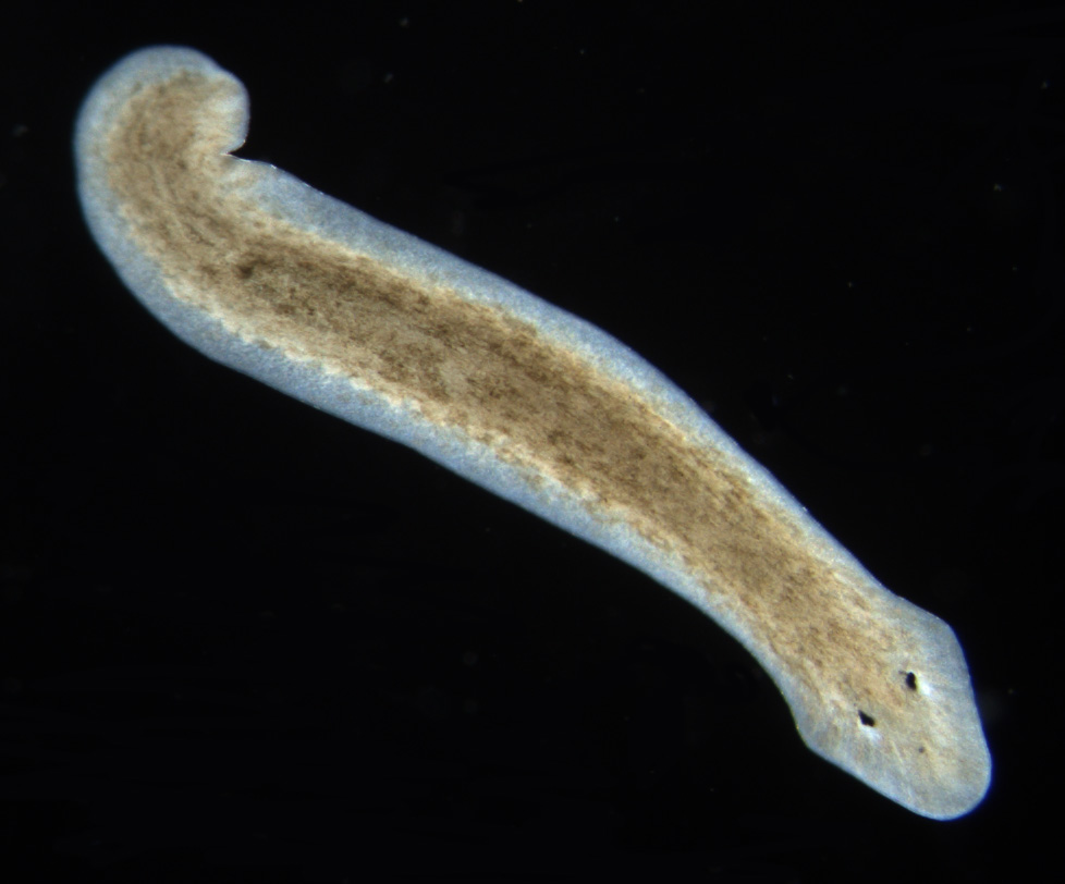
Planarians (triclads) are free-living flatworms of the class Turbellaria,[2][3] order Tricladida,[4] which includes hundreds of species, found in freshwater, marine, and terrestrial habitats.[5] Planarians are characterized by a three-branched intestine, including a single anterior and two posterior branches.[5] Their body is populated by adult stem cells called neoblasts, which planarians use for regenerating missing body parts.[6] Many species are able to regenerate any missing organ, which has made planarians a popular model in research of regeneration and stem cell biology.[7] The genome sequences of several species are available, as are tools for molecular biology analysis.[7][8]

The order Tricladida is split into three suborders, according to their phylogenetic relationships: Maricola, Cavernicola and Continenticola. Formerly, the Tricladida was split according to their habitat: Maricola (marine planarians); Paludicola (freshwater planarian); and Terricola (land planarians).[9]

Planarians move by beating cilia on the ventral dermis, allowing them to glide along on a film of mucus. Some also can move by undulations of the whole body by the contractions of muscles built into the body membrane.[10]

Triclads play an important role in watercourse ecosystems and are often very important as bio-indicators.[11]

Phylogeny and taxonomy

[edit]

Phylogeny

[edit]

Phylogenetic supertree after Sluys et al., 2009:[1]

Tricladida

Taxonomy

[edit]
Sabussowia ronaldi, a Maricola.
Polycelis felina, a planariid.
Platydemus manokwari, a geoplanid.

Linnaean ranks after Sluys et al., 2009:[1]

Anatomy and physiology

[edit]

Planarians are bilaterian flatworms that lack a fluid-filled body cavity, and the space between their organ systems is filled with parenchyma.[5][13] Planarians lack a circulatory system, and absorb oxygen through their body wall. They uptake food to their gut using a muscular pharynx, and nutrients diffuse to internal tissues. A three-branched intestine runs across almost the entire body, and includes a single anterior and two posterior branches. The planarian intestine is a blind sac, having no exit cavity, and therefore planarians uptake food and egest waste through the same orifice, located near the middle of the ventral body surface.[5]

The excretory system is made of many tubes with many flame cells and excretory pores on them. Also, flame cells remove unwanted liquids from the body by passing them through ducts which lead to excretory pores, where waste is released on the dorsal surface of the planarian.

The triclads have an anterior end or head where sense organs, such as eyes and chemoreceptors, are usually found. Some species have auricles that protrude from the margins of the head. The auricles can contain chemical and mechanical sensory receptors.[14]

The number of eyes in the triclads is variable depending on the species. While many species have two eyes (e.g. Dugesia or Microplana), others have many more distributed along the body (e.g. most Geoplaninae). Sometimes, those species with two eyes may present smaller accessory or supernumerary eyes. The subterranean triclads are often eyeless or blind.[14]

The body of the triclads is covered by a ciliated epidermis that contains rhabdites. Between the epidermis and the gastrodermis there is a parenchymatous tissue or mesenchyme.[14]

Nervous system

[edit]
Planaria nervous system

The planarian nervous systems consists of a bilobed shaped cerebral ganglion that is referred to as the planarian brain.[15] Longitudinal ventral nerve chords extend from the brain to the tail. Transverse nerves, commissure, connect the ventral nerve chords forming ladder-like nerve system.[5] The brain has been shown to exhibit spontaneous electrophysiological oscillations,[16] similar to the electroencephalographic (EEG) activity of other animals.

The planarian has a soft, flat, wedge-shaped body that may be black, brown, blue, gray, or white. The blunt, triangular head has two ocelli (eyespots), pigmented areas that are sensitive to light. There are two auricles (earlike projections) at the base of the head, which are sensitive to touch and the presence of certain chemicals. The mouth is located in the middle of the underside of the body, which is covered with hairlike projections (cilia). There are no circulatory or respiratory systems; oxygen enters and carbon dioxide leaves the planarian's body by diffusing through the body wall.

Reproduction

[edit]
Planarian reproductive system

Triclads reproduce sexually and asexually, and different species may be able to reproduce by one or both modes.[5] Planarians are hermaphrodites. In sexual reproduction, the mating generally involves mutual insemination.

Thus, one of their gametes will combine with the gamete of another planarian. Each planarian transports its secretion to the other planarian, giving and receiving sperm. Eggs develop inside the body and are shed in capsules. Weeks later, the eggs hatch and grow into adults. In asexual reproduction, the planarian fissions and each fragment regenerates its missing tissues, generating complete anatomy and restoring functions.[17] Asexual reproduction, similar to regeneration following injury, requires neoblasts, adult stem cells, which proliferate and produce differentiated cells.[17] Some researchers claim that the products derived from bisecting a planarian are similar to the products of planarian asexual reproduction; however, debates about the nature of asexual reproduction in planarians and its effect on the population are ongoing.[18] Some species of planarian are exclusively asexual, whereas some can reproduce both sexually and asexually.[19] In most of the cases the sexual reproduction involve two individuals; auto fecundation has been rarely reported (e.g. in Cura foremanii).[14]

Neoblasts

[edit]

Neoblasts are abundant adult stem cells that are found in the planarian parenchyma across the planarian body.[20] They are small and round cells, 5 to 10 μm, and characterized by a large nucleus, which is surrounded by little cytoplasm.[20] Neoblasts are required for regenerating missing tissues and organs, and they continuously replenish tissues by producing new cells.[17] Neoblasts can self-renew and generate progenitors for different cell types. In contrast to adult vertebrate stem cells (e.g., hematopoietic stem cell), neoblasts are pluripotent (i.e., producing all somatic cell types).[21] Moreover, they give rise to differentiating, post-mitotic, cells directly,[22] and not by producing rapidly-dividing transit amplifying cells.[20] Consequently, neoblasts divide frequently, and apparently lack a large sub-population of dormant or slow-cycling cells.[23]

As a model system in biological and biomedical research

[edit]

The life history of planarians make them a model system for investigating a number of biological processes, many of which may have implications for human health and disease. Advances in molecular genetic technologies has made the study of gene function possible in these animals and scientists are studying them worldwide. Like other invertebrate model organisms, for example C. elegans and D. melanogaster, the relative simplicity of planarians facilitates experimental study.

Planarians have a number of cell types, tissues and simple organs that are homologous to human cells, tissues and organs. However, regeneration has attracted the most attention. Thomas Hunt Morgan was responsible for some of the first systematic studies (that still underpin modern research) before the advent of molecular biology as a discipline.

Planarians are also an emerging model organism for aging research. These animals have an apparently limitless regenerative capacity, and asexual Schmidtea mediterranea has been shown to maintain its telomere length through regeneration.[24]

Live planarians are increasingly used in toxicological research due to their regenerative capabilities, simple anatomy, and sensitivity to environmental changes. Their ability to regenerate lost body parts provides a unique model to study the effects of chemical exposures on cellular processes, while their rapid response to toxins makes them an efficient tool for screening potential environmental and pharmaceutical hazards. An example of this application is a fluorescence-based skin irritability assay, where planaria are exposed to various chemicals, and fluorescence dye is used to evaluate their epithelial damage in response to irritation, providing an effective screening method.[25]

Regeneration

[edit]

Planarian regeneration combines new tissue production with reorganization to the existing anatomy, morphallaxis.[17] The rate of tissue regrowth varies between species, but in frequently used lab species, functional regenerated tissues are available already 7–10 days following tissue amputation.[17] Regeneration starts following an injury that require the growth of a new tissue.[26] Neoblasts localized near the injury site proliferate to generate a structure of differentiating cells called blastema. Neoblasts are required for new cell production, and they therefore provide the cellular basis for planarian regeneration.[27] Cell signaling mechanisms provide positional information that regulates the cell types and tissues that are produced from the neoblasts in regeneration.[28] Many signaling molecules that provide positional information to neoblasts, in regeneration and homeostasis, are expressed in muscle cells.[29] Following injury, muscle cells throughout the body can alter the expression of genes that encode molecules that provide positional information.[29] Therefore, the activities of neoblasts and muscle cells following injuries are essential for successful regeneration.[30]

Historically, planarians have been considered "immortal under the edge of a knife."[31] Very small pieces of the planarian, estimated to be as little as 1/279th of the organism it is cut from, can regenerate back into a complete organism over the course of a few weeks.[32] New tissues can grow due to pluripotent stem cells that have the ability to create all the various cell types.[33] These adult stem cells are called neoblasts, and comprise 20% or more of the cells in the adult animal.[34] They are the only proliferating cells in the worm, and they differentiate into progeny that replace older cells. In addition, existing tissue is remodeled to restore symmetry and proportion of the new planaria that forms from a piece of a cut up organism.[34][17]

The organism itself does not have to be completely cut into separate pieces for the regeneration phenomenon to be witnessed. In fact, if the head of a planarian is cut in half down its center, and each side retained on the organism, it is possible for the planarian to regenerate two heads and continue to live.[35] Researchers, including those from Tufts University in the U.S., sought to determine how microgravity and micro-geomagnetic fields would affect the growth and regeneration of planarian flatworms, Dugesia japonica. They discovered that one of the amputated fragments sent to space regenerated into a double-headed worm. The majority of such amputated worms (95%) did not do so, however. An amputated worm regenerated into a double-head worm after spending five weeks aboard the International Space Station (ISS) – though regeneration of amputated worms as double-headed heteromorphosis is not a rare phenomenon unique to a microgravity environment.[36] Double-headed planaria regenerates can be induced by exposing amputated fragments to electrical fields. Electrical field exposure with opposite polarity can induce a planarian with two tails. Double-headed planaria regenerates can also be induced by treating amputated fragments with pharmacological agents that alter levels of calcium, cyclic AMP, and protein kinase C activity in cells,[37] as well as by genetic expression blocks (interference RNA) to the canonical Wnt/β-Catenin signalling pathway.[28]

Biochemical memory experiments

[edit]

In 1955, Robert Thompson and James V. McConnell conditioned planarian flatworms by pairing a bright light with an electric shock. After repeating this several times they took away the electric shock, and only exposed them to the bright light. The flatworms would react to the bright light as if they had been shocked. Thompson and McConnell found that if they cut the worm in two, and allowed both worms to regenerate, each half would develop the light-shock reaction. In 1963, McConnell repeated the experiment, but instead of cutting the trained flatworms in two he ground them into small pieces and fed them to other flatworms. He reported that the flatworms learned to associate the bright light with a shock much faster than flatworms who had not been fed trained worms.

This experiment intended to test whether memory could be transferred chemically. The experiment was repeated with mice, fish, and rats, but it always failed to produce the same results. The perceived explanation was that rather than memory being transferred to the other animals, it was the hormones in the ingested ground animals that changed the behavior.[38] McConnell believed that this was evidence of a chemical basis for memory, which he identified as memory RNA. McConnell's results are now attributed to observer bias.[39][40] No blinded experiment has ever reproduced his results of planarians scrunching when exposed to light. Subsequent explanations of this scrunching behaviour associated with cannibalism of trained planarian worms were that the untrained flatworms were only following tracks left on the dirty glassware rather than absorbing the memory of their fodder.

In 2012, Tal Shomrat and Michael Levin have shown that planarians exhibit evidence of long-term memory retrieval after regenerating a new head.[41]

Planarian species used for research and education

[edit]

Several planarian species are commonly used for biological research. Popular experimental species are Schmidtea mediterranea, Schmidtea polychroa, and Dugesia japonica,[5] which in addition to excellent regenerative abilities, are easy to culture in the lab. In recent decades, S. mediterranea has emerged as the species of choice for modern molecular biology research, due to its diploid chromosomes and the availability of both asexual and sexual strains.[7]

The most frequently used planarian in high school and first-year college laboratories is the brownish Girardia tigrina. Other common species used are the blackish Planaria maculata and Girardia dorotocephala.

See also

[edit]

References

[edit]
[edit]
Revisions and contributorsEdit on WikipediaRead on Wikipedia
from Grokipedia
Planarians are free-living flatworms belonging to the order Tricladida within the class and phylum Platyhelminthes, renowned for their dorsoventrally flattened, ribbon-like bodies and extraordinary capacity for whole-body regeneration. These acoelomate organisms exhibit bilateral , a ciliated epidermis for locomotion, and a triploblastic structure with three germ layers but no true beyond the gut. Planarians inhabit freshwater, marine, and terrestrial environments. Freshwater thrive in clean, oxygen-rich waters such as , ponds, and lakes, where they serve as indicators of due to their . They are carnivorous or scavenging predators, typically under 1 cm in length, that capture prey using a muscular, eversible connected to a branched, incomplete digestive system lacking an , through which both ingestion and egestion occur. A defining feature of planarians is their regenerative ability, powered by neoblasts—pluripotent stem cells comprising up to 30% of their cells—which enable the regrowth of any body part, including the , from fragments as small as 1/279th of the original body. This process involves formation and rapid , allowing via transverse fission, where the worm splits into head and tail pieces that each regenerate into complete individuals. Planarians also reproduce sexually as simultaneous hermaphrodites, exchanging and laying protective cocoons containing multiple eggs that hatch into juveniles after direct development. Their simple , featuring a pair of anterior cerebral ganglia connected to ventral cords and a diffuse network of sensory structures like eyespots, supports behaviors such as phototaxis and prey detection, while an of cells maintains osmotic balance. Ecologically, planarians contribute to freshwater food webs as both predators of microorganisms and prey for larger aquatic animals, and their regenerative properties position them as vital model organisms for research in , developmental , and .

Taxonomy and evolution

Phylogenetic position

Planarians, commonly known as triclads, are free-living flatworms classified within the phylum Platyhelminthes, the class , and the order Tricladida. In contemporary phylogenetic frameworks, they are situated within the subphylum , which encompasses the majority of platyhelminths excluding the more basal Catenulida. Phylogenetic analyses consistently position Platyhelminthes, including planarians, as a monophyletic clade within the , a major subgroup of the superphylum in . This placement is robustly supported by molecular data, such as gene sequences, which demonstrate close relationships with other lophotrochozoans like annelids and mollusks while resolving internal platyhelminth relationships. Complementary evidence from clusters further corroborates these affinities, highlighting shared developmental patterning genes that align Platyhelminthes with spiralian protostomes and underscore their basal position within , often in the subclade. The acoelomate of planarians, featuring a solid filling the space between the and gut without a coelomic cavity, represents a primitive bilaterian condition retained from early metazoan . This trait, combined with bilateral symmetry and a simple triploblastic organization, reflects adaptations that likely emerged in the common ancestor of platyhelminths, enabling efficient diffusion-based transport in soft-bodied forms. Owing to their soft, non-mineralized bodies, planarians and other Platyhelminthes have no confirmed body fossils, complicating direct paleontological evidence. Molecular divergence estimates, calibrated against metazoan timelines, infer their origin around 600 million years ago in the late period, predating the . Indirect support comes from trace fossils suggestive of free-living activity, aligning with phylogenetic predictions of an ancient emergence. Recent phylogenomic analyses (as of ) continue to support this placement while uncovering cryptic diversity and instances of hybridization within Tricladida, enhancing resolution of internal relationships.

Classification

Planarians, commonly known as triclads, belong to the kingdom Animalia, phylum Platyhelminthes, subphylum , and order Tricladida. This order encompasses free-living flatworms distinguished by their three-branched intestine, a defining morphological feature. The subphylum unites various free-living and some parasitic lineages, excluding the more basal and the highly derived parasitic Neodermata. Within Tricladida, three suborders are recognized: Maricola (marine species), Cavernicola (stygobiont cave-dwellers), and Continenticola (freshwater and terrestrial forms). The Continenticola suborder, which includes most familiar planarians, is further divided into families such as Dendrocoelidae, Planariidae, and Dugesiidae, reflecting differences in reproductive structures and habitat adaptations. Prominent genera within these families include Dugesia (family Dugesiidae), named after the 19th-century French zoologist Alfonso Dugès for his contributions to studies; Schmidtea (family Planariidae), honoring the German zoologist Karl Schmidt; and Polycelis (family Planariidae), derived from Greek roots poly- (many) and kelis (spot), alluding to the variable pigmentation patterns observed in these species. These genera represent cosmopolitan groups, with Dugesia alone comprising over 100 described species distributed across freshwater habitats worldwide. More than 1,500 species of Tricladida have been described as of 2024, distributed across marine, freshwater, and terrestrial environments, with the largest numbers in terrestrial habitats (over 800 species) and freshwater (around 700 species), alongside fewer marine forms. Historically, planarians were classified under the class Turbellaria, a polyphyletic assemblage of free-living platyhelminths separated from the parasitic classes Trematoda (flukes) and Cestoda (tapeworms) primarily on the basis of their non-parasitic, ciliated lifestyles and lack of specialized attachment organs. This traditional division within phylum Platyhelminthes emphasized ecological distinctions, with Turbellaria encompassing diverse free-living forms like triclads. Subsequent molecular phylogenies have restructured the taxonomy, confirming the separation of free-living Rhabditophora (including Tricladida) from the derived parasitic Neodermata, while highlighting the paraphyly of Turbellaria.

Description and ecology

Habitat and distribution

Planarians, belonging to the order Tricladida, predominantly inhabit freshwater environments such as streams, ponds, and lakes, where they occupy benthic zones often under rocks or debris in temperate and subtropical regions worldwide. While most species are freshwater dwellers, a smaller number occur in marine interstitial habitats, such as sandy or muddy sediments along coastlines. These flatworms exhibit a preference for clean, well-oxygenated waters with slow or standing flow, avoiding fast-flowing currents that could dislodge them and polluted conditions to which they are highly sensitive. The geographic distribution of planarians is extensive across continents, excluding , with high diversity in , , and ; for instance, species of the genus are recorded from through to . In , Dugesia tigrina is native to many freshwater bodies but has been introduced to parts of via human activities, expanding its range beyond its original temperate habitats. Ecologically, planarians serve as key predators in benthic communities, feeding on small invertebrates like protozoans, rotifers, and microcrustaceans, thereby influencing community dynamics and nutrient cycling in freshwater ecosystems. Their presence is often indicative of good water quality, as their sensitivity to pollutants helps monitor environmental health.

External morphology

Planarians exhibit an elongated body that is dorsoventrally flattened, measuring typically 3 to 20 mm in length with a width that is proportionally narrower, resulting in a thin, ribbon-like form. This flattened structure lacks a and is characteristic of their free-living lifestyle in aquatic environments. The anterior region features a distinct head, often triangular or wedge-shaped, bearing two lateral auricles resembling ear-like lobes that function in chemosensory detection. The posterior end tapers into a pointed , contributing to the overall streamlined . Many species possess two dorsal eyespots, or ocelli, positioned near the auricles; these consist of pigmented cups surrounding photoreceptor cells with rhabdomeres. The entire body surface is covered by a ciliated , enabling gliding locomotion through coordinated ciliary beating. This also contains rhabdites, rod-shaped secretory structures that may aid in protection or production. Color variations among planarians include brown, black, or translucent appearances, arising from epidermal pigments such as ommochromes and porphyrins.

Anatomy

Internal organs

The digestive system of planarians is a blind, branched gastrovascular cavity that functions in both and distribution. Food is ingested through a muscular, eversible located ventrally near the midpoint of the body, which protrudes to capture prey and then retracts to connect with the intestine. The serves as both and in this incomplete system. From the , the intestine branches into three main lobes: a single anterior lobe extending toward the head and two posterior lobes running along the body sides, with extensive lateral diverticula that facilitate absorption throughout the body. This triclad structure allows for via gastrodermal cells that phagocytose food particles. The consists of protonephridia, a network of tubules distributed throughout the body for and waste removal. These structures include , specialized cells with tufts of cilia that beat rhythmically to filter ultrafiltrate from the body fluids, resembling a flickering flame under . Each connects to proximal tubules via a diaphragm, leading to distal tubules that merge into collecting ducts emptying through pores on the body surface. This system maintains ionic balance in freshwater environments by regulating water and solute . Planarian musculature is organized into a subepidermal body wall layer and additional fibers associated with internal organs, enabling gliding locomotion and body shape maintenance. The body wall comprises outer circular fibers, inner longitudinal fibers running anteroposteriorly, and diagonal fibers arranged in crisscross patterns for torsional movements. Muscle fibers around the and intestine provide contractility for feeding and . These obliquely striated muscles are regenerated from stem cells during injury. Planarians lack specialized circulatory and respiratory organs, relying instead on across their thin body surface for and nutrient transport. Oxygen and move directly through the and intercellular junctions to reach internal tissues, a process sufficient due to the animal's small size and flattened shape.

Nervous system

The planarian nervous system is characterized by a centralized structure that includes a bilobed brain and paired ventral nerve cords, forming the core of its (CNS). The brain consists of two anterior cerebral ganglia connected by a ventral commissure, which integrates sensory inputs and coordinates motor outputs across the body. This arrangement creates an orthogonal , with longitudinal and transverse fibers organized in perpendicular planes to facilitate efficient . Extending from the brain, two longitudinal ventral nerve cords run posteriorly along the body, interconnected by a series of transverse commissures and finer connectives that form a ladder-like configuration. This orthogonal layout allows for distributed processing and rapid communication between anterior and posterior regions, supporting coordinated locomotion and environmental responses. The peripheral nervous system branches from these cords, innervating muscles and sensory structures throughout the parenchyma. Key neurotransmitters in the planarian CNS include serotonin (5-HT) and (DA), which modulate neuromuscular function and behavioral coordination. Serotonin influences ciliary gliding and overall motility, while dopamine regulates locomotor patterns and responses to stimuli, contributing to the system's integrative capabilities. These monoamines operate through specific receptors, enabling fine-tuned neural signaling. The planarian exhibits notable complexity, with the housing several thousand neurons that undergo continuous turnover and rescaling to match body size changes. This high neuronal density supports basic forms of learning and , such as to light or associative conditioning with chemical cues, as evidenced by electrophysiological recordings of neural activity during behavioral tasks.

Physiology and behavior

Locomotion and feeding

Planarians primarily locomote by gliding across substrates, propelled by the coordinated beating of cilia on their ventral epidermal surface over a thin layer of mucus secreted by mucous glands. This ciliary action enables smooth, efficient forward movement, with typical gliding speeds of 1–2 mm/s, equivalent to up to several body lengths per minute for an average 10–20 mm individual. In addition to ciliary gliding, planarians can employ undulating muscular waves involving body-wall muscles to facilitate turning, twisting, or locomotion in varied environments, such as when navigating obstacles or in viscous media. As carnivores and , planarians feed on small aquatic , including protozoans, nematodes, and other soft-bodied prey. They lack jaws and instead capture prey through chemosensory detection, using auricles and distributed chemoreceptors to sense chemical cues from food sources in the environment, which guide oriented toward potential meals. Upon contact, the muscular is everted through the to envelop and suck in the prey, drawing it into the gastrovascular cavity for processing. Digestion in planarians combines extracellular and intracellular processes within the branched gut. Extracellular digestion begins in the gastrovascular cavity, where enzymes such as cysteine and aspartyl proteases secreted by gastrodermal cells break down proteins and other macromolecules into smaller fragments. These nutrients are then absorbed by gastrodermal cells for intracellular digestion in lysosomes, allowing further breakdown and distribution via diffusion to the rest of the body. Undigested remnants are expelled through the pharynx.

Sensory perception

Planarians detect through simple eyespots, or ocelli, located on the dorsal surface of the head. These structures consist of rhabdomeric photoreceptor cells whose microvilli form rhabdoms that capture photons, surrounded by pigmented cup cells that direct and enhance contrast. This organization enables negative phototaxis, where planarians actively avoid illuminated areas to seek shelter in darker environments, a critical for their in aquatic habitats. Chemosensory perception in planarians is mediated by specialized structures in the head region, including auricles and ciliated sensory pits or grooves. The auricles, ear-like projections, house ciliated epithelial cells and sensory neurons that detect dissolved chemical cues, such as those from food sources or potential mates, facilitating . These pits, lined with densely packed cilia, amplify chemical signal detection by increasing surface area and fluid flow, allowing planarians to toward or away from specific gradients in their environment. For mechanoreception, planarians lack statocysts, relying instead on distributed tactile sensors such as ciliated cells and peripheral endings throughout the body. These mechanosensory elements detect physical contact, vibrations, and subtle water movements, enabling responses to mechanical stimuli without specialized balance organs. Planarians also exhibit environmental sensory responses, including geotaxis and rheotaxis, which help navigate streams and aquatic flows. In geotaxis, they display positive geotaxis, tending to orient towards and settle at , mediated by body-wide ciliary and neural sensors. Rheotaxis involves positive orientation into water currents, achieved through auricular cilia that sense flow direction and velocity, aiding in positioning. These responses integrate with central neural processing in the for coordinated .

Reproduction

Asexual reproduction

Planarians primarily reproduce asexually through binary fission, a process in which the worm's body undergoes transverse constriction at a midpoint, typically behind the pharynx, resulting in two fragments: a head piece and a tail piece. Each fragment then regenerates the missing anterior or posterior structures to form complete individuals. This fission is characteristic of asexual strains, such as those of Schmidtea mediterranea and Dugesia species, and is mediated by mechanical forces that tear the body apart. In laboratory cultures, this mode of reproduction is routinely observed under controlled conditions, allowing for rapid propagation of clonal populations without the need for mates. Some planarian species also exhibit as an asexual strategy, where unfertilized eggs develop into viable , often activated by sperm from sexual encounters but without genetic contribution from males. This form is documented in parthenogenetic biotypes of species like Schmidtea polychroa. Following fission, regeneration of the head on the tail fragment and the tail on the head fragment occurs within approximately one week, driven by the proliferation and differentiation of neoblasts, the unique to planarians. These neoblasts migrate to the sites and rebuild the lost structures, ensuring each fragment restores a fully functional . Asexual reproduction via fission and confers advantages such as rapid population expansion in stable habitats, enabling a single individual to found new colonies and avoiding the energetic costs associated with sexual mate-finding. This strategy supports efficient colonization of favorable environments, where genetic uniformity from is less detrimental than in variable conditions.

Sexual reproduction

Planarians are simultaneous hermaphrodites, possessing both male and female reproductive organs that develop concurrently, allowing individuals to function as both sperm donors and recipients during reproduction. This hermaphroditic condition facilitates cross-fertilization, where mating typically involves reciprocal through copulation, with partners exchanging via mutual insertion of their copulatory organs. Internal fertilization occurs as are transferred directly into the partner's reproductive tract, promoting without self-fertilization in most species. Gamete production begins in the gonads: ovaries, located immediately posterior to the and adjacent to the ventral nerve cords, generate oocytes through , while numerous testes distributed along the dorso-lateral body margins produce within testicular follicles via . These s mature independently, with stored in until copulation, and oocytes released into the oviducts for potential fertilization. Following successful copulation, fertilized eggs are encapsulated in protective cocoons formed by secretions from the vitelline glands, which provide nutritive cells to support development. These cocoons, containing multiple zygotes surrounded by , are laid on the substrate as adhesive capsules, offering mechanical protection and nourishment against environmental stressors. Embryonic development in planarians is direct, lacking a free-living larval stage, and proceeds through a series of cleavage and events within the cocoon, culminating in hatchlings that resemble miniature adults. Hatching typically occurs 2-4 weeks after cocoon deposition, depending on species and temperature, with juveniles emerging fully formed and capable of immediate locomotion and feeding.

Cellular biology

Neoblasts

Neoblasts are the adult pluripotent stem cells in planarians, constituting undifferentiated cells that comprise approximately 10-30% of the total body cell population and are characterized by the expression of the piwi-1 gene marker. These cells are the only proliferative somatic cells in the adult planarian, enabling continuous tissue turnover through mitosis and maintaining homeostasis by replacing differentiated cells lost to normal wear or injury. Neoblasts exhibit high sensitivity to ionizing radiation, which selectively ablates them within hours of exposure, halting proliferation and underscoring their essential role in cellular renewal. In terms of lineage potential, neoblasts, particularly the subset known as clonogenic neoblasts (cNeoblasts), possess pluripotent capabilities and can differentiate into nearly all somatic cell types, including those of the epidermis, musculature, digestive system, and , but exclude the germline lineage, which arises from a distinct set of stem cells. This differentiation process involves progression from pluripotent cNeoblasts to lineage-restricted progenitors, ensuring balanced production of diverse cell types without contributing to reproductive cells. Recent advancements in single-cell sequencing have revealed heterogeneity among neoblasts, identifying distinct subtypes such as neural-committed neoblasts (ν-neoblasts) and other fate-biased populations that exhibit varying transcriptional profiles and cycling rates, enhancing understanding of their specialization during homeostasis. Furthermore, post-2020 studies have highlighted neoblasts' involvement in aging processes, where their regenerative activity can rejuvenate tissues by countering age-related declines in cellular composition, such as neuronal loss, through targeted proliferation and differentiation. Neoblasts also play a key role in regeneration by mobilizing to injury sites, though the mechanisms of this deployment are detailed elsewhere.

Regeneration mechanisms

Planarian regeneration is initiated at the wound site through the formation of a , a mass of undifferentiated cells derived primarily from the proliferation and migration of neoblasts toward the injury. Upon wounding, such as or transverse , signals from the wound trigger neoblast activation, leading to their directed migration and subsequent mitotic division within hours to days post-amputation. This proliferative response generates the progenitor cells necessary for replacing lost structures, with the emerging as a proliferative zone that organizes into specific tissues over time. The establishment of anterior-posterior (AP) polarity during regeneration is critically regulated by Wnt/β-catenin signaling, which specifies posterior fates and suppresses anterior identity. In decapitated planarians, the posterior-facing regenerates a due to maintained Wnt activity from pre-existing posterior gradients, while the anterior forms a head through inhibition of Wnt signaling. Transverse experiments demonstrate this polarity decision: the anterior fragment regenerates a and at its posterior cut site, whereas the posterior fragment regenerates a head at its anterior cut site, ensuring proper axis reformation regardless of fragment size. Seminal studies have shown that ectopic Wnt activation posteriorizes anterior blastemas, resulting in two-tailed animals, while Wnt inhibition leads to two-headed phenotypes. Full regeneration in planarians typically completes within 1-2 weeks, depending on the injury type and species; for instance, following , the and head structures reform in about 14 days, with functional recovery of behaviors like occurring progressively. Bisection yields faster initial outgrowth in smaller fragments but similar overall timelines, as both ends coordinate proportional scaling to restore the complete . Regeneration is highly sensitive to inhibitors that disrupt cellular proliferation or patterning cues. Ionizing radiation at doses above 15 Gy ablates neoblasts by inducing DNA damage and , completely blocking blastema formation and regeneration, though sublethal exposures can be tolerated with adaptive responses in some contexts. Additionally, the BMP signaling pathway is essential for dorsoventral (DV) polarity, with BMP ligands expressed dorsally re-specifying and maintaining dorsal identity during blastema differentiation; inhibition of BMP results in ventralized regeneration, such as ectopic dorsal structures on the ventral side.

Research applications

As a model organism

Planarians serve as versatile s in biological research, particularly for investigating regeneration, stem cell biology, and developmental processes, owing to their extraordinary capacity to regenerate entire body parts from small fragments. This regenerative ability, driven by a population of pluripotent known as neoblasts, provides a unique system for studying tissue renewal and positional control . Additionally, planarians offer practical advantages such as simple laboratory maintenance and genetic tractability, making them accessible for experimental manipulation without the ethical and logistical challenges associated with models. Their use as model organisms dates back to the early , when naturalists first explored their regenerative properties and employed them in studies of and development. Early investigations highlighted their "immortal under the edge of the knife" nature, sparking interest in regeneration mechanisms. In the modern era, genetic tools have enhanced their utility; RNA interference (RNAi) was pioneered in the planarian Schmidtea mediterranea through bacterially expressed double-stranded RNA feeding in 2003, enabling efficient . The primary species selected for laboratory research is Schmidtea mediterranea, particularly its asexual strains, which facilitate clonal propagation and consistent experimental conditions. In , japonica is commonly used due to its availability and suitability for similar studies. These choices leverage the species' robust regeneration—allowing complete animals to reform from tissue fragments—as a foundational trait for research. Planarians are maintained in sterile cultures within petri dishes filled with spring water, which is changed daily to prevent contamination and maintain optimal conditions at 21–23°C. They are fed every 2 or 3 days with nutrient sources like beef liver or egg yolk, followed by transfer to to ensure hygiene and health. This low-cost, straightforward protocol supports large-scale experiments while minimizing stress on the organisms.

Historical and current studies

Early studies on planarians focused on their physiological responses to environmental stressors, with C.M. Child conducting pioneering work in the 1910s on susceptibility gradients. Child used species like Planaria dorotocephala to investigate how different body regions varied in tolerance to toxins such as and oxygen deprivation, revealing metabolic gradients that influenced regeneration and survival rates during starvation. These experiments demonstrated that anterior regions were more susceptible, supporting Child's axial gradient theory and laying foundational insights into planarian . In 1901, advanced regeneration research through grafting experiments, including attempts to transplant heads between planarian fragments to test polarity determination. Morgan's work showed that transplanted heads could induce anterior structures in host bodies, but outcomes depended on the host's original polarity, highlighting intrinsic factors controlling head-tail identity rather than simple transplantation success. This contributed to early understanding of regenerative limits and influenced subsequent embryological studies. Mid-20th-century research explored behavioral aspects, notably James V. McConnell's experiments in the 1950s and 1960s on . McConnell trained planarians to associate light with electric shock, then fed cannibalized trained worms to untrained ones, claiming faster learning in recipients suggested RNA-mediated . However, replication failures revealed issues like poor controls and pseudo-conditioning, leading to the experiments' discreditation by the 1970s. Regeneration polarity research progressed in the 2000s with molecular insights, particularly the role of β-catenin. In Schmidtea mediterranea, RNAi knockdown of β-catenin caused posterior fragments to regenerate heads instead of tails, establishing β-catenin as a key switch for anteroposterior identity during both regeneration and . This work, building on Wnt signaling pathways, provided a mechanistic basis for Morgan's earlier observations and advanced planarians as models for axial patterning. Post-2020 studies leverage planarians for translational applications, including models for human regeneration. Neoblast-driven regeneration in aging planarians rejuvenates tissues globally, mirroring potential mechanisms in mammalian stem cells and offering insights for age-related regenerative therapies. A 2025 study showed that regeneration in aging sexual planarians leads to global tissue , underscoring their value in aging research. In neuropharmacology, planarians screen antipsychotics for ; phenotypic profiling distinguishes drug classes like antipsychotics from anxiolytics based on locomotor responses, validating their use as alternatives to models. For , planarians detect pollutant impacts, with behavioral assays revealing sublethal effects of nanoplastics and pesticides on feeding and regeneration, supporting their role in adverse outcome pathway frameworks. Controversies persist around historical experiments, often critiqued as pseudoscientific due to irreproducibility and methodological flaws, which overshadowed legitimate planarian behavioral . Emerging ethical considerations address potential , as planarians exhibit and learning, prompting calls for welfare assessments in studies, though evidence remains insufficient for stringent regulations.

References

Add your contribution
Related Hubs
User Avatar
No comments yet.