Community hub
Recent from talks
Knowledge base stats:
Talk channels stats:
Members stats:
Pharynx
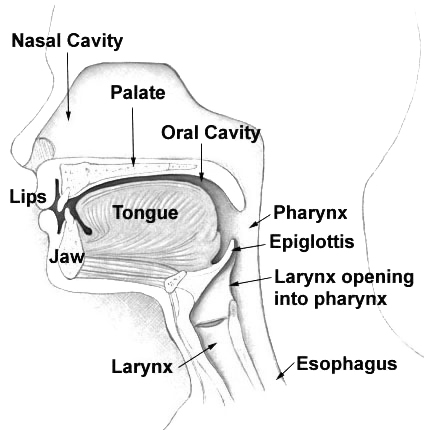
The pharynx (pl.: pharynges) is the part of the throat behind the mouth and nasal cavity, and above the esophagus and trachea (the tubes going down to the stomach and the lungs respectively). It is found in vertebrates and invertebrates, though its structure varies across species. The pharynx carries food to the esophagus and air to the larynx. The flap of cartilage called the epiglottis stops food from entering the larynx.
In humans, the pharynx is part of the digestive system and the conducting zone of the respiratory system. (The conducting zone—which also includes the nostrils of the nose, the larynx, trachea, bronchi, and bronchioles—filters, warms, and moistens air and conducts it into the lungs). The human pharynx is conventionally divided into three sections: the nasopharynx, oropharynx, and laryngopharynx (hypopharynx).
In humans, two sets of pharyngeal muscles form the pharynx and determine the shape of its lumen. They are arranged as an inner layer of longitudinal muscles, and an outer circular layer of pharyngeal constrictor muscles.
The upper portion of the pharynx, the nasopharynx, extends from the base of the skull to the upper surface of the soft palate. It includes the space between the internal nares and the soft palate and lies above the oral cavity. The adenoids, also known as the pharyngeal tonsils, are lymphoid tissue structures located in the posterior wall of the nasopharynx. Waldeyer's tonsillar ring is an annular arrangement of lymphoid tissue in both the nasopharynx and oropharynx. The nasopharynx is lined by respiratory epithelium that is pseudostratified, columnar, and ciliated.
Polyps or mucus can obstruct the nasopharynx, as can congestion due to an upper respiratory infection. The auditory tube, which connects the middle ear to the pharynx, opens into the nasopharynx at the pharyngeal opening of the auditory tube. The opening and closing of the auditory tubes serves to equalize the barometric pressure in the middle ear with that of the ambient atmosphere.
The anterior aspect of the nasopharynx communicates through the choanae with the nasal cavities. On its lateral wall is the pharyngeal opening of the auditory tube, somewhat triangular in shape and bounded behind by a firm prominence, the torus tubarius or cushion, caused by the medial end of the cartilage of the tube that elevates the mucous membrane. Two folds arise from the cartilaginous opening:
The oropharynx lies behind the oral cavity, extending from the uvula to the level of the hyoid bone. It opens anteriorly, through the isthmus faucium, into the mouth, while in its lateral wall, between the palatoglossal arch and the palatopharyngeal arch, is the palatine tonsil. The anterior wall consists of the base of the tongue and the epiglottic vallecula; the lateral wall is made up of the tonsil, tonsillar fossa, and tonsillar (faucial) pillars; the superior wall consists of the inferior surface of the soft palate and the uvula. Because both food and air pass through the pharynx, a flap of connective tissue called the epiglottis closes over the glottis when food is swallowed to prevent aspiration. The oropharynx is lined by non-keratinized squamous stratified epithelium.
The HACEK organisms (Haemophilus, Actinobacillus actinomycetemcomitans, Cardiobacterium hominis, Eikenella corrodens, Kingella) are part of the normal oropharyngeal flora, which grow slowly, prefer a carbon dioxide-enriched atmosphere, and share an enhanced capacity to produce endocardial infections, especially in young children. Fusobacterium is a pathogen.
Hub AI
Pharynx AI simulator
(@Pharynx_simulator)
Pharynx
The pharynx (pl.: pharynges) is the part of the throat behind the mouth and nasal cavity, and above the esophagus and trachea (the tubes going down to the stomach and the lungs respectively). It is found in vertebrates and invertebrates, though its structure varies across species. The pharynx carries food to the esophagus and air to the larynx. The flap of cartilage called the epiglottis stops food from entering the larynx.
In humans, the pharynx is part of the digestive system and the conducting zone of the respiratory system. (The conducting zone—which also includes the nostrils of the nose, the larynx, trachea, bronchi, and bronchioles—filters, warms, and moistens air and conducts it into the lungs). The human pharynx is conventionally divided into three sections: the nasopharynx, oropharynx, and laryngopharynx (hypopharynx).
In humans, two sets of pharyngeal muscles form the pharynx and determine the shape of its lumen. They are arranged as an inner layer of longitudinal muscles, and an outer circular layer of pharyngeal constrictor muscles.
The upper portion of the pharynx, the nasopharynx, extends from the base of the skull to the upper surface of the soft palate. It includes the space between the internal nares and the soft palate and lies above the oral cavity. The adenoids, also known as the pharyngeal tonsils, are lymphoid tissue structures located in the posterior wall of the nasopharynx. Waldeyer's tonsillar ring is an annular arrangement of lymphoid tissue in both the nasopharynx and oropharynx. The nasopharynx is lined by respiratory epithelium that is pseudostratified, columnar, and ciliated.
Polyps or mucus can obstruct the nasopharynx, as can congestion due to an upper respiratory infection. The auditory tube, which connects the middle ear to the pharynx, opens into the nasopharynx at the pharyngeal opening of the auditory tube. The opening and closing of the auditory tubes serves to equalize the barometric pressure in the middle ear with that of the ambient atmosphere.
The anterior aspect of the nasopharynx communicates through the choanae with the nasal cavities. On its lateral wall is the pharyngeal opening of the auditory tube, somewhat triangular in shape and bounded behind by a firm prominence, the torus tubarius or cushion, caused by the medial end of the cartilage of the tube that elevates the mucous membrane. Two folds arise from the cartilaginous opening:
The oropharynx lies behind the oral cavity, extending from the uvula to the level of the hyoid bone. It opens anteriorly, through the isthmus faucium, into the mouth, while in its lateral wall, between the palatoglossal arch and the palatopharyngeal arch, is the palatine tonsil. The anterior wall consists of the base of the tongue and the epiglottic vallecula; the lateral wall is made up of the tonsil, tonsillar fossa, and tonsillar (faucial) pillars; the superior wall consists of the inferior surface of the soft palate and the uvula. Because both food and air pass through the pharynx, a flap of connective tissue called the epiglottis closes over the glottis when food is swallowed to prevent aspiration. The oropharynx is lined by non-keratinized squamous stratified epithelium.
The HACEK organisms (Haemophilus, Actinobacillus actinomycetemcomitans, Cardiobacterium hominis, Eikenella corrodens, Kingella) are part of the normal oropharyngeal flora, which grow slowly, prefer a carbon dioxide-enriched atmosphere, and share an enhanced capacity to produce endocardial infections, especially in young children. Fusobacterium is a pathogen.