Hubbry Logo
search
logo
1912177

Pyoderma gangrenosum

logo
Community Hub0 Subscribers
Read side by side
from Wikipedia
Pyoderma gangrenosum
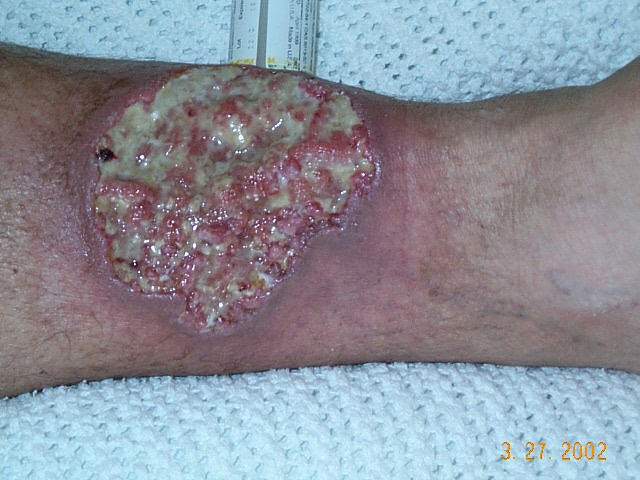
Pyoderma gangrenosum on the leg of a person with ulcerative colitis.
SpecialtyDermatology Edit this on Wikidata
Usual onset40s or 50s[1]
TreatmentCorticosteroids, ciclosporin, infliximab, canakinumab[2]

Pyoderma gangrenosum is a rare, inflammatory skin disease where painful pustules or nodules become ulcers that progressively grow.[3] Pyoderma gangrenosum is not infectious.[3]

Treatments may include corticosteroids, ciclosporin, infliximab, or canakinumab.[2]

The disease was identified in 1908. It affects approximately 1 person in 100,000 in the population. Though it can affect people of any age, it mostly affects people in their 40s and 50s.[1]

Types

[edit]
Pyoderma gangrenosum

There are two main types of pyoderma gangrenosum:[1]

  • the 'typical' ulcerative form, which occurs in the legs
  • an 'atypical' form that is more superficial and occurs in the hands and other parts of the body

Other variations are:[4]

  • Peristomal pyoderma gangrenosum comprises 15% of all cases of pyoderma
  • Bullous pyoderma gangrenosum
  • Pustular pyoderma gangrenosum[5]
  • Vegetative pyoderma gangrenosum[6]

Presentation

[edit]

Associations

[edit]

The following are conditions commonly associated with pyoderma gangrenosum:[7][8]

A rare[10] syndromic association called pyogenic arthritis, pyoderma gangrenosum and acne syndrome (PAPA syndrome), a type of autoinflammatory disorder, is associated with mutations in the proline-serine-threonine phosphatase-interacting 1 gene (PSTPIP1).[10][11]

Causes

[edit]

Though the cause is not well understood, the disease is thought to be due to immune system dysfunction, and particularly improper functioning of neutrophils. In support of an immune cause, a variety of immune mediators such as interleukin (IL)-8, IL-1β, IL-6, interferon (IFN)-γ, granulocyte colony-stimulating factor, tumor necrosis factor alpha, matrix metalloproteinase (MMP)-9, MMP10, and elafin have all been reported to be elevated in patients with pyoderma gangrenosum.[12]

Also in support of an immune cause is the finding that at least half of all pyoderma gangrenosum patients suffer from immune-mediated diseases.[1] For instance, ulcerative colitis, rheumatoid arthritis,[4] and monoclonal gammopathies[13] have all been associated with pyoderma gangrenosum. It can also be part of autoinflammatory syndromes such as PAPA syndrome.[10][11] Marzano et al. (2017) identified a variety of single-nucleotide polymorphisms (SNPs) linked to autoinflammation that were carried, singly or in combination, in subsets of patients with pyoderma gangrenosum, acne and suppurative hidradenitis syndrome (PASH syndrome) or isolated pyoderma gangrenosum of the ulcerative subtype.[14]

One hallmark of pyoderma gangrenosum is pathergy, which is the appearance of new lesions at sites of trauma.[15]

Diagnosis

[edit]

Diagnosis of PG is challenging owing to its variable presentation, clinical overlap with other conditions, association with several systemic diseases, and absence of defining histopathologic or laboratory findings. Misdiagnosis and delayed diagnosis are common. It has been shown that up to 39% of patients who initially received a diagnosis of PG have an alternative diagnosis.[16] In light of this, validated diagnostic criteria have recently been developed for ulcerative pyoderma gangrenosum.[17]

Diagnostic criteria

[edit]

In addition to a biopsy demonstrating a neutrophilic infiltrate, patients must have at least 4 minor criteria to meet diagnostic criteria.[17] These criteria are based on histology, history, clinical examination, and treatment.[citation needed]

  • Histology: Exclusion of infection (including histologically indicated stains and tissue cultures)
  • Pathergy (ulcer occurring at sites of trauma, with the ulcer extending past the area of trauma)
  • Personal history of inflammatory bowel disease or inflammatory arthritis
  • History of papule, pustule, or vesicle that rapidly ulcerated
  • Clinical examination (or photographic evidence) of peripheral erythema, undermining border, and tenderness at the site of ulceration
  • Multiple ulcerations (at least 1 occurring on an anterior lower leg)
  • Cribriform or “wrinkled paper” scars at sites of healed ulcers
  • Decrease in ulcer size within 1 month of initiating immunosuppressive medications

Treatment

[edit]

First-line therapy for disseminated or localized instances of pyoderma gangrenosum is systemic treatment with corticosteroids and ciclosporin. Topical application of clobetasol, mupirocin, and gentamicin alternated with tacrolimus can be effective. Pyoderma gangrenosum ulcers demonstrate pathergy, that is, a worsening in response to minor trauma or surgical debridement. Significant care should be taken with dressing changes to prevent potentially rapid wound growth. Many patients respond differently to different types of treatment, for example, some benefit from a moist environment, so treatment should be carefully evaluated at each stage.[citation needed]

If ineffective, alternative therapeutic procedures include systemic treatment with corticosteroids and mycophenolate mofetil; mycophenolate mofetil and ciclosporin; tacrolimus; thalidomide; infliximab; or plasmapheresis.[18]

See also

[edit]

References

[edit]
[edit]
Revisions and contributorsEdit on WikipediaRead on Wikipedia
from Grokipedia
Pyoderma gangrenosum is a rare, noninfectious inflammatory skin disorder classified within the neutrophilic dermatoses, characterized by the rapid progression of painful, necrolytic ulcers with undermined, violaceous borders, most commonly affecting the lower extremities.[1][2] It typically begins as a small, tender papule, pustule, or nodule that evolves within days into a enlarging open sore with a ragged edge, often exhibiting pathergy—an exaggerated inflammatory response to minor skin trauma such as surgery or injury.[3][2] The condition is not contagious and affects individuals of all ages, though it is uncommon in children.[3] The estimated incidence of pyoderma gangrenosum is approximately 0.63 cases per 100,000 population annually, with a median age of onset around 59 years and a variable sex distribution that ranges from equal to predominantly female (up to 76%).[2] Up to 50% of cases are associated with underlying systemic conditions, including inflammatory bowel disease (such as ulcerative colitis in 5-12% and Crohn's disease in 1-2%), rheumatoid arthritis (about 10%), hematologic malignancies (5-20%), and other autoimmune or inflammatory disorders.[2][3] The exact etiology remains unknown but involves immune dysregulation, neutrophil dysfunction, and possible genetic factors, with no infectious agent identified despite the name suggesting a bacterial origin.[3] Risk factors include a history of autoimmune diseases and skin trauma, which can precipitate lesions in susceptible individuals.[1] Diagnosis is primarily clinical and by exclusion, requiring correlation of history, physical examination, and histopathology showing sterile neutrophilic infiltrates in the dermis, while ruling out mimics such as infections, vasculitis, venous ulcers, or malignancies.[2][3] Proposed diagnostic criteria, such as those by Su et al. or Maverakis et al., emphasize rapid progression of a painful ulcer and exclusion of other causes, often supported by elevated inflammatory markers like C-reactive protein.[2] Management focuses on wound care, pain control, and systemic immunosuppression; first-line therapies include corticosteroids (0.5-1 mg/kg/day) or cyclosporine, with biologics like infliximab showing response rates up to 69% in refractory cases.[2][3] Surgical interventions should be avoided due to pathergy risk.[3] Prognosis varies but often involves recurrence in about 30-50% of cases, with potential complications including secondary bacterial infections, significant scarring, and hyper- or hypopigmentation, particularly in individuals with brown or Black skin.[1][2] Early diagnosis and multidisciplinary care involving dermatologists, wound specialists, and rheumatologists are essential to improve outcomes and prevent progression.[3]

Overview and Epidemiology

Definition and Characteristics

Pyoderma gangrenosum is a rare, sterile inflammatory skin disorder classified as a neutrophilic dermatosis, characterized by the rapid development of painful ulcers with undermined, violaceous borders.[4] Unlike infectious conditions, it involves no identifiable pathogens and is diagnosed primarily by exclusion of other ulcerative diseases.[3] The term "pyoderma gangrenosum" was coined in 1930 by Brunsting and colleagues, who initially described it as resembling a gangrenous pyoderma, erroneously suggesting a bacterial etiology despite the absence of infection.[4] Key characteristics include the formation of sterile ulcers that expand rapidly due to neutrophilic infiltration, often beginning as a small pustule or nodule before progressing to a larger lesion with irregular, overhanging edges and surrounding erythema.[3] Upon healing, these ulcers typically leave cribriform or "worm-eaten" scarring, reflecting the destructive inflammatory process.[5] Although primarily cutaneous, pyoderma gangrenosum can exhibit extracutaneous involvement, such as aseptic pulmonary nodules in the lungs or scleritis and corneal ulceration in the eyes.[5] The condition has an estimated annual incidence of 3 to 10 cases per million population, underscoring its rarity.[4] It is frequently associated with underlying systemic diseases, including inflammatory bowel disease and hematologic malignancies, though the precise mechanisms linking these remain under investigation.[3]

Epidemiology

Pyoderma gangrenosum (PG) is a rare dermatological condition, with a global incidence estimated at 3 to 10 cases per million population per year.[6] This translates to approximately 0.3 to 1 case per 100,000 individuals annually, underscoring its infrequent occurrence in clinical practice.[7] Epidemiological data indicate higher reported rates in Northern European populations, where adjusted incidence figures reach 0.63 per 100,000 person-years, potentially reflecting improved diagnostic awareness or regional predispositions.[7] The disease predominantly affects adults, with peak onset between the ages of 40 and 60 years, aligning with the third to sixth decades of life.[8] Pediatric cases are exceptionally uncommon, comprising less than 5% of all diagnoses, often presenting unique challenges in early detection.[9] Gender distribution shows approximate parity overall, though multiple studies report a slight female predominance, with ratios exceeding 1.8:1 in some cohorts.[10] Geographic variations may contribute to disparities in reported prevalence.[11]

Clinical Presentation

Signs and Symptoms

Pyoderma gangrenosum typically begins with the development of tender, erythematous papules, nodules, or pustules on the skin, often resembling a spider bite or furuncle. These initial lesions rapidly progress, breaking down into painful ulcers within days.[1][12][3] The characteristic ulcers have an irregular, jagged shape with purple-blue or violaceous, undermined edges that extend beneath the surrounding skin. The ulcer base is often necrotic, purulent, or covered with yellowish exudate, and the lesions demonstrate rapid enlargement, frequently expanding by several centimeters over days to weeks. Multiple ulcers may coalesce, leading to larger defects.[13][12][3] Severe pain is a hallmark symptom, often disproportionate to the apparent size or extent of the lesion, and can significantly impair daily activities. In extensive cases, patients may experience associated systemic symptoms such as fever and malaise. Extracutaneous involvement is uncommon but can include arthralgias in some instances.[1][13][3]

Types and Variants

Pyoderma gangrenosum (PG) is classified into several morphological and clinical variants, with the classic ulcerative type being the most prevalent, accounting for approximately 80% of cases. This form typically presents as deep, painful ulcers with irregular, violaceous, and undermined borders, most commonly affecting the lower legs.[14][3] Other variants include the atypical form, characterized by superficial and eccentric ulcers that often occur on unusual sites such as the hands or upper extremities, differing from the deeper lesions of the classic type.[15][3] The bullous variant features tense, hemorrhagic blisters that may rupture to form superficial erosions, resembling bullous leukemia cutis and frequently involving the upper body, face, or arms; it is often associated with underlying hematologic malignancies.[13][16] The pustular variant manifests as multiple sterile pustules on the trunk or extensor surfaces, which can resolve spontaneously or evolve into ulcerative lesions, particularly in patients with inflammatory bowel disease.[13][16] The vegetative variant presents as indolent, granulomatous plaques or nodules with less aggressive undermining and minimal ulceration, typically on the trunk or extremities.[3][15] A key feature across variants, particularly the classic form, is the pathergy phenomenon, an exaggerated inflammatory response to minor skin trauma that occurs in 25-50% of cases. This response induces lesion formation through dysregulated neutrophil activation and cytokine release (e.g., increased IL-8 and TNF-α), leading to rapid tissue necrosis and ulceration at the site of injury, such as after venipuncture or incision.[16][3] Lesion sites vary by variant: classic ulcers predominate on the lower legs, while bullous lesions favor the upper body and pustular or vegetative forms often appear on the trunk or proximal extremities.[15][13]

Pathogenesis

Etiology

Pyoderma gangrenosum (PG) is a rare inflammatory skin disorder with an unclear etiology, and approximately 50% of cases are idiopathic, meaning no underlying cause can be identified despite extensive evaluation.[17] Despite its name suggesting a bacterial or pyogenic origin, no infectious agent has been consistently identified as causative; early assumptions of streptococcal or other microbial involvement have been disproven through histopathological and microbiological studies showing sterile neutrophilic infiltrates.[3] In the remaining cases, PG is often associated with systemic diseases such as inflammatory bowel disease, rheumatoid arthritis, or hematologic malignancies, though these associations do not imply direct causation and are explored further in related sections.[17] Genetic factors play a role in a subset of PG cases, particularly in rare familial or syndromic presentations. Mutations in the PSTPIP1 gene, which encodes proline-serine-threonine phosphatase-interacting protein 1, are linked to pyogenic arthritis, pyoderma gangrenosum, and acne (PAPA) syndrome, an autosomal dominant autoinflammatory disorder characterized by recurrent sterile joint inflammation, skin ulcers, and severe acne.[18] These mutations disrupt normal immune regulation, leading to enhanced inflammatory responses, but such genetic associations account for only a small fraction of PG occurrences, with most cases lacking identifiable hereditary patterns.[19] Environmental triggers are implicated in initiating or exacerbating PG lesions, especially in individuals exhibiting pathergy—a hyperreactive response to minor skin injury. Common inciting events include trivial trauma, surgical procedures, or subcutaneous injections, which can precipitate rapid ulcer formation at the site of injury in pathergy-positive patients.[3] This phenomenon underscores PG as a hypersensitivity reaction rather than a primary infectious or neoplastic process, with immune dysregulation central to the aberrant response.[20]

Pathophysiology

Pyoderma gangrenosum (PG) is characterized by a complex interplay of dysregulated innate and adaptive immune responses, leading to inflammation and subsequent tissue destruction in the skin. The condition is classified as a neutrophilic dermatosis, where aberrant activation of the immune system results in sterile inflammation without identifiable infectious agents. While traditionally central to this process is the recruitment and dysfunction of neutrophils, which infiltrate the dermis and release proteolytic enzymes and reactive oxygen species, causing collagenolysis and extracellular matrix degradation that culminates in ulceration, recent histopathological studies as of 2025 indicate that neutrophil-predominant inflammation is uncommon.[3][21] Neutrophil-mediated inflammation plays a pivotal role where present, with hyperactive neutrophils exhibiting delayed apoptosis and impaired clearance, perpetuating tissue damage. These cells accumulate at the ulcer edge, forming a dense infiltrate that undermines the wound border and drives rapid lesion expansion through the release of matrix metalloproteinases and elastases. Emerging mechanisms include enhanced neutrophil extracellular traps (NETs) and low-density granulocytes (LDGs), contributing to tissue damage, particularly in syndromic forms. Inflammasome activation, such as via NLRP3 and AIM2, further amplifies this response by promoting neutrophil chemotaxis and survival through IL-1β release, contributing to the hallmark necrotic ulceration observed in PG. The complement cascade, including C5a, also promotes neutrophil recruitment.[22] Cytokine dysregulation exacerbates neutrophilic infiltration, with elevated levels of interleukin-8 (IL-8), IL-23, and tumor necrosis factor-alpha (TNF-α) detected in lesional skin and serum of affected patients. IL-8 acts as a potent chemoattractant, drawing neutrophils to the site and sustaining their activation, while TNF-α amplifies proinflammatory signaling by upregulating adhesion molecules and further cytokine production. IL-23, in turn, stimulates T-helper 17 cells to produce IL-17, which reinforces the neutrophilic response and promotes chronic inflammation; studies have shown significantly higher serum concentrations of these cytokines correlating with disease activity and lesion severity.[23][24][25] Immune complex deposition in dermal vessels contributes to the inflammatory cascade, with immunohistochemical analyses revealing deposits of immunoglobulins and complement components along vessel walls, suggesting an immune-mediated vasculopathy. This deposition triggers local complement activation and neutrophil recruitment without evidence of systemic immune complex disease. Concurrently, clonal expansion of T-cells, particularly at the ulcer periphery, indicates an adaptive immune component that sustains chronicity; expanded T-cell receptor clones have been identified in both peripheral blood and lesional skin, implying antigen-driven proliferation that maintains the inflammatory milieu.[26][27] The histopathological appearance includes vasculitis-like changes, such as perivascular neutrophilic infiltration and fibrinoid necrosis of vessel walls, yet true leukocytoclastic vasculitis is absent, with no fibrinoid degeneration or significant endothelial damage. This pseudovasculitic pattern leads to ischemic sterile necrosis through microvascular occlusion by neutrophil aggregates and thrombi, rather than direct vessel wall destruction, distinguishing PG from primary vasculitides.[3]

Associated Conditions

Systemic Diseases

Pyoderma gangrenosum (PG) is associated with a variety of systemic diseases in approximately 50-60% of cases, highlighting the importance of screening for underlying conditions to guide management.[28][9] Among these, inflammatory bowel disease (IBD) represents one of the strongest links, occurring in 17.6% of PG patients overall.[28] Specifically, PG develops in 1-5% of individuals with ulcerative colitis and 1-2% of those with Crohn's disease, often preceding or coinciding with gastrointestinal flares.[29][30] Rheumatic disorders, particularly rheumatoid arthritis and seronegative spondyloarthropathies, are implicated in about 12.8% of PG cases, with rheumatoid arthritis frequently preceding skin involvement by years.[28] These associations underscore shared inflammatory pathways, though the exact mechanisms remain under investigation. In some instances, treating the underlying arthritis can improve PG lesions, but this is addressed in detail elsewhere. Hematologic conditions, including monoclonal gammopathies and leukemias, are linked to PG in roughly 8-9% of cases for hematologic malignancies and 7-9% for monoclonal gammopathies of undetermined significance, often involving IgA paraproteins.[28][31] Approximately 20% of PG cases overall may connect to malignancies, either hematologic or solid, necessitating vigilant oncologic evaluation.[32] Other systemic illnesses, such as hidradenitis suppurativa and sarcoidosis, occur less frequently but are well-documented comorbidities in PG, potentially forming part of broader autoinflammatory syndromes.[33][34] These rarer associations emphasize the heterogeneous nature of PG's systemic ties.

Triggers and Risk Factors

Pyoderma gangrenosum (PG) lesions can be precipitated or exacerbated by minor trauma, a phenomenon known as pathergy, in which an exaggerated inflammatory response occurs at the site of injury. This affects approximately 30% of patients with PG and may manifest following procedures such as surgery, skin biopsies, or injections, leading to rapid ulceration or worsening of existing wounds.[35][3][36] Certain medications have been implicated in rare cases of drug-induced PG, with granulocyte colony-stimulating factor (G-CSF) associated with the development of bullous lesions at sites of prior skin conditions. Other drugs, including tyrosine kinase inhibitors and propylthiouracil, may trigger PG through mechanisms involving dysregulated neutrophil function and inflammatory responses.[37][38][39] Obesity is a recognized risk factor for peristomal PG in patients with inflammatory bowel disease, potentially due to increased skin irritation and impaired wound healing. Smoking has been linked to a higher incidence of extraintestinal manifestations, including PG, in Crohn's disease, suggesting it may increase disease severity in susceptible individuals.[40][41] In some pediatric cases, PG has followed upper respiratory tract infections, as observed in an infant who developed lesions after an episode accompanied by cervical lymphadenopathy. These post-infectious triggers appear uncommon but highlight the role of acute inflammatory events in initiating disease in children.

Diagnosis

Clinical Evaluation

The clinical evaluation of pyoderma gangrenosum begins with a detailed history-taking to identify key features suggestive of the condition. Patients often report a rapid onset of lesions, typically progressing from a small pustule or nodule to a large ulcer within days to weeks.[39] Severe pain at the site is a hallmark symptom, frequently described as disproportionate to the visible injury.[15] Inquiry into trauma history is crucial, as minor injuries or surgical procedures may precede lesion development due to pathergy, a hyperreactive response to trauma.[39] Systemic symptoms such as fever, arthralgias, or gastrointestinal complaints should be assessed to uncover potential underlying diseases, while family history may reveal clustering in relatives, indicating a possible genetic predisposition.[39] Physical examination focuses on thorough inspection of the lesion and surrounding skin. Ulcers typically exhibit irregular, undermined edges with a characteristic violaceous or erythematous border, often accompanied by a necrotic base covered in purulent exudate.[42] The lesions are markedly tender to palpation, and satellite pustules or nodules may be present at the periphery, particularly in early stages or pustular variants.[15] A full-body examination is essential to identify additional lesions or signs of extracutaneous involvement, such as oral ulcers or joint swelling.[39] Biopsy plays a supportive role in evaluation, primarily to exclude alternative diagnoses rather than confirm pyoderma gangrenosum definitively, as histopathology is nonspecific. A superficial shave or incisional biopsy from the active edge is preferred to demonstrate a dense neutrophilic infiltrate in the dermis, often with epidermal necrosis and leukocytoclasia.[42] Deep punch biopsies should be avoided due to the risk of pathergy, which can exacerbate the lesion and worsen ulceration.[39] Patients must be informed of this potential complication prior to any procedure.[39] Laboratory tests are employed to evaluate for inflammation and underlying systemic conditions. Basic blood work includes a complete blood count (CBC) to detect leukocytosis or anemia, erythrocyte sedimentation rate (ESR), and C-reactive protein (CRP) to quantify inflammatory activity, which is often elevated.[42] These initial assessments help guide further investigation while excluding infection or malignancy mimics.[15]

Diagnostic Criteria

The diagnosis of pyoderma gangrenosum (PG) relies on established criteria that emphasize clinical features, histopathological findings, and exclusion of alternative etiologies, as no single test is pathognomonic.[43] The 2004 criteria proposed by Su et al. require fulfillment of both major criteria and at least two minor criteria for confirmation.[44] The major criteria include: (1) rapid progression of a painful, necrolytic ulcer with irregular violaceous and undermined borders, and (2) exclusion of other causes such as infection, malignancy, or other inflammatory conditions through appropriate investigations.[44] The minor criteria consist of: (1) history of pathergy or cribriform scarring, (2) association with a systemic disease linked to PG (e.g., inflammatory bowel disease, arthritis, IgA gammopathy, or malignancy), (3) histopathological evidence of neutrophilic dermal infiltrate without specific features of other conditions, and (4) rapid response to systemic steroid treatment (50% improvement in 1 month).[44] An updated international consensus from 2018, developed via a Delphi process involving expert dermatologists, refined these guidelines specifically for ulcerative PG, the most common variant.[43] This framework specifies one major criterion—biopsy of the ulcer edge showing a neutrophilic infiltrate—and requires at least four of eight minor criteria for diagnosis.[43] The minor criteria include: (1) exclusion of infection; (2) pathergy; (3) history of inflammatory bowel disease or inflammatory arthritis; (4) history of papule, pustule, or vesicle ulcerating within 4 days of appearing; (5) peripheral erythema, undermining border, and tenderness at ulceration site; (6) multiple ulcerations, at least 1 on an anterior lower leg; (7) cribriform or “wrinkled paper” scar(s) at healed ulcer sites; (8) decreased ulcer size within 1 month of initiating immunosuppressive medication(s).[43] These criteria underscore a multimodal assessment, integrating clinical, laboratory, and histopathological data while prioritizing exclusion of mimics.[43] Microbiological cultures from wound swabs or tissue biopsies are essential to rule out bacterial, fungal, or mycobacterial infections, with negative results supporting PG.[45] Imaging modalities, such as ultrasound or MRI, may be employed adjunctively to exclude deep-seated infections or underlying osteomyelitis, particularly in lesions near bone, though they are not diagnostic for PG itself.[42] In atypical presentations, such as bullous, pustular, or vegetative variants, the criteria exhibit lower specificity, often necessitating multidisciplinary input from dermatologists, rheumatologists, and pathologists to integrate clinical context and serial evaluations for accurate diagnosis.[46]

Differential Diagnosis

Pyoderma gangrenosum (PG) must be differentiated from other causes of cutaneous ulceration, as it is a diagnosis of exclusion without pathognomonic features, relying on clinical presentation, history, and targeted investigations to rule out mimics.[3] Key distinctions often involve microbiological cultures, vascular imaging, serological tests, and biopsy to identify alternative etiologies that may present with painful, rapidly enlarging ulcers similar to PG's characteristic violaceous border and undermined edges.[4] Infectious causes include bacterial infections such as ecthyma, which typically arises from streptococcal or staphylococcal sources in immunocompromised individuals, as well as mycobacterial infections (e.g., tuberculosis or atypical mycobacteria) and deep fungal ulcers (e.g., sporotrichosis or chromoblastomycosis). These are distinguished from PG by positive wound cultures, tissue staining, or PCR demonstrating pathogens, along with a history of exposure or systemic symptoms like fever, whereas PG ulcers are sterile.[3][4][15] Vascular conditions encompass arterial and venous ulcers, often located on the lower extremities due to ischemia or stasis, and vasculitides such as ANCA-associated vasculitis (e.g., granulomatosis with polyangiitis). Differentiation involves ankle-brachial index or Doppler ultrasound to confirm reduced perfusion in vascular ulcers, and serological tests like ANCA positivity or biopsy showing vessel wall inflammation in vasculitis, contrasting with PG's lack of true vascular occlusion.[4][15][3] Neoplastic disorders include cutaneous T-cell lymphoma (e.g., mycosis fungoides with ulcerated plaques) and metastatic malignancies (e.g., from breast or lung cancer), which may erode the skin to form ulcers. These are ruled out by skin biopsy revealing atypical lymphocytes or malignant cells, often with systemic staging via imaging or bone marrow evaluation, unlike PG's neutrophilic infiltrate without malignancy.[4][15] Other mimics comprise neutrophilic dermatoses like Sweet syndrome, which features tender plaques or nodules rather than deep ulcers but can progress to ulceration; halogenodermas (e.g., iododerma or bromoderma) induced by halide exposure; and factitial dermatitis from self-inflicted trauma. Distinction relies on clinical history (e.g., drug exposure for halogenoderma or psychiatric factors for factitial lesions), biopsy showing sparse neutrophilic infiltrates in Sweet syndrome without ulceration primacy, and absence of pathergy in non-PG conditions.[3][4][15]

Management

Pharmacological Treatment

The pharmacological treatment of pyoderma gangrenosum primarily aims to control the underlying neutrophilic inflammation through systemic and topical agents, with selection influenced by lesion severity, number, and associated systemic diseases.[46][47] For mild or localized disease, alternative oral therapies such as dapsone (100-150 mg/day) or minocycline (100 mg twice daily) may be considered, with reported response rates of approximately 50-70% in small studies.[48] Systemic corticosteroids serve as first-line therapy for rapid disease control in moderate to severe cases, typically administered as oral prednisone at 0.5–1 mg/kg/day (maximum 60 mg/day) or intravenous methylprednisolone at 1 g/day for 1–5 days in acute settings.[46][49] These agents stabilize lesions within 2–3 days and achieve clinical response in 40–50% of patients, though complete remission occurs in about 40% and relapse in 20% of multi-lesional cases; monitoring for adverse effects such as infections (reported in 13% of cases) is essential, with short-term use preferred to minimize risks like osteoporosis and hyperglycemia.[46][49] To facilitate steroid tapering, steroid-sparing immunosuppressants such as cyclosporine (2.5–5 mg/kg/day) or mycophenolate mofetil (1–3 g/day) are commonly added, showing comparable healing rates to corticosteroids (e.g., 47% with cyclosporine) and improvement in 85% of cases with mycophenolate, respectively; cyclosporine requires monitoring for nephrotoxicity and hypertension, while mycophenolate necessitates gastrointestinal and hematologic surveillance.[46][49][47] For refractory cases, biologic agents targeting inflammatory cytokines represent a key escalation, with anti-tumor necrosis factor (TNF) inhibitors like infliximab (administered intravenously at 5 mg/kg at weeks 0, 2, and 6, then every 8 weeks) and adalimumab (subcutaneous 40 mg every 1–2 weeks) demonstrating high efficacy, including complete response rates of 67% and partial response in 87% across meta-analyses of over 350 patients.[49][50] These agents are particularly effective in 70–80% of steroid- or immunosuppressant-refractory pyoderma gangrenosum, supported by a randomized controlled trial for infliximab and observational data for adalimumab showing 55% complete healing at 26 weeks; potential side effects include infections and infusion reactions, requiring screening for tuberculosis and hepatitis.[49][46] Emerging biologics such as interleukin (IL) inhibitors, including ustekinumab (subcutaneous 90 mg every 8 weeks after initial doses), have gained traction since the early 2020s for refractory disease, with complete response rates of 71% in systematic reviews of case series, offering a favorable safety profile in patients with comorbid inflammatory bowel disease. As of 2025, ongoing clinical trials are evaluating additional agents, such as the IL-36 inhibitor spesolimab, for refractory cases.[49][51][52] Topical therapies are reserved for mild, localized lesions smaller than 2 cm², utilizing high-potency corticosteroids (e.g., clobetasol propionate 0.05% ointment applied twice daily) or calcineurin inhibitors like tacrolimus (0.1% ointment twice daily), which promote healing in 42.6% of cases within 6 months without systemic exposure.[46][49] These agents require monitoring for local skin atrophy with prolonged corticosteroid use and potential absorption-related immunosuppression with tacrolimus, though evidence is limited to observational studies lacking randomized trials.[46][47]

Wound Care and Supportive Measures

Wound care in pyoderma gangrenosum emphasizes conservative strategies to minimize trauma and promote healing while avoiding exacerbation of the condition through pathergy. Surgical debridement is generally contraindicated due to the risk of pathergy, which can lead to rapid worsening of ulcers; instead, gentle mechanical or autolytic debridement methods, such as hydrogels for eschar removal, are preferred when necessary.[53][54] Wounds should be cleansed gently with sterile saline or lukewarm water to reduce irritation without causing further tissue damage.[46][55] Appropriate dressings play a critical role in maintaining a moist healing environment and preventing adherence that could provoke pathergy. Non-adherent options, such as hydrocolloids, foams, or polymeric membrane dressings, are recommended based on wound characteristics like depth, exudate level, and granulation status; for instance, alginates or hydrofibers suit exudative lesions, while hydrogels benefit superficial or eschar-covered areas.[53][12] These dressings should be changed as needed to manage moisture without frequent disruption, and topical antiseptics are limited to cases of critical colonization to avoid unnecessary irritation.[56] Pain management is essential given the severe discomfort associated with pyoderma gangrenosum ulcers, often requiring a multidisciplinary approach involving dermatologists, wound care specialists, and pain experts. Opioids may be used for acute control, though their application demands caution to mitigate dependency risks, while gabapentinoids like gabapentin or pregabalin address neuropathic components by stabilizing nerve membranes.[57][58] Psychological support, including counseling, is integrated to address the emotional burden of chronic pain and disfigurement.[59] Surgical interventions, such as skin grafting or negative pressure wound therapy, are typically avoided during active disease phases due to pathergy risks but may be considered cautiously in rare cases of healed or stable ulcers under close monitoring. Overall, supportive measures prioritize non-traumatic local care alongside patient education on avoiding injury to foster gradual epithelialization.[15][53]

Prognosis and Complications

Prognosis

Patients with pyoderma gangrenosum have an increased all-cause mortality risk compared to the general population, with 5-year survival rates of approximately 84.5% as of data up to 2022.[60] With appropriate treatment, approximately 40-60% of patients with pyoderma gangrenosum achieve remission within 6 months. The STOP GAP randomized controlled trial, which compared cyclosporine and prednisolone as first-line therapies, found equivalent healing outcomes between the two agents, with 47% of ulcers healed at 6 months in each group and no significant difference in speed of healing.[3][61] Recurrence rates for pyoderma gangrenosum range from 30% to 50%, with studies reporting figures such as 39% requiring inpatient management and 48% overall in long-term follow-up. These rates are higher in patients with associated systemic diseases, such as inflammatory bowel disease or arthritis, due to ongoing underlying inflammation. Early diagnosis and prompt initiation of therapy significantly improve healing trajectories and reduce the likelihood of prolonged disease course. Pediatric cases, which represent less than 5% of all instances, often exhibit a more favorable response to treatment, including systemic corticosteroids.[62][63][15][3] In the long term, mild cases of pyoderma gangrenosum may resolve spontaneously with conservative wound care, though this is uncommon. Some cases become chronic, necessitating ongoing immunosuppression and monitoring to prevent relapses triggered by pathergy or comorbidities. Treatment strategies outlined in management guidelines, such as systemic immunosuppressants, play a key role in achieving sustained remission.[3][64]

Complications

Pyoderma gangrenosum lesions often heal with characteristic local complications, including cribriform atrophic scarring that resembles thin "cigarette paper" due to the destructive nature of the ulcerative process.[65] In some cases, healing can lead to contractures, particularly when extensive ulceration affects joints or mobile areas, resulting in functional limitations.[66] Secondary bacterial superinfections are a notable risk, occurring not uncommonly in open wounds and potentially exacerbating tissue damage, with pathogens such as Staphylococcus aureus frequently implicated.[67][68] Systemic complications arise primarily in cases of extensive or poorly controlled disease. Sepsis can develop from widespread lesions or secondary infections, sometimes mimicking severe conditions like necrotizing fasciitis and leading to life-threatening systemic inflammatory responses.[69][70] Chronic severe pain associated with the ulcers frequently necessitates long-term use of opioids or other controlled analgesics, raising the risk of dependence in affected patients.[65][1] Treatment-related complications are common given the reliance on immunosuppressive therapies. Systemic corticosteroids, often used at high doses (0.5–1 mg/kg/day) for rapid control, can induce side effects such as osteoporosis, hyperglycemia leading to diabetes, weight gain, and mood disturbances with prolonged administration.[65][47] Immunosuppressive agents like cyclosporine or biologics further increase susceptibility to opportunistic infections, compounding the risk of secondary bacterial or viral complications in already compromised skin.[65][5] The psychological burden of pyoderma gangrenosum is significant, with disfiguring scars and persistent pain contributing to high rates of depression and anxiety among patients. Studies indicate that depressed mood is a common comorbidity, often exacerbated by sleep disruption, social isolation, and embarrassment from visible lesions, leading to reduced quality of life.[71][59] Anxiety may intensify due to uncertainty about disease recurrence and dependence on others for daily activities.[72][73]

References

User Avatar
No comments yet.