Hubbry Logo
KeratodermaKeratodermaMain
Open search
Keratoderma
Community hub
Keratoderma
logo
8 pages, 0 posts
0 subscribers
Be the first to start a discussion here.
Keratoderma
Keratoderma
from Wikipedia
Keratoderma
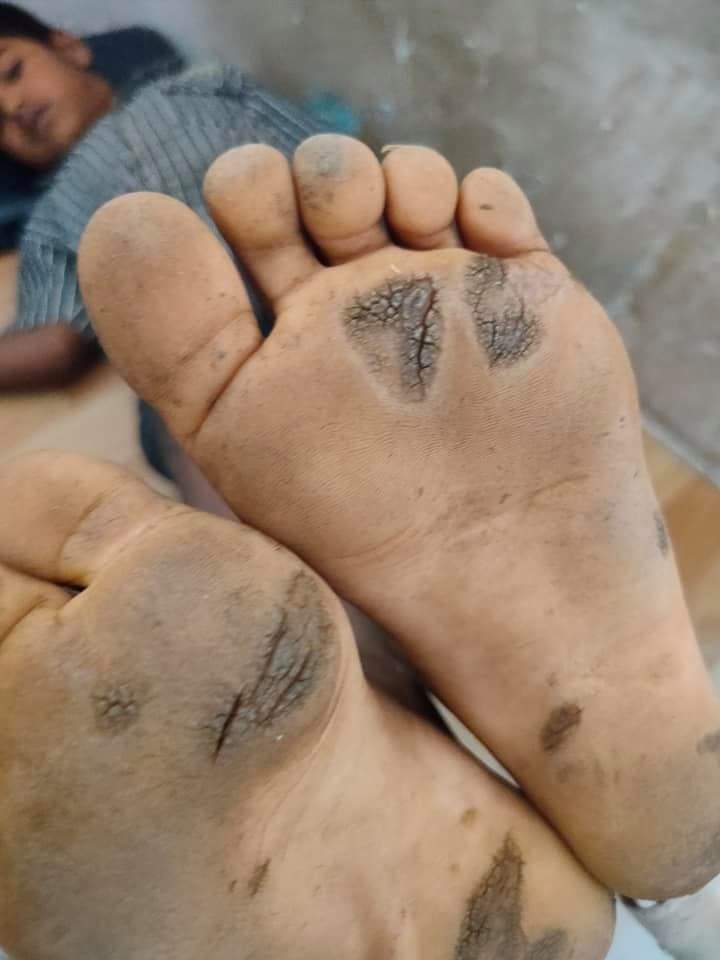
Diffuse keratodermas affect most of the palms and soles.
SpecialtyDermatology Edit this on Wikidata

Keratoderma is a local or general thickening of the horny layer of the epidermis.[1]

Classification

[edit]

The keratodermas are classified into the following subgroups:[2]: 506 

Congenital

[edit]

Acquired

[edit]

See also

[edit]

References

[edit]
[edit]
Revisions and contributorsEdit on WikipediaRead on Wikipedia
from Grokipedia
Keratoderma refers to a diverse group of skin disorders characterized by abnormal and excessive thickening () of the , the outermost layer of the skin, most commonly affecting the palms and soles. These conditions, often termed palmoplantar keratodermas when involving the palms and soles, can manifest as diffuse, focal, striate, or punctate patterns of and may occur in isolation or as part of broader syndromes. Hereditary forms are rare genetic disorders typically presenting in infancy or childhood, while acquired forms develop later in life secondary to other conditions. The prevalence of hereditary palmoplantar keratodermas varies by subtype but is generally estimated at 1 in 25,000 to 1 in 100,000 individuals worldwide; acquired forms are more common, though exact incidence is unknown and often associated with underlying diseases. Clinically, they present with thickened, scaly skin on the palms and soles, potentially causing pain or complications, and diagnosis involves clinical evaluation, , and where indicated. Management aims at symptom control and addressing any underlying causes.

Overview

Definition

Keratoderma refers to a heterogeneous group of disorders characterized by abnormal thickening, or , of the , the outermost layer of the , resulting in rough, hardened plaques. This pathological process involves excessive accumulation of , leading to localized or diffuse areas of indurated that can impair function and cause discomfort. While the condition can affect various body sites, it primarily manifests with involvement of the palms and soles, often termed , though non-palmoplantar variants such as tylosis and focal keratoses are also recognized within the spectrum. The term "keratoderma" derives from the Greek words "keras," meaning horn, and "derma," meaning skin, reflecting the horn-like thickening observed clinically. It was first described in medical literature during the 19th century, with early reports documenting hereditary forms affecting the extremities. Keratoderma is distinguished from ichthyosis, which features generalized scaling across the body surface due to defective epidermal barrier function, whereas keratoderma typically presents with more localized hyperkeratotic plaques. In contrast to calluses, which represent non-pathological, reactive thickening from mechanical friction, keratoderma arises from underlying genetic or acquired abnormalities in keratinization.

Epidemiology

Keratoderma encompasses a group of rare dermatological disorders characterized by hyperkeratotic thickening of the skin, primarily affecting the palms and soles, with hereditary forms being rare and subtype prevalences estimated at 1-9 per 100,000 in certain populations (e.g., 1.17 to 3.3 per 100,000 for punctate palmoplantar keratoderma in Croatia and Slovenia), while Mal de Meleda has a worldwide prevalence of approximately 1 per 100,000. Acquired forms are more common than hereditary ones but remain uncommon overall, with exact prevalence rates not well-established due to underreporting and diagnostic challenges. Incidence of congenital keratodermas typically manifests at birth or during , reflecting their genetic basis, whereas acquired variants predominantly occur in adults, particularly those over 50 years old, often in association with underlying comorbidities such as malignancies or nutritional deficiencies. Hereditary forms exhibit an equal male-to-female ratio, though certain subtypes like unilateral linear punctate show a male predominance (approximately 65%). Acquired keratodermas are more frequently observed in elderly populations and individuals with paraneoplastic syndromes. Geographic variations highlight higher incidences of specific hereditary subtypes in isolated populations due to founder effects, such as elevated rates of Mal de Meleda in Mediterranean and North African communities, Nagashima-type palmoplantar keratosis in Japanese populations, and Bothnian-type diffuse in Northern European groups like those in . These disorders remain rare, with no reported epidemics.

Etiology and Pathogenesis

Genetic Factors

Keratodermas often arise from hereditary that disrupt the structural integrity of the , particularly in . in genes, such as KRT1 and KRT9, are central to epidermolytic palmoplantar keratoderma (EPPK), where they impair the assembly of intermediate filaments, leading to cytoskeletal instability and epidermal fragility under mechanical stress. These typically affect the highly conserved rod domains of the keratins, causing dominant-negative effects that prevent proper filament and result in clumping of aggregates within suprabasal . Beyond keratins, mutations in other genes contribute to specific syndromic forms of keratoderma. For instance, mutations in the connexin 26 gene (GJB2) underlie Vohwinkel syndrome, where altered function disrupts intercellular communication in the . Similarly, mutations in desmoglein 1 (DSG1), a desmosomal protein, cause striate by weakening cell-cell attachments in the suprabasal layers. In Olmsted syndrome, gain-of-function mutations in TRPV3 lead to hyperactivity of the transient receptor potential vanilloid 3 , promoting excessive proliferation and inflammation. Inheritance patterns of genetic keratodermas vary, with autosomal dominant transmission being the most prevalent, as seen in Unna-Thost type , characterized by high but variable expressivity influenced by modifying genetic or environmental factors. Autosomal recessive forms are less common but occur, such as in Nagashima-type due to biallelic mutations in SERPINB7, which affect inhibition and epidermal barrier formation. These patterns often exhibit incomplete , leading to phenotypic heterogeneity even within families. The pathogenic pathways involve disrupted desmosomal adhesion, as in DSG1 and GJB2 mutations, which compromise epidermal cohesion and trigger compensatory . Abnormal cornification results from mutations that induce models, where misfolded filaments form tonofilament clumps, mimicking aggregates in other filamentopathies and exacerbating barrier dysfunction through impaired processing and water retention in the . This leads to overall epidermal barrier defects, increasing susceptibility to mechanical trauma and in palmoplantar regions. Recent studies as of 2024 have expanded the known genetic variants associated with , highlighting the importance of for accurate diagnosis. As of 2025, over 50 palmoplantar epidermal differentiation disorders have been reported, with a new -based nomenclature proposed to guide targeted therapies, and emerging approaches show promise for monogenic forms.

Acquired Factors

Acquired keratoderma refers to non-hereditary forms of palmoplantar that develop in adulthood, often asymmetrically, and may resolve upon addressing the underlying trigger. These conditions arise from external or secondary factors that disrupt normal epidermal turnover, leading to excessive accumulation on the palms and soles. Unlike genetic , acquired keratodermas lack inherited mutations and are typically linked to environmental exposures, inflammatory processes, infections, medications, or systemic illnesses. Reactive forms of acquired keratoderma commonly result from chronic mechanical friction or irritation, particularly in occupational settings involving manual labor, where repeated pressure on the palms and soles produces linear or striate hyperkeratotic plaques. For instance, construction workers or farmers may develop focal thickening due to prolonged tool handling or foot pressure, manifesting as callosities that mimic diffuse keratoderma. Inflammatory dermatoses also contribute significantly, with presenting as scaly, erythematous plaques on pressure-bearing areas, often with pustular variants like palmoplantar pustulosis. Eczema, including atopic or contact types, induces hyperkeratosis through persistent irritation and barrier disruption, while can cause violaceous, polygonal papules with overlying scale on acral sites. These reactive changes are driven by localized trauma or immune-mediated epidermal proliferation. Paraneoplastic keratoderma represents a rare but critical association with underlying malignancies, where skin changes often precede tumor detection by months to years. Acrokeratosis paraneoplastica, or Bazex syndrome, exemplifies this, featuring psoriasiform on the ears, nose, hands, and feet, most frequently linked to squamous cell carcinomas of the upper aerodigestive tract, such as bronchial or esophageal cancers. The acral distribution and nail dystrophy distinguish it, with resolution possible after tumor treatment, underscoring its role as a warning sign for systemic evaluation. Infectious etiologies include viral and fungal agents that provoke verrucous or hyperkeratotic responses. Human papillomavirus (HPV), particularly types causing verruca vulgaris, can form confluent, exuberant masses on palms and soles, termed verrucous keratoderma, especially in immunocompromised individuals where lesions mimic diffuse thickening. Fungal infections, such as from species, lead to moccasin-type hyperkeratosis on soles and interdigital spaces, with scaling and fissuring due to epidermal invasion and inflammatory response. Bacterial causes like or may also induce secondary through granulomatous reactions. Drug-induced acquired keratoderma arises as an adverse reaction to various medications, often reversible upon discontinuation. Lithium therapy, used in , has been associated with palmoplantar pustulosis and hyperkeratotic plaques, possibly through exacerbation of psoriasiform changes. Interferon-alpha (IFN-alpha) therapy, administered for or malignancies, can trigger psoriasiform eruptions with palmoplantar involvement, including keratoderma-like thickening. Other agents, such as BRAF inhibitors (e.g., ), cause dose-dependent in approximately 70-74% of cases, while chemotherapy like is associated with in about 2-3% of cases. Systemic diseases contribute to generalized or localized keratoderma through metabolic or circulatory alterations. and associated lead to doughy, hyperkeratotic palms and soles due to mucopolysaccharide deposition and reduced epidermal turnover, with improvement following thyroid hormone replacement. , particularly , manifests as follicular hyperkeratosis (phrynoderma) extending to palmoplantar areas, resulting from impaired keratinization and epithelial integrity; from vitamin C deficiency can compound this with additional scaling. Other associations include diabetes mellitus with neurotrophic changes and chronic promoting secondary thickening. The pathogenic mechanisms in acquired keratoderma primarily involve chronic inflammation without genetic mutations, where external triggers upregulate proliferation and differentiation. Cytokines such as interleukin-1 (IL-1) and tumor necrosis factor-alpha (TNF-alpha) play central roles, released by activated and immune cells in response to irritation, infection, or drugs, promoting hyperproliferation via and MAPK pathways. In inflammatory or reactive forms, these cytokines sustain a cycle of epidermal and impaired , leading to clinical thickening.

Classification

Congenital Keratodermas

In recent years, as of 2024, experts have proposed a gene-based reclassification of inherited palmoplantar keratodermas (PPKs) under the umbrella term palmoplantar epidermal differentiation disorders (pEDD), emphasizing mutations in genes involved in epidermal structure and differentiation to guide targeted therapies. Traditional eponymous subtypes remain useful for clinical description. Congenital keratodermas, also known as inherited PPKs, are a group of rare genetic disorders characterized by abnormal thickening of the skin on the palms and soles, typically presenting from birth or early infancy. These conditions arise from mutations in genes encoding proteins essential for epidermal structure and function, such as keratins or other structural elements, leading to persistent that can range from mild to severe and mutilating. While most forms manifest at birth or within the first few years of life, some may begin with neonatal erythroderma—a widespread redness and scaling—that evolves into localized by adulthood, often exacerbated by and . These conditions are rare, with prevalence estimates varying by subtype and population; for example, diffuse forms may affect 1–5 per 100,000 individuals in certain regions, while rarer subtypes occur in fewer than 1 per 100,000. Diffuse forms of congenital PPK involve uniform hyperkeratosis across the entire palmoplantar surface. Unna-Thost disease represents a non-epidermolytic variant (associated with KRT1 mutations), featuring mild, symmetric, yellowish thickening without blistering, often accompanied by and secondary infections; it typically onset in infancy and remains stable into adulthood without progression to autoamputation. In contrast, Vörner disease is epidermolytic (primarily KRT9 mutations, sometimes KRT1), characterized by more pronounced brown-yellow with fissuring and blistering upon trauma, starting in and showing limited transgredient spread to dorsal aspects in some cases. Both subtypes are autosomal dominant and relatively common among hereditary PPKs, with onset by age 3-4 years and gradual thickening over time. Striate or focal congenital keratodermas present with linear or localized hyperkeratotic lesions rather than diffuse involvement. Bart-Pumphrey syndrome exemplifies a striate form, with longitudinal palmar streaks, , and , often linked to ; it begins in infancy, progresses slowly with weight-bearing, and stems from GJB2 gene mutations in an autosomal dominant pattern. Richner-Hanhart syndrome, a focal variant tied to type II, features painful yellowish-white papules on pressure points and corneal erosions, with onset in early infancy or , potential in up to 60% of cases, and autosomal recessive inheritance via TAT gene defects; lesions worsen with friction but stabilize without intervention. These rarer subtypes highlight the focal nature, with plantar changes often preceding palmar ones. Syndromic congenital keratodermas integrate PPK with extracutaneous features, underscoring their multisystem impact. Pachyonychia congenita involves focal, painful plantar alongside hypertrophic nail and oral leukokeratosis, affecting over 90% of patients with nail and plantar issues; it emerges in infancy or , intensifies with ambulation, and arises from autosomal dominant in KRT6A, KRT6B, KRT16, or KRT17 genes. Mal de Meleda, prevalent in Mediterranean populations, manifests as severe diffuse PPK with maceration, malodor, and pseudoainhum-like constrictions leading to digit mutilation; onset at birth progresses markedly by age 4, with nail and contractures, due to autosomal recessive SLURP1 . These conditions often start with erythrodermic features in neonates, evolving to chronic . Among rare congenital forms, loricrin keratoderma presents a Vohwinkel-like with mild diffuse , honeycomb-patterned palms, and ichthyosiform erythroderma, sometimes initiating with a membrane at birth; it is non-progressive, autosomal dominant, and caused by LOR gene mutations, without associated or mutilation. Overall, these keratodermas exhibit variable progression from initial inflammatory phases to stable adult , influenced by mechanical stress. Recent genetic studies have identified additional genes, such as variants in some nonsyndromic and syndromic forms.

Acquired Keratodermas

Acquired keratodermas represent a group of non-hereditary palmoplantar hyperkeratotic disorders that develop secondary to external triggers or underlying systemic conditions, distinguishing them from congenital forms by their later onset and potential for reversibility. These conditions typically manifest in adulthood, often with asymmetric or focal distribution on the palms and soles, and frequently resolve upon addressing the primary , such as through eradication or tumor resection. Unlike inherited variants, acquired keratodermas emphasize the role of reactive, neoplastic, infectious, or toxic processes in pathogenesis. Reactive keratodermas arise in response to inflammatory dermatoses, exemplifying how chronic irritation or immune-mediated skin changes can induce localized . A representative subtype is keratoderma associated with (PRP), characterized by annular and circinate plaques on the palms and soles, a rare papulosquamous disorder featuring reddish-orange scaly plaques and follicular . In PRP, the palmoplantar involvement often presents as diffuse or patchy thickening with a gritty texture, reflecting epidermal proliferation driven by inflammatory cytokines. Treatment of the underlying PRP, such as with systemic retinoids, can lead to improvement in these keratotic features. Malignant-associated keratodermas serve as paraneoplastic markers, signaling occult internal neoplasms through exaggerated and . Acanthosis palmaris, also known as tripe palms, manifests as velvety, ridged thickening of the palms resembling bovine mucosa, frequently linked to gastric as a of tumor-secreted growth factors like transforming growth factor-alpha. This subtype typically emerges rapidly in older adults, with symmetric palmar involvement and possible extension to soles or digits, and has been reported alongside in approximately 70–90% of cases. Resection or successful management of the underlying often results in regression of the skin changes, underscoring their reversible nature. Infectious keratodermas result from microbial invasion leading to reactive epidermal , often mimicking idiopathic forms but resolving with targeted therapy. Hyperkeratotic , caused by human papillomavirus (HPV) subtypes such as HPV-1 or HPV-2, present as compact, verrucous plaques on weight-bearing areas of the soles or palms, with endophytic growth and potential for mosaicism in chronic cases. Similarly, tinea-induced thickening, particularly from dermatophytes like , produces diffuse on the soles with scaling and fissuring, exacerbated by occlusive environments. These lesions are usually focal or unilateral, reflecting the site of inoculation, and demonstrate marked improvement or complete clearance following or antiviral interventions. Drug- and toxin-induced keratodermas highlight environmental exposures as precipitants of toxic epidermal responses, with chronicity tied to cumulative insult. Arsenic exposure, often from contaminated water or historical medications, induces diffuse punctate or confluent palmar keratoses resembling Bowen's disease, featuring hyperkeratotic papules with atypical and increased risk. These changes typically onset after years of exposure, predominantly affecting palms and soles in a symmetric pattern, and may partially regress with cessation of exposure and , though residual lesions can persist. Other agents, such as certain chemotherapeutics, similarly provoke transient that abates upon discontinuation.

Clinical Features

Symptoms

Patients with keratoderma often report significant and discomfort, particularly in areas such as the heels and balls of the feet, where cracking and fissuring of the thickened can lead to tenderness and exacerbate discomfort during movement. This is a prominent feature in hereditary forms like palmoplantar keratodermas, where it arises from mechanical stress on the hyperkeratotic plaques and can be challenging to manage. Functional impairment is a common subjective complaint, with individuals experiencing difficulty walking due to painful fissures and thickened skin on the soles, as well as challenges gripping objects or wearing shoes because of rigid plaques on the palms. These limitations can severely restrict daily activities, such as standing for prolonged periods or performing manual tasks, leading to frustration and reduced independence. Associated sensations include itching, or pruritus, which affects a majority of patients with palmoplantar variants and is often more pronounced in inflammatory or acquired types; additionally, an unpleasant may arise from secondary bacterial infections in fissured areas. In cases like aquagenic , patients describe burning alongside pruritus upon water exposure. In syndromic congenital forms, such as those associated with pachyonychia congenita, patients may experience recurrent infections that can lead to fever, contributing to overall fatigue. The psychological impact on is profound, with many patients reporting embarrassment and social withdrawal due to the visible and malodorous nature of the skin changes, alongside that leads to emotional distress and reduced . These subjective experiences can result in isolation and heightened anxiety about daily interactions.

Physical Signs

Keratoderma manifests primarily through abnormal thickening of the on the palms and soles, presenting as hyperkeratotic plaques that vary in color, texture, and distribution. These plaques often appear yellowish-brown and can be diffuse, covering the entire palm or sole, or focal, localized to pressure points such as the heels or metatarsal arches. In some cases, the hyperkeratosis exhibits a transgrediens pattern, extending beyond the acral areas to involve the dorsal surfaces of the digits or hands. The texture of these lesions ranges from smooth and waxy to verrucous or scaly, frequently accompanied by an erythematous border and painful fissures, particularly in diffuse forms. Patterns may include epidermolytic changes, characterized by blistering upon minor trauma, contrasting with non-epidermolytic variants that show dry, scaling without formation. Severe presentations, such as in Olmsted syndrome, can feature pseudoainhum, with constricting fibrous bands around the digits leading to potential autoamputation. Nail involvement is common in certain subtypes, including pachyonychia congenita, where dystrophic changes such as ridging, thickening, and subungual are observed. Mucosal surfaces may show , particularly oral leukokeratosis in syndromic forms. While primarily acral, distribution can become generalized in ichthyosiform variants, affecting broader skin areas with ichthyosis-like scaling. Lesions often evolve over time, beginning as erythematous patches or mild thickening in infancy for hereditary types, progressing to more pronounced warty in adulthood, with worsening fissures and cracking. Acquired forms may present similarly but typically onset later, with symmetric involvement and possible extension to dorsal aspects.

Diagnosis

Clinical Assessment

Clinical assessment of keratoderma begins with a detailed to determine the and guide further evaluation. Key elements include the age of onset, which distinguishes congenital forms typically presenting in infancy or from acquired types emerging in adulthood. Family is crucial, as hereditary palmoplantar keratodermas often follow autosomal dominant or recessive patterns, such as in epidermolytic palmoplantar keratoderma due to KRT1 mutations. Occupational and environmental exposures should be explored, particularly for acquired cases linked to chronic friction, chemicals, or irritants like in contact dermatitis-related hyperkeratosis. Associated symptoms warrant screening for underlying conditions, including hyperhidrosis, pain, blistering, or signs of malignancy such as weight loss or gastrointestinal disturbances in paraneoplastic variants. Physical examination involves systematic and to characterize the lesions. Distribution patterns are evaluated for and extent: diffuse involvement suggests types like Vorner , while focal or striate forms localize to pressure areas or flexural lines. assesses texture, revealing waxy thickening or painful fissuring that predisposes to secondary infections. are examined for dystrophy, such as ridging or in pachyonychia congenita, and mucosa for rare transgrediens spread or oral leukokeratosis. Dermoscopy aids in identifying subtle patterns and vascular structures to differentiate from inflammatory mimics. Differential diagnosis relies on distinguishing keratoderma from similar hyperkeratotic conditions through history and exam findings. presents with well-defined scaling plaques and nail pitting, often with joint involvement, unlike the uniform thickening in diffuse keratoderma. Eczema features vesicles, oozing, and pruritus in a background of , contrasting the dry, adherent scales of keratoderma. Secondary may mimic with symmetric palmoplantar eruptions, but typically includes generalized rash and constitutional symptoms resolvable with . Red flags prompt urgent referral or further investigation. Rapid onset in adulthood raises suspicion for paraneoplastic keratoderma associated with internal malignancies, necessitating oncologic screening. Syndromic features, such as sensorineural deafness in Vohwinkel syndrome due to GJB2 mutations, indicate genetic consultation to identify multi-system involvement.

Laboratory and Imaging Tests

Skin serves as a cornerstone confirmatory test for keratoderma, revealing characteristic histological features that distinguish it from other hyperkeratotic disorders. In most cases, biopsies demonstrate orthokeratotic , acanthosis, and variable degrees of epidermolysis, with non-specific thickening of the and epidermal observed across both congenital and acquired forms. For genetic subtypes such as epidermolytic , electron may identify tonofilament clumping within , aiding in the differentiation of epidermolytic from non-epidermolytic variants. These findings are essential for excluding mimics like or , though is not always required if clinical features are . Genetic testing is pivotal for confirming hereditary keratodermas, particularly in syndromic or familial presentations. Targeted next-generation sequencing (NGS) panels analyze in key genes such as KRT1, KRT9 for epidermolytic forms, GJB2 for Vohwinkel syndrome, and connexins or keratins in palmoplantar keratodermas (PPK). These panels, often including 20-50 genes associated with PPK and related ectodermal dysplasias, enable precise subtyping and guide counseling on patterns, with diagnostic yields of up to 83% in suspected genetic cases as reported in a 2024 study of 76 probands. For non-syndromic isolated PPK, sequencing of AAGAB or LOR may identify punctate or striate variants. Blood tests are employed to investigate underlying systemic etiologies in acquired keratoderma. (TFTs), including TSH and free T4 levels, help rule out hypothyroidism-associated forms, while and blood glucose assessments identify metabolic contributors like or hepatic disease. In suspected paraneoplastic keratoderma, tumor markers such as (CEA) or cancer antigen 19-9 (CA 19-9) may be measured alongside a comprehensive workup, though their specificity is limited. For tyrosinemia type II (Richner-Hanhart syndrome), plasma amino acid analysis reveals elevated levels (>500 µmol/L), confirming the diagnosis in cases with painful palmoplantar . Imaging modalities are infrequently utilized in routine keratoderma evaluation but play a role in assessing associated complications or underlying malignancies. Computed tomography (CT) or magnetic resonance imaging (MRI) of the chest, abdomen, or pelvis is indicated in paraneoplastic suspicions to detect occult tumors, such as carcinomas linked to acquired hyperkeratosis. In syndromic keratodermas involving ectodermal dysplasias, sweat tests—such as starch-iodine or gravimetric assessments—quantify hypohidrosis and map functional sweat glands, supporting diagnoses like hypohidrotic ectodermal dysplasia with palmoplantar features. Additional supportive tests exclude infectious differentials. (KOH) preparation of skin scrapings is performed to rule out or candidal s mimicking keratoderma, with revealing hyphae in positive cases. Bacterial cultures may be obtained if secondary is suspected, though these are tailored to clinical rather than routine.

Management

Pharmacological Treatments

Pharmacological treatments for keratoderma primarily aim to reduce and improve skin barrier function through topical and systemic agents, with selection based on the type, severity, and underlying of the condition. Topical keratolytics, such as in concentrations of 10-40% creams or at 6-20%, are first-line therapies for softening and exfoliating hyperkeratotic plaques in both congenital and acquired forms, including (PPK). These agents disrupt desmosomal connections and promote , often applied under occlusion for enhanced penetration, though they may cause skin irritation, , or burning in sensitive areas. Emollients like petrolatum or glycerin-based formulations complement keratolytics by providing hydration and preventing fissuring, particularly in dry, thickened skin lesions. Topical corticosteroids, such as potent formulations (e.g., ), are used for inflammatory variants of keratoderma to reduce associated and pruritus, typically in short courses to minimize . For more severe or hereditary keratodermas, such as or non-epidermolytic PPK, systemic retinoids like at doses of 0.5-1 mg/kg/day offer significant reduction in scaling and thickening by modulating differentiation and proliferation. Topical retinoids, including gel, are effective for milder PPK cases, applied once daily to affected areas, with improvements in lesion size and symptoms observed within 3 months. Patients on systemic retinoids require monitoring for , mucocutaneous dryness, and teratogenicity, necessitating contraception in women of childbearing potential due to the drug's long . In psoriasis-associated keratoderma, (typically 7.5-25 mg weekly) serves as an option to control hyperproliferation, often combined with topicals for palmoplantar involvement. Emerging targeted therapies include biologics such as anti-IL-17 inhibitors (e.g., or ixekizumab) for psoriatic , which inhibit inflammatory cytokines driving and achieve clear or nearly clear skin in moderate-to-severe cases. Preclinical research on gene editing, including / approaches targeting KRT1 mutations in epidermolytic forms, shows promise for correcting aggregation but remains investigational without established clinical trials. Overall, treatment responses vary by , with combination therapies often optimizing outcomes while balancing side effects like injection-site reactions from biologics or gastrointestinal upset from .

Surgical and Supportive Interventions

Surgical interventions for keratoderma are typically reserved for severe cases where hyperkeratotic tissue causes significant functional impairment, pain, or complications such as constricting bands, and these are often unresponsive to conservative measures. or paring of hyperkeratotic plaques involves mechanical removal of thickened skin layers down to the to alleviate pressure and improve mobility, though recurrence is common within months without adjunctive care. For conditions like pseudoainhum or constricting bands seen in variants such as loricrin or Vohwinkel syndrome, surgical excision of the fibrotic ring is performed, sometimes combined with flaps, local tissue rearrangement, or to restore digital circulation and prevent autoamputation. using (CO2) lasers has shown efficacy in treating focal palmoplantar lesions by vaporizing hyperkeratotic tissue with minimal scarring, providing symptomatic in cases. Supportive interventions focus on reducing , preventing fissures, and maintaining integrity to manage daily symptoms. Custom , such as cushioned insoles or pressure-relieving inserts, are recommended to distribute weight evenly across the plantar surface, minimizing from thickened during ambulation. Protective gloves or padded hand wear can similarly reduce mechanical irritation on palmar surfaces, particularly in occupational settings. Podiatric care, including regular of fissures and calluses, helps prevent secondary infections and promotes healing in areas. Non-invasive supportive measures include daily soaking routines, such as foot baths with salts () in lukewarm water for 10-15 minutes, which soften hyperkeratotic tissue and soothe inflammation without pharmacological agents. Avoidance of irritants like harsh soaps or tight is essential to prevent exacerbation, alongside meticulous wound care for any breaks in the skin to mitigate risk. These approaches are indicated for patients with ongoing functional limitations despite initial therapies. Stem cell therapies, including autologous epidermal grafts derived from revertant in mosaic cases, are under investigation for , with case studies demonstrating potential epidermal regeneration in limited patients. Gene-corrected approaches remain preclinical for epidermolytic forms of keratoderma.

Prognosis and Complications

Long-Term Outcomes

Congenital forms of keratoderma, such as hereditary palmoplantar keratodermas, typically follow a lifelong trajectory characterized by persistent that begins in infancy or and remains manageable with ongoing . Acquired forms, in contrast, often remit upon addressing the underlying cause, with complete resolution reported in cases following interventions like tumor excision or nutritional correction. Response to consistent therapy varies, with marked improvement observed in approximately 50% of patients treated with oral retinoids, though overall rates range from 33% to 83% depending on the agent used. Genetic forms exhibit variable severity; for instance, Unna-Thost presents as mild diffuse , while Olmsted syndrome is debilitating, often leading to mutilation and periorificial involvement. Long-term monitoring includes annual follow-ups to assess progression and screen for associated features, alongside for hereditary cases to inform . Recent advances up to 2025, including refined genetic classifications and emerging CRISPR-based gene editing in preclinical models, promise improved management and outcomes for refractory forms. Survival is generally excellent for isolated keratodermas, but syndromic or paraneoplastic variants reduce ; for example, RHBDF2-associated carries a 95% of esophageal by age 65, and paraneoplastic cases have a dependent on the underlying ; for example, those associated with mixed serous neuroendocrine pancreatic tumors have an overall 5-year of approximately 40%.

Associated Risks

Keratoderma, particularly palmoplantar forms, predisposes individuals to secondary bacterial infections such as , often arising from painful fissures and cracks in the hyperkeratotic skin that serve as entry points for pathogens. These infections can escalate to systemic involvement, including , especially in severe or untreated cases. In syndromic variants of keratoderma, systemic associations include sensorineural , as seen in conditions like with deafness syndrome caused by GJB2 mutations, and congenital cataracts reported in Mal de Meleda (SLURP1-related). Acquired keratoderma linked to (severe ) carries risks of cardiovascular events, including , , and due to the underlying dysfunction. Additionally, certain hereditary forms, such as those involving desmoplakin (DSP) or plakoglobin (JUP) mutations, are associated with , heightening the risk of sudden cardiac death. Functional risks encompass from hyperkeratotic plaques and fissures, which can impair mobility and lead to joint stiffness over time. The persistent physical discomfort and visible changes contribute to a significant psychological burden, with studies indicating an elevated of anxiety and depression; for instance, patients with cornification disorders like keratoderma show over four times higher odds of moderate-to-high trait anxiety compared to controls. Malignancy risks are notable in specific subtypes, including a markedly elevated incidence of in the affected skin of chronic , with up to a 100-fold increase reported in mutilating forms like Vohwinkel syndrome. In Howel-Evans syndrome (tylosis), there is a very high lifetime risk of esophageal , estimated at 95% by age 65. Huriez syndrome further heightens the risk of development in palmoplantar lesions. Preventive measures for associated risks focus on infection prophylaxis through regular skin hygiene and monitoring for fissures to mitigate secondary complications.

References

Add your contribution
Related Hubs
User Avatar
No comments yet.